A Deep Learning Approach to Multi-Fiber Parameter Estimation and Uncertainty Quantification in Diffusion MRI
2405.13655
0
0
Abstract
Diffusion MRI (dMRI) is the primary imaging modality used to study brain microstructure in vivo. Reliable and computationally efficient parameter inference for common dMRI biophysical models is a challenging inverse problem, due to factors such as variable dimensionalities (reflecting the unknown number of distinct white matter fiber populations in a voxel), low signal-to-noise ratios, and non-linear forward models. These challenges have led many existing methods to use biologically implausible simplified models to stabilize estimation, for instance, assuming shared microstructure across all fiber populations within a voxel. In this work, we introduce a novel sequential method for multi-fiber parameter inference that decomposes the task into a series of manageable subproblems. These subproblems are solved using deep neural networks tailored to problem-specific structure and symmetry, and trained via simulation. The resulting inference procedure is largely amortized, enabling scalable parameter estimation and uncertainty quantification across all model parameters. Simulation studies and real imaging data analysis using the Human Connectome Project (HCP) demonstrate the advantages of our method over standard alternatives. In the case of the standard model of diffusion, our results show that under HCP-like acquisition schemes, estimates for extra-cellular parallel diffusivity are highly uncertain, while those for the intra-cellular volume fraction can be estimated with relatively high precision.
Create account to get full access
Overview
- This paper presents a deep learning approach for estimating multi-fiber parameters and quantifying uncertainty in diffusion MRI.
- The proposed method uses a neural network to capture the complex nonlinear relationship between the diffusion-weighted MRI signal and the underlying tissue microstructure.
- The authors also introduce a novel uncertainty quantification technique to provide reliable estimates of the parameter uncertainty.
Plain English Explanation
Diffusion MRI is a powerful medical imaging technique that can provide insights into the structure and organization of the brain and other tissues. However, accurately modeling the complex diffusion patterns in the presence of multiple fiber orientations within a single imaging voxel is a challenging task.
The researchers in this paper tackle this problem by developing a deep learning-based method. Their approach uses a neural network to learn the relationship between the diffusion-weighted MRI signal and the underlying tissue microstructure, including the presence of multiple fiber orientations. This allows the method to estimate the parameters of these fiber bundles, such as their orientation and density, more accurately than traditional analytical techniques.
Additionally, the researchers introduce a novel way to quantify the uncertainty in these parameter estimates. This is important because it helps clinicians and researchers better understand the reliability of the results, which is crucial for making informed decisions based on the data.
The proposed method has the potential to significantly improve the interpretation of diffusion MRI data, particularly in areas with complex fiber architecture, such as the brain's white matter. This could lead to better diagnosis and monitoring of neurological conditions, as well as a deeper understanding of the brain's structure and function.
Technical Explanation
The paper presents a deep learning approach for multi-fiber parameter estimation and uncertainty quantification in diffusion MRI. The method uses a convolutional neural network (CNN) to capture the complex nonlinear relationship between the diffusion-weighted MRI signal and the underlying tissue microstructure, including the presence of multiple fiber orientations within a single imaging voxel.
The CNN architecture consists of several convolutional and pooling layers, followed by fully connected layers that output the estimated fiber parameters, such as the orientation and volume fraction of each fiber bundle. The authors also introduce a novel uncertainty quantification technique based on Monte Carlo dropout, which provides reliable estimates of the parameter uncertainty.
The performance of the proposed method is evaluated on both simulated and in vivo diffusion MRI data, and the results are compared to traditional analytical techniques. The deep learning approach is shown to outperform the analytical methods in terms of fiber parameter estimation accuracy and uncertainty quantification.
The authors also discuss the potential clinical applications of their method, such as improving the diagnosis and monitoring of neurological conditions by providing a more detailed and reliable characterization of the brain's white matter structure.
Critical Analysis
The paper presents a well-designed and comprehensive study, with a clear motivation and a robust evaluation of the proposed method. The deep learning approach appears to be a promising solution for the challenging problem of multi-fiber parameter estimation in diffusion MRI.
One potential limitation of the study is the reliance on simulated data for a significant portion of the evaluation. While the authors do include some in vivo experiments, it would be beneficial to see more extensive validation on real-world clinical data to better understand the method's performance in practical scenarios.
Additionally, the paper does not delve deeply into the interpretability of the neural network's internal representations or the factors that contribute to the improved performance over analytical techniques. Providing more insights into the network's decision-making process could further strengthen the understanding and trustworthiness of the method.
It would also be interesting to see the authors explore the application of their uncertainty quantification technique to other deep learning-based methods in diffusion MRI, such as those presented in papers like DeepMPMRI: Tensor Decomposition Regularized Learning for Fast and High-Resolution Multi-Parametric MRI, Structural-Based Uncertainty in Deep Learning Across Anatomical Scales, and PinQI: An End-to-End Physics-Informed Approach for Accelerated MRI Reconstruction. This could further demonstrate the versatility and broader applicability of their uncertainty quantification technique.
Conclusion
The presented deep learning approach for multi-fiber parameter estimation and uncertainty quantification in diffusion MRI represents a significant advancement in the field. By leveraging the powerful nonlinear modeling capabilities of neural networks, the method can provide more accurate and reliable estimates of the underlying tissue microstructure, particularly in regions with complex fiber architecture.
The proposed uncertainty quantification technique is a valuable addition, as it allows clinicians and researchers to better understand the reliability of the estimated parameters, which is crucial for making informed decisions based on the data. The potential clinical applications of this work, such as improved diagnosis and monitoring of neurological conditions, make it an important contribution to the field of medical imaging.
Overall, this paper demonstrates the potential of deep learning to tackle complex problems in diffusion MRI and serves as a stepping stone towards more accurate and interpretable models for tissue microstructure analysis.
This summary was produced with help from an AI and may contain inaccuracies - check out the links to read the original source documents!
Related Papers
DeepMpMRI: Tensor-decomposition Regularized Learning for Fast and High-Fidelity Multi-Parametric Microstructural MR Imaging
Wenxin Fan, Jian Cheng, Cheng Li, Xinrui Ma, Jing Yang, Juan Zou, Ruoyou Wu, Zan Chen, Yuanjing Feng, Hairong Zheng, Shanshan Wang
0
0
Deep learning has emerged as a promising approach for learning the nonlinear mapping between diffusion-weighted MR images and tissue parameters, which enables automatic and deep understanding of the brain microstructures. However, the efficiency and accuracy in the multi-parametric estimations are still limited since previous studies tend to estimate multi-parametric maps with dense sampling and isolated signal modeling. This paper proposes DeepMpMRI, a unified framework for fast and high-fidelity multi-parametric estimation from various diffusion models using sparsely sampled q-space data. DeepMpMRI is equipped with a newly designed tensor-decomposition-based regularizer to effectively capture fine details by exploiting the correlation across parameters. In addition, we introduce a Nesterov-based adaptive learning algorithm that optimizes the regularization parameter dynamically to enhance the performance. DeepMpMRI is an extendable framework capable of incorporating flexible network architecture. Experimental results demonstrate the superiority of our approach over 5 state-of-the-art methods in simultaneously estimating multi-parametric maps for various diffusion models with fine-grained details both quantitatively and qualitatively, achieving 4.5 - 22.5$times$ acceleration compared to the dense sampling of a total of 270 diffusion gradients.
5/7/2024
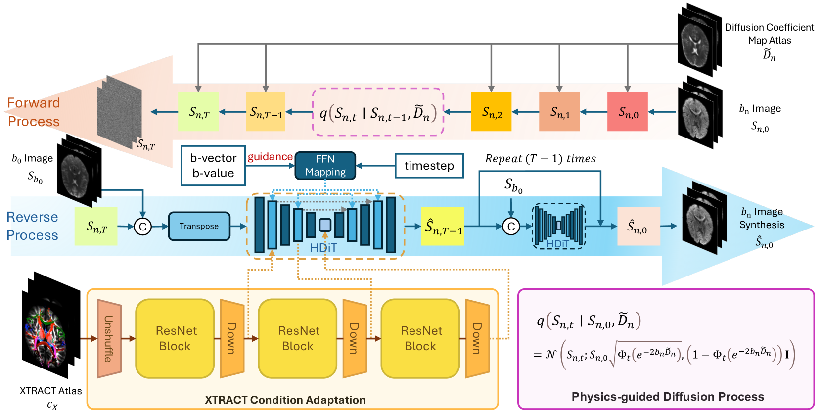
Phy-Diff: Physics-guided Hourglass Diffusion Model for Diffusion MRI Synthesis
Juanhua Zhang, Ruodan Yan, Alessandro Perelli, Xi Chen, Chao Li
0
0
Diffusion MRI (dMRI) is an important neuroimaging technique with high acquisition costs. Deep learning approaches have been used to enhance dMRI and predict diffusion biomarkers through undersampled dMRI. To generate more comprehensive raw dMRI, generative adversarial network based methods are proposed to include b-values and b-vectors as conditions, but they are limited by unstable training and less desirable diversity. The emerging diffusion model (DM) promises to improve generative performance. However, it remains challenging to include essential information in conditioning DM for more relevant generation, i.e., the physical principles of dMRI and white matter tract structures. In this study, we propose a physics-guided diffusion model to generate high-quality dMRI. Our model introduces the physical principles of dMRI in the noise evolution in the diffusion process and introduce a query-based conditional mapping within the difussion model. In addition, to enhance the anatomical fine detials of the generation, we introduce the XTRACT atlas as prior of white matter tracts by adopting an adapter technique. Our experiment results show that our method outperforms other state-of-the-art methods and has the potential to advance dMRI enhancement.
6/6/2024
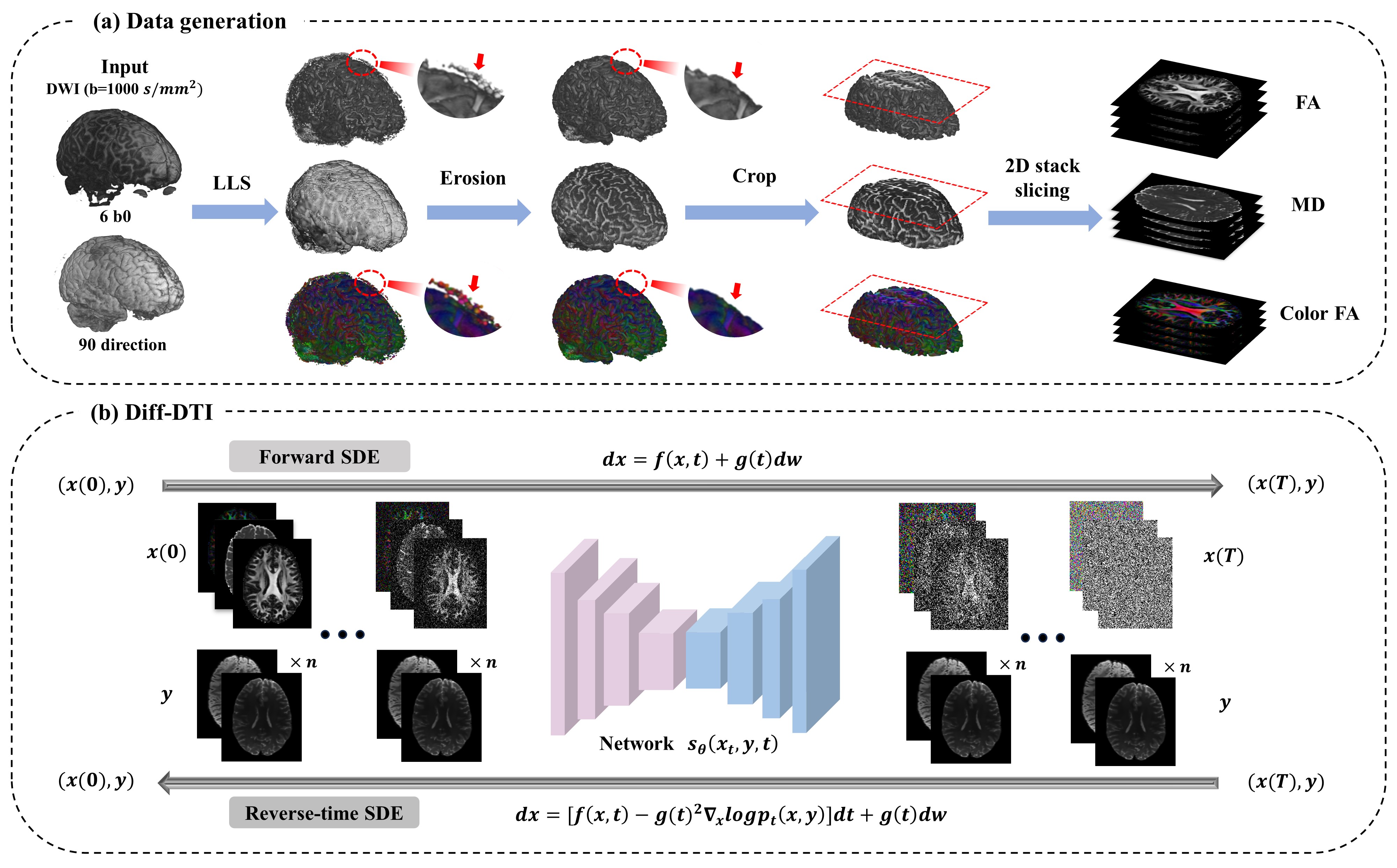
Diff-DTI: Fast Diffusion Tensor Imaging Using A Feature-Enhanced Joint Diffusion Model
Lang Zhang, Jinling He, Dong Liang, Hairong Zheng, Yanjie Zhu
0
0
Magnetic resonance diffusion tensor imaging (DTI) is a critical tool for neural disease diagnosis. However, long scan time greatly hinders the widespread clinical use of DTI. To accelerate image acquisition, a feature-enhanced joint diffusion model (Diff-DTI) is proposed to obtain accurate DTI parameter maps from a limited number of diffusion-weighted images (DWIs). Diff-DTI introduces a joint diffusion model that directly learns the joint probability distribution of DWIs with DTI parametric maps for conditional generation. Additionally, a feature enhancement fusion mechanism (FEFM) is designed and incorporated into the generative process of Diff-DTI to preserve fine structures in the generated DTI maps. A comprehensive evaluation of the performance of Diff-DTI was conducted on the Human Connectome Project dataset. The results demonstrate that Diff-DTI outperforms existing state-of-the-art fast DTI imaging methods in terms of visual quality and quantitative metrics. Furthermore, Diff-DTI has shown the ability to produce high-fidelity DTI maps with only three DWIs, thus overcoming the requirement of a minimum of six DWIs for DTI.
5/28/2024
🤿
Structural-Based Uncertainty in Deep Learning Across Anatomical Scales: Analysis in White Matter Lesion Segmentation
Nataliia Molchanova, Vatsal Raina, Andrey Malinin, Francesco La Rosa, Adrien Depeursinge, Mark Gales, Cristina Granziera, Henning Muller, Mara Graziani, Meritxell Bach Cuadra
0
0
This paper explores uncertainty quantification (UQ) as an indicator of the trustworthiness of automated deep-learning (DL) tools in the context of white matter lesion (WML) segmentation from magnetic resonance imaging (MRI) scans of multiple sclerosis (MS) patients. Our study focuses on two principal aspects of uncertainty in structured output segmentation tasks. Firstly, we postulate that a good uncertainty measure should indicate predictions likely to be incorrect with high uncertainty values. Second, we investigate the merit of quantifying uncertainty at different anatomical scales (voxel, lesion, or patient). We hypothesize that uncertainty at each scale is related to specific types of errors. Our study aims to confirm this relationship by conducting separate analyses for in-domain and out-of-domain settings. Our primary methodological contributions are (i) the development of novel measures for quantifying uncertainty at lesion and patient scales, derived from structural prediction discrepancies, and (ii) the extension of an error retention curve analysis framework to facilitate the evaluation of UQ performance at both lesion and patient scales. The results from a multi-centric MRI dataset of 334 patients demonstrate that our proposed measures more effectively capture model errors at the lesion and patient scales compared to measures that average voxel-scale uncertainty values. We provide the UQ protocols code at https://github.com/Medical-Image-Analysis-Laboratory/MS_WML_uncs.
4/29/2024