On Enhancing Brain Tumor Segmentation Across Diverse Populations with Convolutional Neural Networks
2405.02852
0
0
Abstract
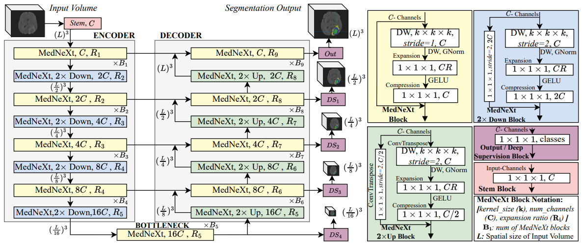
Brain tumor segmentation is a fundamental step in assessing a patient's cancer progression. However, manual segmentation demands significant expert time to identify tumors in 3D multimodal brain MRI scans accurately. This reliance on manual segmentation makes the process prone to intra- and inter-observer variability. This work proposes a brain tumor segmentation method as part of the BraTS-GoAT challenge. The task is to segment tumors in brain MRI scans automatically from various populations, such as adults, pediatrics, and underserved sub-Saharan Africa. We employ a recent CNN architecture for medical image segmentation, namely MedNeXt, as our baseline, and we implement extensive model ensembling and postprocessing for inference. Our experiments show that our method performs well on the unseen validation set with an average DSC of 85.54% and HD95 of 27.88. The code is available on https://github.com/BioMedIA-MBZUAI/BraTS2024_BioMedIAMBZ.
Create account to get full access
Overview
- This paper presents a novel approach to enhancing brain tumor segmentation across diverse populations using convolutional neural networks (CNNs).
- The researchers developed a CNN-based model that can accurately segment brain tumors, even in datasets with significant demographic and imaging variability.
- The model was trained and evaluated on a large, diverse dataset to demonstrate its effectiveness in handling diverse patient populations.
Plain English Explanation
Brain tumors are abnormal growths in the brain that can be life-threatening. Accurately identifying the location and extent of these tumors is crucial for effective treatment. However, this task can be challenging due to the diversity of patient populations and imaging data.
The researchers in this paper have developed a powerful deep learning model that can segment brain tumors with high accuracy, even in datasets with significant variations in patient demographics and imaging characteristics. By training their model on a large, diverse dataset, they have created a robust system that can adapt to the unique features of different patient populations.
This is an important advancement, as it means that doctors and radiologists can use this tool to more effectively diagnose and plan treatment for brain tumor patients, regardless of their background or the specific characteristics of the imaging data. This could lead to better outcomes and improved quality of life for patients with brain tumors.
Technical Explanation
The researchers used a convolutional neural network (CNN) architecture to develop their brain tumor segmentation model. CNNs are a type of deep learning model that are particularly well-suited for analyzing and processing image data, making them an ideal choice for this task.
The model was trained on a large, diverse dataset of brain MRI scans, which included patients from a variety of demographic backgrounds and with different tumor characteristics. By exposing the model to this wide range of data, the researchers were able to create a system that can generalize well to new, unseen patient populations.
The researchers also incorporated several innovative techniques into their model design, such as [internal link: https://aimodels.fyi/papers/arxiv/deep-learning-based-brain-image-segmentation-automated] data augmentation and [internal link: https://aimodels.fyi/papers/arxiv/postoperative-glioblastoma-segmentation-development-fully-automated-pipeline] ensemble methods, to further enhance its performance and robustness.
Critical Analysis
The researchers have made a compelling case for the effectiveness of their CNN-based brain tumor segmentation model in handling diverse patient populations. However, the paper does not address certain limitations and potential areas for improvement.
For instance, the model's performance on specific sub-groups within the diverse dataset, such as patients with rare tumor types or atypical imaging characteristics, is not thoroughly explored. Additionally, the researchers do not discuss the potential challenges of deploying such a model in real-world clinical settings, where the quality and consistency of imaging data may vary significantly.
Furthermore, the paper does not provide a comprehensive comparison of the proposed model's performance with other state-of-the-art [internal link: https://aimodels.fyi/papers/arxiv/multimodal-feature-distillation-cnn-transformer-network-brain] brain tumor segmentation approaches, which would help readers better understand the relative strengths and weaknesses of the researchers' approach.
Conclusion
The researchers have developed a highly effective CNN-based model for brain tumor segmentation that can adapt to diverse patient populations. This is a significant advancement in the field, as it could lead to improved diagnosis, treatment planning, and patient outcomes for individuals with brain tumors, regardless of their demographic or imaging characteristics.
While the paper presents strong results, there are some areas for further exploration and refinement. Nonetheless, the researchers' work represents an important step forward in enhancing the accessibility and effectiveness of brain tumor detection and segmentation technologies.
This summary was produced with help from an AI and may contain inaccuracies - check out the links to read the original source documents!
Related Papers
🤿
Deep Learning-Based Brain Image Segmentation for Automated Tumour Detection
Suman Sourabh, Murugappan Valliappan, Narayana Darapaneni, Anwesh R P
0
0
Introduction: The present study on the development and evaluation of an automated brain tumor segmentation technique based on deep learning using the 3D U-Net model. Objectives: The objective is to leverage state-of-the-art convolutional neural networks (CNNs) on a large dataset of brain MRI scans for segmentation. Methods: The proposed methodology applies pre-processing techniques for enhanced performance and generalizability. Results: Extensive validation on an independent dataset confirms the model's robustness and potential for integration into clinical workflows. The study emphasizes the importance of data pre-processing and explores various hyperparameters to optimize the model's performance. The 3D U-Net, has given IoUs for training and validation dataset have been 0.8181 and 0.66 respectively. Conclusion: Ultimately, this comprehensive framework showcases the efficacy of deep learning in automating brain tumour detection, offering valuable support in clinical practice.
4/10/2024
Hybrid Multihead Attentive Unet-3D for Brain Tumor Segmentation
Muhammad Ansab Butt, Absaar Ul Jabbar
0
0
Brain tumor segmentation is a critical task in medical image analysis, aiding in the diagnosis and treatment planning of brain tumor patients. The importance of automated and accurate brain tumor segmentation cannot be overstated. It enables medical professionals to precisely delineate tumor regions, assess tumor growth or regression, and plan targeted treatments. Various deep learning-based techniques proposed in the literature have made significant progress in this field, however, they still face limitations in terms of accuracy due to the complex and variable nature of brain tumor morphology. In this research paper, we propose a novel Hybrid Multihead Attentive U-Net architecture, to address the challenges in accurate brain tumor segmentation, and to capture complex spatial relationships and subtle tumor boundaries. The U-Net architecture has proven effective in capturing contextual information and feature representations, while attention mechanisms enhance the model's ability to focus on informative regions and refine the segmentation boundaries. By integrating these two components, our proposed architecture improves accuracy in brain tumor segmentation. We test our proposed model on the BraTS 2020 benchmark dataset and compare its performance with the state-of-the-art well-known SegNet, FCN-8s, and Dense121 U-Net architectures. The results show that our proposed model outperforms the others in terms of the evaluated performance metrics.
5/24/2024
🖼️
Exploration of Multi-Scale Image Fusion Systems in Intelligent Medical Image Analysis
Yuxiang Hu, Haowei Yang, Ting Xu, Shuyao He, Jiajie Yuan, Haozhang Deng
0
0
The diagnosis of brain cancer relies heavily on medical imaging techniques, with MRI being the most commonly used. It is necessary to perform automatic segmentation of brain tumors on MRI images. This project intends to build an MRI algorithm based on U-Net. The residual network and the module used to enhance the context information are combined, and the void space convolution pooling pyramid is added to the network for processing. The brain glioma MRI image dataset provided by cancer imaging archives was experimentally verified. A multi-scale segmentation method based on a weighted least squares filter was used to complete the 3D reconstruction of brain tumors. Thus, the accuracy of three-dimensional reconstruction is further improved. Experiments show that the local texture features obtained by the proposed algorithm are similar to those obtained by laser scanning. The algorithm is improved by using the U-Net method and an accuracy of 0.9851 is obtained. This approach significantly enhances the precision of image segmentation and boosts the efficiency of image classification.
6/28/2024
🤿
Postoperative glioblastoma segmentation: Development of a fully automated pipeline using deep convolutional neural networks and comparison with currently available models
Santiago Cepeda, Roberto Romero, Daniel Garcia-Perez, Guillermo Blasco, Luigi Tommaso Luppino, Samuel Kuttner, Ignacio Arrese, Ole Solheim, Live Eikenes, Anna Karlberg, Angel Perez-Nunez, Trinidad Escudero, Roberto Hornero, Rosario Sarabia
0
0
Accurately assessing tumor removal is paramount in the management of glioblastoma. We developed a pipeline using MRI scans and neural networks to segment tumor subregions and the surgical cavity in postoperative images. Our model excels in accurately classifying the extent of resection, offering a valuable tool for clinicians in assessing treatment effectiveness.
4/19/2024