A label-free and data-free training strategy for vasculature segmentation in serial sectioning OCT data
2405.13757
0
0

Abstract
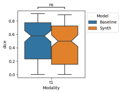
Serial sectioning Optical Coherence Tomography (sOCT) is a high-throughput, label free microscopic imaging technique that is becoming increasingly popular to study post-mortem neurovasculature. Quantitative analysis of the vasculature requires highly accurate segmentation; however, sOCT has low signal-to-noise-ratio and displays a wide range of contrasts and artifacts that depend on acquisition parameters. Furthermore, labeled data is scarce and extremely time consuming to generate. Here, we leverage synthetic datasets of vessels to train a deep learning segmentation model. We construct the vessels with semi-realistic splines that simulate the vascular geometry and compare our model with realistic vascular labels generated by constrained constructive optimization. Both approaches yield similar Dice scores, although with very different false positive and false negative rates. This method addresses the complexity inherent in OCT images and paves the way for more accurate and efficient analysis of neurovascular structures.
Create account to get full access
Overview
- This paper presents a new approach for robust stroke segmentation using synthetic data
- The authors develop a novel technique for automating the segmentation of blood vessels in medical imaging of the heart and brain
- The paper introduces a "snake-shifted window" method for learning to adapt vessel segmentation to different contexts
- The researchers provide guidelines for managing imperfect annotations in cerebrovascular segmentation tasks
- A new dataset is introduced for measuring the performance of blood vessel segmentation algorithms
Plain English Explanation
The paper on synthetic data for robust stroke segmentation describes a new way to accurately identify strokes in medical images. The authors created artificial training data to help the algorithm learn how to find stroke-affected areas, even in complex or low-quality images. This can be useful for quickly diagnosing stroke patients.
The paper on automating vessel segmentation introduces a technique to automatically identify blood vessels in medical scans of the heart and brain. This could help doctors more easily analyze images and detect potential issues like blockages or damage.
The paper on the "snake-shifted window" method describes a way for vessel segmentation algorithms to adapt to different imaging contexts. This flexibility could allow the same tool to be used on a variety of medical scans without needing to be retrained for each new application.
The guidelines paper provides recommendations for working with imperfect or incomplete data when developing cerebrovascular (brain blood vessel) segmentation algorithms. This can help researchers build more robust and reliable tools.
Finally, the new dataset paper introduces a benchmark to evaluate the performance of blood vessel segmentation algorithms. This can help advance the field by allowing for better comparisons between different techniques.
Technical Explanation
The paper on synthetic data for robust stroke segmentation describes a deep learning approach that uses synthetic training data to improve the segmentation of strokes in medical images. The authors generate realistic synthetic scans with simulated stroke lesions and use these to train a convolutional neural network. Experiments show this technique outperforms models trained on real data alone, particularly for challenging cases with low contrast or anatomical variations.
The paper on automating vessel segmentation introduces an automated pipeline for segmenting blood vessels in cardiac and cerebrovascular MRI scans. The method combines multiple deep learning models to detect vessel centerlines, boundaries, and hierarchical relationships. Evaluations on public datasets demonstrate the approach can accurately segment vessels in both the heart and brain.
The paper on the "snake-shifted window" method presents a novel technique for adapting vessel segmentation to different imaging contexts. The key idea is to use a sliding "window" that can learn to deform and shift to match the local vessel structure. This allows a single model to handle a wide variety of vessel morphologies without the need for extensive retraining.
The guidelines paper provides recommendations for cerebrovascular segmentation in the face of imperfect training data annotations. The authors analyze common issues like inconsistent labeling, missing ground truth, and contextual variations. They propose strategies such as uncertainty-aware loss functions and data augmentation to improve model robustness.
Finally, the new dataset paper introduces a benchmark dataset for evaluating blood vessel segmentation algorithms. The dataset includes a variety of cardiac and brain MRI scans with expert-annotated vessel masks. Comprehensive evaluation metrics are provided to assess performance on different vessel types and branching patterns.
Critical Analysis
The paper on synthetic data for robust stroke segmentation presents a promising approach, but the authors acknowledge that the synthetic data may not fully capture the complexity of real-world stroke cases. Further validation on larger clinical datasets would be needed to demonstrate the method's real-world applicability.
The automated vessel segmentation paper makes significant advances, but the authors note that segmentation accuracy can still be limited by image quality, anatomical variations, and pathological changes. Incorporating more advanced techniques like few-shot learning or domain adaptation may help improve robustness.
The "snake-shifted window" paper presents an interesting and flexible segmentation approach, but the authors do not provide extensive comparisons to other adaptive methods. Further benchmarking against state-of-the-art techniques would help contextualize the contributions.
The guidelines paper offers valuable insights, but the proposed strategies have not been thoroughly validated. Applying these recommendations in real-world segmentation projects and reporting on their efficacy would strengthen the practical utility of the guidelines.
The new dataset paper introduces an important resource, but the scope is limited to cardiac and cerebrovascular imaging. Expanding the dataset to include a wider range of vascular anatomy and pathologies would further enhance its usefulness for the research community.
Conclusion
This collection of papers advances the state of the art in medical image segmentation, particularly for blood vessels and stroke detection. The novel techniques, guidelines, and benchmark datasets presented can significantly improve the robustness, adaptability, and evaluation of such algorithms.
These contributions have the potential to positively impact clinical practice by enabling more accurate and efficient analysis of medical scans. Improved stroke segmentation can lead to faster diagnosis and treatment, while automated vessel segmentation can aid in the detection of cardiovascular and neurological diseases.
Overall, this body of work demonstrates the valuable role that machine learning and computer vision can play in enhancing medical imaging capabilities and, ultimately, improving patient outcomes.
This summary was produced with help from an AI and may contain inaccuracies - check out the links to read the original source documents!
Related Papers
Synthetic Data for Robust Stroke Segmentation
Liam Chalcroft, Ioannis Pappas, Cathy J. Price, John Ashburner
0
0
Deep learning-based semantic segmentation in neuroimaging currently requires high-resolution scans and extensive annotated datasets, posing significant barriers to clinical applicability. We present a novel synthetic framework for the task of lesion segmentation, extending the capabilities of the established SynthSeg approach to accommodate large heterogeneous pathologies with lesion-specific augmentation strategies. Our method trains deep learning models, demonstrated here with the UNet architecture, using label maps derived from healthy and stroke datasets, facilitating the segmentation of both healthy tissue and pathological lesions without sequence-specific training data. Evaluated against in-domain and out-of-domain (OOD) datasets, our framework demonstrates robust performance, rivaling current methods within the training domain and significantly outperforming them on OOD data. This contribution holds promise for advancing medical imaging analysis in clinical settings, especially for stroke pathology, by enabling reliable segmentation across varied imaging sequences with reduced dependency on large annotated corpora. Code and weights available at https://github.com/liamchalcroft/SynthStroke.
4/3/2024
🤿
Automating Vessel Segmentation in the Heart and Brain: A Trend to Develop Multi-Modality and Label-Efficient Deep Learning Techniques
Nazik Elsayed, Yousuf Babiker M. Osman, Cheng Li, Jiong Zhang, Shanshan Wang
0
0
Cardio-cerebrovascular diseases are the leading causes of mortality worldwide, whose accurate blood vessel segmentation is significant for both scientific research and clinical usage. However, segmenting cardio-cerebrovascular structures from medical images is very challenging due to the presence of thin or blurred vascular shapes, imbalanced distribution of vessel and non-vessel pixels, and interference from imaging artifacts. These difficulties make manual or semi-manual segmentation methods highly time-consuming, labor-intensive, and prone to errors with interobserver variability, where different experts may produce different segmentations from a variety of modalities. Consequently, there is a growing interest in developing automated algorithms. This paper provides an up-to-date survey of deep learning techniques, for cardio-cerebrovascular segmentation. It analyzes the research landscape, surveys recent approaches, and discusses challenges such as the scarcity of accurately annotated data and variability. This paper also illustrates the urgent needs for developing multi-modality label-efficient deep learning techniques. To the best of our knowledge, this paper is the first comprehensive survey of deep learning approaches that effectively segment vessels in both the heart and brain. It aims to advance automated segmentation techniques for cardio-cerebrovascular diseases, benefiting researchers and healthcare professionals.
4/3/2024
Snake with Shifted Window: Learning to Adapt Vessel Pattern for OCTA Segmentation
Xinrun Chen, Mei Shen, Haojian Ning, Mengzhan Zhang, Chengliang Wang, Shiying Li
0
0
Segmenting specific targets or structures in optical coherence tomography angiography (OCTA) images is fundamental for conducting further pathological studies. The retinal vascular layers are rich and intricate, and such vascular with complex shapes can be captured by the widely-studied OCTA images. In this paper, we thus study how to use OCTA images with projection vascular layers to segment retinal structures. To this end, we propose the SSW-OCTA model, which integrates the advantages of deformable convolutions suited for tubular structures and the swin-transformer for global feature extraction, adapting to the characteristics of OCTA modality images. Our model underwent testing and comparison on the OCTA-500 dataset, achieving state-of-the-art performance. The code is available at: https://github.com/ShellRedia/Snake-SWin-OCTA.
4/30/2024
Guidelines for Cerebrovascular Segmentation: Managing Imperfect Annotations in the context of Semi-Supervised Learning
Pierre Roug'e, Pierre-Henri Conze, Nicolas Passat, Odyss'ee Merveille
0
0
Segmentation in medical imaging is an essential and often preliminary task in the image processing chain, driving numerous efforts towards the design of robust segmentation algorithms. Supervised learning methods achieve excellent performances when fed with a sufficient amount of labeled data. However, such labels are typically highly time-consuming, error-prone and expensive to produce. Alternatively, semi-supervised learning approaches leverage both labeled and unlabeled data, and are very useful when only a small fraction of the dataset is labeled. They are particularly useful for cerebrovascular segmentation, given that labeling a single volume requires several hours for an expert. In addition to the challenge posed by insufficient annotations, there are concerns regarding annotation consistency. The task of annotating the cerebrovascular tree is inherently ambiguous. Due to the discrete nature of images, the borders and extremities of vessels are often unclear. Consequently, annotations heavily rely on the expert subjectivity and on the underlying clinical objective. These discrepancies significantly increase the complexity of the segmentation task for the model and consequently impair the results. Consequently, it becomes imperative to provide clinicians with precise guidelines to improve the annotation process and construct more uniform datasets. In this article, we investigate the data dependency of deep learning methods within the context of imperfect data and semi-supervised learning, for cerebrovascular segmentation. Specifically, this study compares various state-of-the-art semi-supervised methods based on unsupervised regularization and evaluates their performance in diverse quantity and quality data scenarios. Based on these experiments, we provide guidelines for the annotation and training of cerebrovascular segmentation models.
4/3/2024