Multimodal MRI-based Detection of Amyloid Status in Alzheimer's Disease Continuum
2406.13305
0
0

Abstract
Amyloid-$beta$ (A$beta$) plaques in conjunction with hyperphosphorylated tau proteins in the form of neurofibrillary tangles are the two neuropathological hallmarks of Alzheimer's disease (AD). In particular, the accumulation of A$beta$ plaques, as evinced by the A/T/N (amyloid/tau/neurodegeneration) framework, marks the initial stage. Thus, the identification of individuals with A$beta$ positivity could enable early diagnosis and potentially lead to more effective interventions. Deep learning methods relying mainly on amyloid PET images have been employed to this end. However, PET imaging has some disadvantages, including the need of radiotracers and expensive acquisitions. Hence, in this work, we propose a novel multimodal approach that integrates information from structural, functional, and diffusion MRI data to discriminate A$beta$ status in the AD continuum. Our method achieved an accuracy of $0.762pm0.04$. Furthermore, a textit{post-hoc} explainability analysis (guided backpropagation) was performed to retrieve the brain regions that most influenced the model predictions. This analysis identified some key regions that were common across modalities, some of which were well-established AD-discriminative biomarkers and related to A$beta$ deposition, such as the hippocampus, thalamus, precuneus, and cingulate gyrus. Hence, our study demonstrates the potential viability of MRI-based characterization of A$beta$ status, paving the way for further research in this domain.
Create account to get full access
Overview
- This paper presents a multimodal MRI-based approach for detecting amyloid status in Alzheimer's disease (AD) continuum.
- The researchers used machine learning models to analyze MRI data from patients at different stages of Alzheimer's disease, including those with normal cognition, mild cognitive impairment, and Alzheimer's dementia.
- The goal was to develop a non-invasive and accessible way to detect amyloid pathology, which is a hallmark of Alzheimer's disease, using only MRI scans.
Plain English Explanation
Alzheimer's disease is a progressive brain disorder that leads to memory loss and cognitive decline. One of the key features of Alzheimer's is the buildup of a protein called amyloid in the brain. Detecting amyloid can help doctors diagnose Alzheimer's and monitor its progression, but the current gold standard method, a PET scan, is expensive and not widely available.
In this study, the researchers explored whether they could use a more common and affordable imaging technique, MRI, to detect amyloid in the brain. They trained machine learning models to analyze MRI data from people at different stages of Alzheimer's disease, including those with normal cognition, mild cognitive impairment, and Alzheimer's dementia. The goal was to find patterns in the MRI scans that could reliably indicate the presence of amyloid, without needing a PET scan.
The researchers found that their multimodal MRI-based approach was able to accurately detect amyloid status in the study participants. This suggests that MRI scans could potentially be used as a non-invasive and more accessible way to screen for Alzheimer's disease in the future, which could help with earlier diagnosis and treatment.
Technical Explanation
The researchers used a multimodal MRI-based approach to detect amyloid status in the Alzheimer's disease continuum. They collected structural, functional, and diffusion MRI data from 174 participants, including those with normal cognition, mild cognitive impairment, and Alzheimer's dementia. The participants also underwent PET scans to assess their amyloid status, which served as the ground truth.
The researchers then used machine learning models, specifically support vector machines and random forests, to analyze the MRI data and predict the participants' amyloid status. They explored various feature engineering techniques, including selecting the most informative MRI-derived metrics, and compared the performance of the models with different input modalities (structural, functional, and diffusion MRI).
The results showed that the multimodal MRI-based approach achieved high accuracy, sensitivity, and specificity in detecting amyloid status, outperforming models that used only a single MRI modality. The researchers also found that incorporating both structural and functional MRI features led to the best performance, highlighting the complementary information provided by these different MRI modalities.
Critical Analysis
The researchers acknowledge several limitations of their study, including the relatively small sample size and the cross-sectional nature of the data, which does not allow for longitudinal analysis of Alzheimer's disease progression. Additionally, the study was conducted in a single center, and the generalizability of the findings to other populations and settings needs to be further explored.
Furthermore, while the multimodal MRI-based approach shows promise in detecting amyloid status, it is not yet clear how this information can be directly used for clinical decision-making or patient management. Additional research is needed to understand the relationship between MRI-based amyloid detection and other clinical and cognitive measures, as well as the potential impact on patient outcomes.
Conclusion
This study demonstrates the potential of using multimodal MRI data, combined with machine learning techniques, to non-invasively detect amyloid status in the Alzheimer's disease continuum. If validated in larger, longitudinal studies, this approach could provide a more accessible and affordable alternative to PET scans for the early detection and monitoring of Alzheimer's disease. However, further research is needed to fully understand the clinical utility and implications of this approach.
This summary was produced with help from an AI and may contain inaccuracies - check out the links to read the original source documents!
Related Papers

An interpretable generative multimodal neuroimaging-genomics framework for decoding Alzheimer's disease
Giorgio Dolci (Department of Engineering for Innovation Medicine, University of Verona, Verona, Italy, Tri-Institutional Center for Translational Research in Neuroimaging and Data Science), Federica Cruciani (Department of Engineering for Innovation Medicine, University of Verona, Verona, Italy), Md Abdur Rahaman (Tri-Institutional Center for Translational Research in Neuroimaging and Data Science), Anees Abrol (Tri-Institutional Center for Translational Research in Neuroimaging and Data Science), Jiayu Chen (Tri-Institutional Center for Translational Research in Neuroimaging and Data Science), Zening Fu (Tri-Institutional Center for Translational Research in Neuroimaging and Data Science), Ilaria Boscolo Galazzo (Department of Engineering for Innovation Medicine, University of Verona, Verona, Italy), Gloria Menegaz (Department of Engineering for Innovation Medicine, University of Verona, Verona, Italy), Vince D. Calhoun (Tri-Institutional Center for Translational Research in Neuroimaging and Data Science)
0
0
Alzheimer's disease (AD) is the most prevalent form of dementia with a progressive decline in cognitive abilities. The AD continuum encompasses a prodormal stage known as Mild Cognitive Impairment (MCI), where patients may either progress to AD or remain stable. In this study, we leveraged structural and functional MRI to investigate the disease-induced grey matter and functional network connectivity changes. Moreover, considering AD's strong genetic component, we introduce SNPs as a third channel. Given such diverse inputs, missing one or more modalities is a typical concern of multimodal methods. We hence propose a novel deep learning-based classification framework where generative module employing Cycle GANs was adopted to impute missing data within the latent space. Additionally, we adopted an Explainable AI method, Integrated Gradients, to extract input features relevance, enhancing our understanding of the learned representations. Two critical tasks were addressed: AD detection and MCI conversion prediction. Experimental results showed that our model was able to reach the SOA in the classification of CN/AD reaching an average test accuracy of $0.926pm0.02$. For the MCI task, we achieved an average prediction accuracy of $0.711pm0.01$ using the pre-trained model for CN/AD. The interpretability analysis revealed significant grey matter modulations in cortical and subcortical brain areas well known for their association with AD. Moreover, impairments in sensory-motor and visual resting state network connectivity along the disease continuum, as well as mutations in SNPs defining biological processes linked to amyloid-beta and cholesterol formation clearance and regulation, were identified as contributors to the achieved performance. Overall, our integrative deep learning approach shows promise for AD detection and MCI prediction, while shading light on important biological insights.
6/21/2024
🤯
Three-Dimensional Amyloid-Beta PET Synthesis from Structural MRI with Conditional Generative Adversarial Networks
Fernando Vega, Abdoljalil Addeh, M. Ethan MacDonald
0
0
Motivation: Alzheimer's Disease hallmarks include amyloid-beta deposits and brain atrophy, detectable via PET and MRI scans, respectively. PET is expensive, invasive and exposes patients to ionizing radiation. MRI is cheaper, non-invasive, and free from ionizing radiation but limited to measuring brain atrophy. Goal: To develop an 3D image translation model that synthesizes amyloid-beta PET images from T1-weighted MRI, exploiting the known relationship between amyloid-beta and brain atrophy. Approach: The model was trained on 616 PET/MRI pairs and validated with 264 pairs. Results: The model synthesized amyloid-beta PET images from T1-weighted MRI with high-degree of similarity showing high SSIM and PSNR metrics (SSIM>0.95&PSNR=28). Impact: Our model proves the feasibility of synthesizing amyloid-beta PET images from structural MRI ones, significantly enhancing accessibility for large-cohort studies and early dementia detection, while also reducing cost, invasiveness, and radiation exposure.
5/6/2024
🧠
Analysing heterogeneity in Alzheimer Disease using multimodal normative modelling on ATN biomarkers
Sayantan Kumara, Thomas Earnest, Braden Yang, Deydeep Kothapalli, Tammie L. S. Benzinger, Brian A. Gordon, Philip Payne, Aristeidis Sotiras
0
0
Alzheimer Disease (AD) is a multi-faceted disorder, with each modality providing unique and complementary info about AD. In this study, we used a deep-learning based multimodal normative model to assess the heterogeneity in regional brain patterns for ATN (amyloid-tau-neurodegeneration) biomarkers. We selected discovery (n = 665) and replication (n = 430) cohorts with simultaneous availability of ATN biomarkers: Florbetapir amyloid, Flortaucipir tau and T1-weighted MRI (magnetic resonance imaging) imaging. A multimodal variational autoencoder (conditioned on age and sex) was used as a normative model to learn the multimodal regional brain patterns of a cognitively unimpaired (CU) control group. The trained model was applied on individuals on the ADS (AD Spectrum) to estimate their deviations (Z-scores) from the normative distribution, resulting in a Z-score regional deviation map per ADS individual per modality. ADS individuals with moderate or severe dementia showed higher proportion of regional outliers for each modality as well as more dissimilarity in modality-specific regional outlier patterns compared to ADS individuals with early or mild dementia. DSI was associated with the progressive stages of dementia, (ii) showed significant associations with neuropsychological composite scores and (iii) related to the longitudinal risk of CDR progression. Findings were reproducible in both discovery and replication cohorts. Our is the first study to examine the heterogeneity in AD through the lens of multiple neuroimaging modalities (ATN), based on distinct or overlapping patterns of regional outlier deviations. Regional MRI and tau outliers were more heterogenous than regional amyloid outliers. DSI has the potential to be an individual patient metric of neurodegeneration that can help in clinical decision making and monitoring patient response for anti-amyloid treatments.
4/10/2024
Trustworthy Enhanced Multi-view Multi-modal Alzheimer's Disease Prediction with Brain-wide Imaging Transcriptomics Data
Shan Cong, Zhoujie Fan, Hongwei Liu, Yinghan Zhang, Xin Wang, Haoran Luo, Xiaohui Yao
0
0
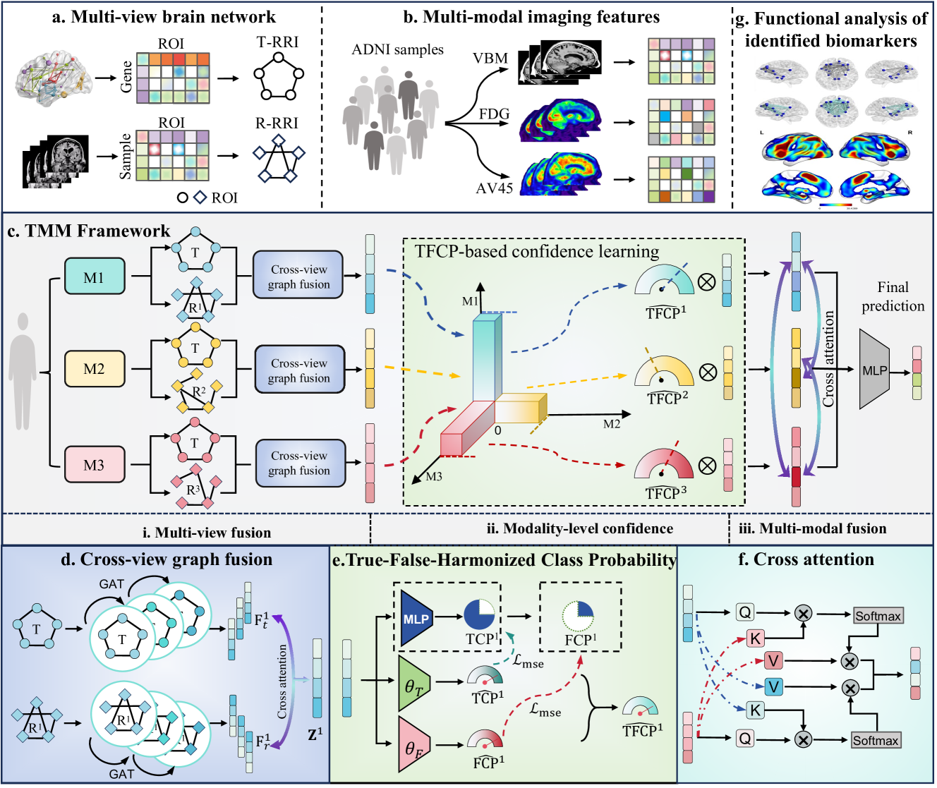
Brain transcriptomics provides insights into the molecular mechanisms by which the brain coordinates its functions and processes. However, existing multimodal methods for predicting Alzheimer's disease (AD) primarily rely on imaging and sometimes genetic data, often neglecting the transcriptomic basis of brain. Furthermore, while striving to integrate complementary information between modalities, most studies overlook the informativeness disparities between modalities. Here, we propose TMM, a trusted multiview multimodal graph attention framework for AD diagnosis, using extensive brain-wide transcriptomics and imaging data. First, we construct view-specific brain regional co-function networks (RRIs) from transcriptomics and multimodal radiomics data to incorporate interaction information from both biomolecular and imaging perspectives. Next, we apply graph attention (GAT) processing to each RRI network to produce graph embeddings and employ cross-modal attention to fuse transcriptomics-derived embedding with each imagingderived embedding. Finally, a novel true-false-harmonized class probability (TFCP) strategy is designed to assess and adaptively adjust the prediction confidence of each modality for AD diagnosis. We evaluate TMM using the AHBA database with brain-wide transcriptomics data and the ADNI database with three imaging modalities (AV45-PET, FDG-PET, and VBM-MRI). The results demonstrate the superiority of our method in identifying AD, EMCI, and LMCI compared to state-of-the-arts. Code and data are available at https://github.com/Yaolab-fantastic/TMM.
6/24/2024