SegmentAnything helps microscopy images based automatic and quantitative organoid detection and analysis
2309.04190
0
0
🔎
Abstract
Organoids are self-organized 3D cell clusters that closely mimic the architecture and function of in vivo tissues and organs. Quantification of organoid morphology helps in studying organ development, drug discovery, and toxicity assessment. Recent microscopy techniques provide a potent tool to acquire organoid morphology features, but manual image analysis remains a labor and time-intensive process. Thus, this paper proposes a comprehensive pipeline for microscopy analysis that leverages the SegmentAnything to precisely demarcate individual organoids. Additionally, we introduce a set of morphological properties, including perimeter, area, radius, non-smoothness, and non-circularity, allowing researchers to analyze the organoid structures quantitatively and automatically. To validate the effectiveness of our approach, we conducted tests on bright-field images of human induced pluripotent stem cells (iPSCs) derived neural-epithelial (NE) organoids. The results obtained from our automatic pipeline closely align with manual organoid detection and measurement, showcasing the capability of our proposed method in accelerating organoids morphology analysis.
Create account to get full access
Overview
- Organoids are 3D cell clusters that closely mimic real tissues and organs
- Quantifying organoid morphology is crucial for studying organ development, drug discovery, and toxicity assessment
- Existing microscopy techniques can acquire organoid morphology, but manual image analysis is labor-intensive
- This paper proposes an automated pipeline for organoid morphology analysis using the SegmentAnything model
Plain English Explanation
Organoids are miniature, self-organized 3D structures that closely resemble the actual architecture and function of organs and tissues inside the human body. Analyzing the shape and size of organoids, known as their "morphology," can provide valuable insights into how organs develop, how drugs might affect them, and whether certain substances could be toxic. Recent advancements in microscopy have made it possible to capture detailed images of organoids, but manually analyzing all these images is a time-consuming and labor-intensive process.
To address this challenge, the researchers in this paper have developed an automated pipeline that can quickly and accurately measure various morphological properties of organoids, such as their perimeter, area, radius, smoothness, and circularity. The key innovation is the use of a powerful AI model called SegmentAnything, which can precisely outline the boundaries of individual organoids in the microscopy images.
The researchers tested this automated pipeline on images of neural-epithelial organoids derived from human induced pluripotent stem cells (iPSCs). They found that the results from their automated system closely matched the measurements obtained through manual organoid detection and analysis, demonstrating the effectiveness of their approach in accelerating organoid morphology research.
Technical Explanation
This paper presents a comprehensive pipeline for automated microscopy analysis of organoid morphology. The key components of the pipeline are:
- Leveraging the SegmentAnything model to precisely demarcate the boundaries of individual organoids in microscopy images.
- Extracting a set of morphological properties for each organoid, including perimeter, area, radius, non-smoothness, and non-circularity.
- Automating the quantitative analysis of organoid structures based on these morphological features.
To validate the effectiveness of their approach, the researchers conducted tests on bright-field images of human induced pluripotent stem cells (iPSCs) derived neural-epithelial (NE) organoids. They found that the results from their automated pipeline closely aligned with manual organoid detection and measurement, demonstrating the pipeline's capability in accelerating organoid morphology analysis.
Critical Analysis
The paper presents a robust and innovative approach to automating the quantification of organoid morphology, which is a crucial aspect of organ development, drug discovery, and toxicity assessment research. The use of the SegmentAnything model to precisely demarcate individual organoids is a particularly noteworthy contribution, as it addresses the limitations of manual image analysis.
However, the paper does not explicitly discuss potential limitations or areas for further research. For instance, it would be valuable to understand how the pipeline's performance might be affected by variations in organoid size, shape, or imaging conditions. Additionally, the researchers could consider expanding the set of morphological features analyzed, or exploring the integration of their pipeline with other analytical techniques, such as synthetic data generation for data augmentation.
Conclusion
This paper presents a comprehensive pipeline for automated microscopy analysis of organoid morphology, a crucial aspect of organ development, drug discovery, and toxicity assessment research. The key innovation is the use of the SegmentAnything model to precisely demarcate individual organoids, enabling the extraction and quantitative analysis of various morphological properties. The researchers demonstrated the effectiveness of their approach through tests on human iPSCs-derived neural-epithelial organoids, showing close alignment with manual detection and measurement. This automated pipeline has the potential to significantly accelerate organoid morphology research, allowing for more efficient and scalable studies in the field.
This summary was produced with help from an AI and may contain inaccuracies - check out the links to read the original source documents!
Related Papers
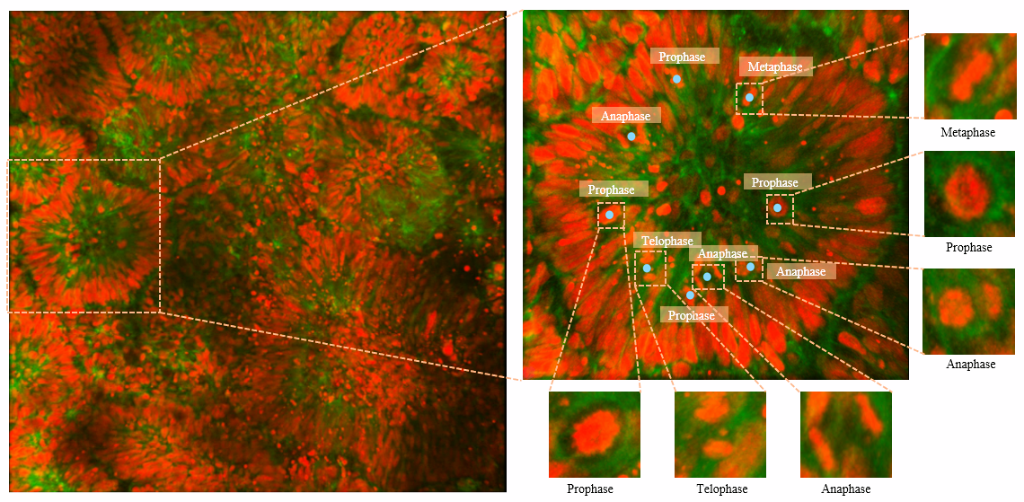
BOrg: A Brain Organoid-Based Mitosis Dataset for Automatic Analysis of Brain Diseases
Muhammad Awais, Mehaboobathunnisa Sahul Hameed, Bidisha Bhattacharya, Orly Reiner, Rao Muhammad Anwer
0
0
Recent advances have enabled the study of human brain development using brain organoids derived from stem cells. Quantifying cellular processes like mitosis in these organoids offers insights into neurodevelopmental disorders, but the manual analysis is time-consuming, and existing datasets lack specific details for brain organoid studies. We introduce BOrg, a dataset designed to study mitotic events in the embryonic development of the brain using confocal microscopy images of brain organoids. BOrg utilizes an efficient annotation pipeline with sparse point annotations and techniques that minimize expert effort, overcoming limitations of standard deep learning approaches on sparse data. We adapt and benchmark state-of-the-art object detection and cell counting models on BOrg for detecting and analyzing mitotic cells across prophase, metaphase, anaphase, and telophase stages. Our results demonstrate these adapted models significantly improve mitosis analysis efficiency and accuracy for brain organoid research compared to existing methods. BOrg facilitates the development of automated tools to quantify statistics like mitosis rates, aiding mechanistic studies of neurodevelopmental processes and disorders. Data and code are available at https://github.com/awaisrauf/borg.
7/1/2024
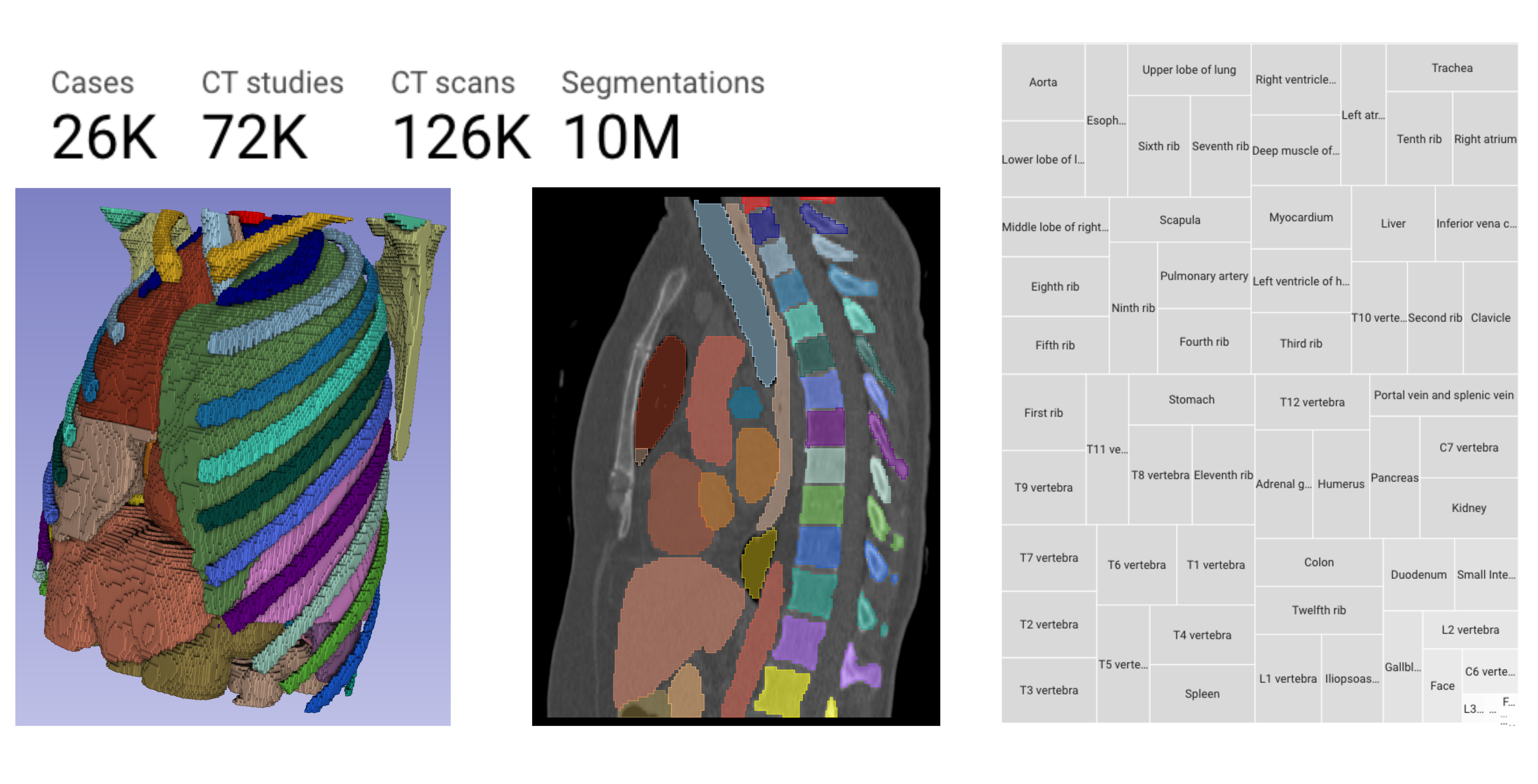
Rule-based outlier detection of AI-generated anatomy segmentations
Deepa Krishnaswamy, Vamsi Krishna Thiriveedhi, Cosmin Ciausu, David Clunie, Steve Pieper, Ron Kikinis, Andrey Fedorov
0
0
There is a dire need for medical imaging datasets with accompanying annotations to perform downstream patient analysis. However, it is difficult to manually generate these annotations, due to the time-consuming nature, and the variability in clinical conventions. Artificial intelligence has been adopted in the field as a potential method to annotate these large datasets, however, a lack of expert annotations or ground truth can inhibit the adoption of these annotations. We recently made a dataset publicly available including annotations and extracted features of up to 104 organs for the National Lung Screening Trial using the TotalSegmentator method. However, the released dataset does not include expert-derived annotations or an assessment of the accuracy of the segmentations, limiting its usefulness. We propose the development of heuristics to assess the quality of the segmentations, providing methods to measure the consistency of the annotations and a comparison of results to the literature. We make our code and related materials publicly available at https://github.com/ImagingDataCommons/CloudSegmentatorResults and interactive tools at https://huggingface.co/spaces/ImagingDataCommons/CloudSegmentatorResults.
6/21/2024

Real Time Multi Organ Classification on Computed Tomography Images
Halid Ziya Yerebakan, Yoshihisa Shinagawa, Gerardo Hermosillo Valadez
0
0
Organ segmentation is a fundamental task in medical imaging, and it is useful for many clinical automation pipelines. Typically, the process involves segmenting the entire volume, which can be unnecessary when the points of interest are limited. In those cases, a classifier could be used instead of segmentation. However, there is an inherent trade-off between the context size and the speed of classifiers. To address this issue, we propose a new method that employs a data selection strategy with sparse sampling across a wide field of view without image resampling. This sparse sampling strategy makes it possible to classify voxels into multiple organs in real time without using accelerators. Although our method is an independent classifier, it can generate full segmentation by querying grid locations at any resolution. We have compared our method with existing segmentation techniques, demonstrating its potential for superior runtime in practical applications in medical imaging.
4/30/2024
🖼️
Image segmentation of treated and untreated tumor spheroids by Fully Convolutional Networks
Matthias Streller, Sov{n}a Michl'ikov'a, Willy Ciecior, Katharina Lonnecke, Leoni A. Kunz-Schughart, Steffen Lange, Anja Voss-Bohme
0
0
Multicellular tumor spheroids (MCTS) are advanced cell culture systems for assessing the impact of combinatorial radio(chemo)therapy. They exhibit therapeutically relevant in-vivo-like characteristics from 3D cell-cell and cell-matrix interactions to radial pathophysiological gradients related to proliferative activity and nutrient/oxygen supply, altering cellular radioresponse. State-of-the-art assays quantify long-term curative endpoints based on collected brightfield image time series from large treated spheroid populations per irradiation dose and treatment arm. Here, spheroid control probabilities are documented analogous to in-vivo tumor control probabilities based on Kaplan-Meier curves. This analyses require laborious spheroid segmentation of up to 100.000 images per treatment arm to extract relevant structural information from the images, e.g., diameter, area, volume and circularity. While several image analysis algorithms are available for spheroid segmentation, they all focus on compact MCTS with clearly distinguishable outer rim throughout growth. However, treated MCTS may partly be detached and destroyed and are usually obscured by dead cell debris. We successfully train two Fully Convolutional Networks, UNet and HRNet, and optimize their hyperparameters to develop an automatic segmentation for both untreated and treated MCTS. We systematically validate the automatic segmentation on larger, independent data sets of spheroids derived from two human head-and-neck cancer cell lines. We find an excellent overlap between manual and automatic segmentation for most images, quantified by Jaccard indices at around 90%. For images with smaller overlap of the segmentations, we demonstrate that this error is comparable to the variations across segmentations from different biological experts, suggesting that these images represent biologically unclear or ambiguous cases.
5/3/2024