Accelerated MR Cholangiopancreatography with Deep Learning-based Reconstruction

0
🤿
Sign in to get full access
Overview
- This study aims to accelerate MR cholangiopancreatography (MRCP) acquisitions using deep learning-based (DL) reconstruction at different magnetic field strengths.
- The researchers trained a variational network (VN) using retrospectively six-fold undersampled data obtained at 3T, and then evaluated their method against standard techniques like parallel imaging (PI) and compressed sensing (CS).
- They also tested their method in a prospective accelerated scenario and evaluated its adaptability to MRCP at 0.55T.
Plain English Explanation
The researchers in this study wanted to find a way to speed up the process of acquiring MRCP images, which are used to examine the bile and pancreatic ducts. MRCP scans can take a long time, so the researchers tried using a deep learning algorithm to reconstruct the images from less data, which would allow for faster scan times.
They trained their deep learning model, called a variational network (VN), using MRCP data that had been intentionally undersampled, meaning not all the data was collected. They then compared the quality of the images reconstructed by their VN method to the standard techniques of parallel imaging (PI) and compressed sensing (CS).
The researchers also tested their VN method in a real-world scenario where the MRCP scans were accelerated, and they looked at how well it worked at a lower magnetic field strength of 0.55T, in addition to the standard 3T.
Overall, the VN method was able to significantly reduce the MRCP scan time, from around 10 minutes down to just 4 or 3 minutes, while still maintaining high image quality. This could be very useful for improving the efficiency and accessibility of MRCP scans in clinical settings.
Technical Explanation
The researchers trained a variational network (VN) using retrospectively six-fold undersampled MRCP data obtained at 3T. They then evaluated this VN method against standard techniques like parallel imaging (PI) and compressed sensing (CS), using metrics like peak signal-to-noise ratio (PSNR) and structural similarity (SSIM).
Additionally, the researchers included a self-supervised DL reconstruction (SSDU) method in their evaluations, as fully-sampled MRCP data is impractical to acquire.
The VN method demonstrated a remarkable reduction in average acquisition time, from 599/542 seconds down to 255/180 seconds for MRCP at 3T/0.55T. In both retrospective and prospective undersampling scenarios, the VN outperformed PI, CS, and SSDU in terms of PSNR and SSIM, while also preserving important image quality features like sharpness and duct visibility.
Importantly, the VN method was also able to produce high-quality reconstructions at the lower 0.55T field strength, resulting in the highest PSNR and SSIM among the tested techniques.
Critical Analysis
The researchers acknowledge that their study was limited to healthy volunteers and did not evaluate the VN method's performance on MRCP images from patients with known pathologies. Further research would be needed to assess the generalizability of the VN approach to clinical MRCP scenarios.
Additionally, the paper does not provide detailed information about the training and optimization of the VN model, which could be important for others seeking to replicate or build upon this work. More transparency around the model architecture, hyperparameters, and training process would be beneficial.
While the VN method demonstrated impressive results in terms of acquisition time reduction and image quality preservation, it would be useful to understand the computational and memory requirements of the model, as well as any potential limitations in terms of deployment in a clinical setting.
Conclusion
This study presents a deep learning-based approach, the variational network (VN), that can significantly accelerate MR cholangiopancreatography (MRCP) acquisitions while maintaining high image quality. The VN method was able to reduce MRCP scan times by over 50% at both 3T and 0.55T field strengths, outperforming standard techniques like parallel imaging and compressed sensing.
The ability to acquire high-quality MRCP images in a shorter time frame could have important implications for improving the efficiency and accessibility of this diagnostic imaging modality in clinical practice. Further research is needed to evaluate the VN method's performance on patient data and to better understand its practical deployment considerations.
This summary was produced with help from an AI and may contain inaccuracies - check out the links to read the original source documents!
Related Papers
🤿
0
Accelerated MR Cholangiopancreatography with Deep Learning-based Reconstruction
Jinho Kim, Marcel Dominik Nickel, Florian Knoll
This study accelerates MR cholangiopancreatography (MRCP) acquisitions using deep learning-based (DL) reconstruction at 3T and 0.55T. Thirty healthy volunteers underwent conventional two-fold MRCP scans at field strengths of 3T or 0.55T. We trained a variational network (VN) using retrospectively six-fold undersampled data obtained at 3T. We then evaluated our method against standard techniques such as parallel imaging (PI) and compressed sensing (CS), focusing on peak signal-to-noise ratio (PSNR) and structural similarity (SSIM) as metrics. Furthermore, considering acquiring fully-sampled MRCP is impractical, we added a self-supervised DL reconstruction (SSDU) to the evaluating group. We also tested our method in a prospective accelerated scenario to reflect real-world clinical applications and evaluated its adaptability to MRCP at 0.55T. Our method demonstrated a remarkable reduction of average acquisition time from 599/542 to 255/180 seconds for MRCP at 3T/0.55T. In both retrospective and prospective undersampling scenarios, the PSNR and SSIM of VN were higher than those of PI, CS, and SSDU. At the same time, VN preserved the image quality of undersampled data, i.e., sharpness and the visibility of hepatobiliary ducts. In addition, VN also produced high quality reconstructions at 0.55T resulting in the highest PSNR and SSIM. In summary, VN trained for highly accelerated MRCP allows to reduce the acquisition time by a factor of 2.4/3.0 at 3T/0.55T while maintaining the image quality of the conventional acquisition.
Read more5/8/2024
🌐
0
Paired Conditional Generative Adversarial Network for Highly Accelerated Liver 4D MRI
Di Xu, Xin Miao, Hengjie Liu, Jessica E. Scholey, Wensha Yang, Mary Feng, Michael Ohliger, Hui Lin, Yi Lao, Yang Yang, Ke Sheng
Purpose: 4D MRI with high spatiotemporal resolution is desired for image-guided liver radiotherapy. Acquiring densely sampling k-space data is time-consuming. Accelerated acquisition with sparse samples is desirable but often causes degraded image quality or long reconstruction time. We propose the Reconstruct Paired Conditional Generative Adversarial Network (Re-Con-GAN) to shorten the 4D MRI reconstruction time while maintaining the reconstruction quality. Methods: Patients who underwent free-breathing liver 4D MRI were included in the study. Fully- and retrospectively under-sampled data at 3, 6 and 10 times (3x, 6x and 10x) were first reconstructed using the nuFFT algorithm. Re-Con-GAN then trained input and output in pairs. Three types of networks, ResNet9, UNet and reconstruction swin transformer, were explored as generators. PatchGAN was selected as the discriminator. Re-Con-GAN processed the data (3D+t) as temporal slices (2D+t). A total of 48 patients with 12332 temporal slices were split into training (37 patients with 10721 slices) and test (11 patients with 1611 slices). Results: Re-Con-GAN consistently achieved comparable/better PSNR, SSIM, and RMSE scores compared to CS/UNet models. The inference time of Re-Con-GAN, UNet and CS are 0.15s, 0.16s, and 120s. The GTV detection task showed that Re-Con-GAN and CS, compared to UNet, better improved the dice score (3x Re-Con-GAN 80.98%; 3x CS 80.74%; 3x UNet 79.88%) of unprocessed under-sampled images (3x 69.61%). Conclusion: A generative network with adversarial training is proposed with promising and efficient reconstruction results demonstrated on an in-house dataset. The rapid and qualitative reconstruction of 4D liver MR has the potential to facilitate online adaptive MR-guided radiotherapy for liver cancer.
Read more5/22/2024
0
TC-KANRecon: High-Quality and Accelerated MRI Reconstruction via Adaptive KAN Mechanisms and Intelligent Feature Scaling
Ruiquan Ge, Xiao Yu, Yifei Chen, Fan Jia, Shenghao Zhu, Guanyu Zhou, Yiyu Huang, Chenyan Zhang, Dong Zeng, Changmiao Wang, Qiegen Liu, Shanzhou Niu
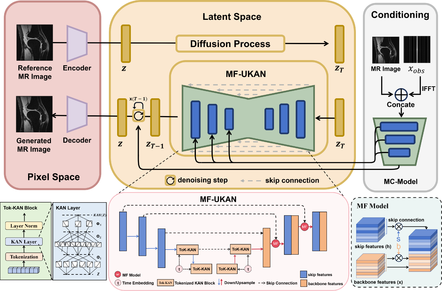
Magnetic Resonance Imaging (MRI) has become essential in clinical diagnosis due to its high resolution and multiple contrast mechanisms. However, the relatively long acquisition time limits its broader application. To address this issue, this study presents an innovative conditional guided diffusion model, named as TC-KANRecon, which incorporates the Multi-Free U-KAN (MF-UKAN) module and a dynamic clipping strategy. TC-KANRecon model aims to accelerate the MRI reconstruction process through deep learning methods while maintaining the quality of the reconstructed images. The MF-UKAN module can effectively balance the tradeoff between image denoising and structure preservation. Specifically, it presents the multi-head attention mechanisms and scalar modulation factors, which significantly enhances the model's robustness and structure preservation capabilities in complex noise environments. Moreover, the dynamic clipping strategy in TC-KANRecon adjusts the cropping interval according to the sampling steps, thereby mitigating image detail loss typically caused by traditional cropping methods and enriching the visual features of the images. Furthermore, the MC-Model module incorporates full-sampling k-space information, realizing efficient fusion of conditional information, enhancing the model's ability to process complex data, and improving the realism and detail richness of reconstructed images. Experimental results demonstrate that the proposed method outperforms other MRI reconstruction methods in both qualitative and quantitative evaluations. Notably, TC-KANRecon method exhibits excellent reconstruction results when processing high-noise, low-sampling-rate MRI data. Our source code is available at https://github.com/lcbkmm/TC-KANRecon.
Read more8/13/2024

0
Adaptive Self-Supervised Consistency-Guided Diffusion Model for Accelerated MRI Reconstruction
Mojtaba Safari, Zach Eidex, Shaoyan Pan, Richard L. J. Qiu, Xiaofeng Yang
Purpose: To propose a self-supervised deep learning-based compressed sensing MRI (DL-based CS-MRI) method named Adaptive Self-Supervised Consistency Guided Diffusion Model (ASSCGD) to accelerate data acquisition without requiring fully sampled datasets. Materials and Methods: We used the fastMRI multi-coil brain axial T2-weighted (T2-w) dataset from 1,376 cases and single-coil brain quantitative magnetization prepared 2 rapid acquisition gradient echoes (MP2RAGE) T1 maps from 318 cases to train and test our model. Robustness against domain shift was evaluated using two out-of-distribution (OOD) datasets: multi-coil brain axial postcontrast T1 -weighted (T1c) dataset from 50 cases and axial T1-weighted (T1-w) dataset from 50 patients. Data were retrospectively subsampled at acceleration rates R in {2x, 4x, 8x}. ASSCGD partitions a random sampling pattern into two disjoint sets, ensuring data consistency during training. We compared our method with ReconFormer Transformer and SS-MRI, assessing performance using normalized mean squared error (NMSE), peak signal-to-noise ratio (PSNR), and structural similarity index (SSIM). Statistical tests included one-way analysis of variance (ANOVA) and multi-comparison Tukey's Honesty Significant Difference (HSD) tests. Results: ASSCGD preserved fine structures and brain abnormalities visually better than comparative methods at R = 8x for both multi-coil and single-coil datasets. It achieved the lowest NMSE at R in {4x, 8x}, and the highest PSNR and SSIM values at all acceleration rates for the multi-coil dataset. Similar trends were observed for the single-coil dataset, though SSIM values were comparable to ReconFormer at R in {2x, 8x}. These results were further confirmed by the voxel-wise correlation scatter plots. OOD results showed significant (p << 10^-5 ) improvements in undersampled image quality after reconstruction.
Read more6/26/2024