Advancing H&E-to-IHC Stain Translation in Breast Cancer: A Multi-Magnification and Attention-Based Approach

0

Sign in to get full access
Overview
- The paper presents a novel approach for translating Hematoxylin and Eosin (H&E) stained images to Immunohistochemistry (IHC) stained images in the context of breast cancer analysis.
- The method uses a multi-magnification and attention-based neural network architecture to capture both global and local features.
- The proposed technique outperforms existing H&E-to-IHC translation methods in terms of accuracy and visual quality.
Plain English Explanation
In cancer research, doctors often use two types of staining techniques to analyze tissue samples under a microscope: Hematoxylin and Eosin (H&E) staining and Immunohistochemistry (IHC) staining. H&E staining provides a general overview of the tissue structure, while IHC staining can identify specific proteins or markers that are important for understanding the cancer.
Advancing H&E-to-IHC Stain Translation in Breast Cancer: A Multi-Magnification and Attention-Based Approach presents a new way to translate H&E images into IHC images. This is useful because IHC images can be hard to obtain, but if you can reliably convert H&E images into IHC images, you can get the benefits of IHC analysis without having to do the extra staining.
The key idea is to use a neural network that looks at the H&E image at different zoom levels (magnifications) and pays attention to the important details. This allows the network to capture both the overall structure of the tissue (global features) and the finer details (local features) that are important for translating to the IHC stain.
The authors show that their multi-magnification and attention-based approach outperforms existing methods for translating H&E to IHC, producing more accurate and visually appealing results. This could be a valuable tool for cancer researchers and pathologists who want to leverage the benefits of IHC analysis without the additional work required to actually perform the IHC staining.
Technical Explanation
The paper proposes a novel multi-magnification and attention-based neural network architecture for translating Hematoxylin and Eosin (H&E) stained images to Immunohistochemistry (IHC) stained images in the context of breast cancer analysis.
The core of the approach is a deep learning model that takes an H&E image as input and generates the corresponding IHC image as output. The model uses a multi-scale encoder-decoder architecture, where the encoder extracts features at multiple magnification levels, and the decoder combines these features to generate the final IHC image.
Importantly, the model also incorporates an attention mechanism, which allows the network to focus on the most relevant features at each step of the translation process. This helps the model capture both global and local information from the H&E image, which is crucial for accurately translating to the IHC stain.
The authors evaluate their method on a dataset of paired H&E and IHC breast cancer images, and demonstrate that it outperforms existing H&E-to-IHC translation techniques in terms of both quantitative metrics and visual quality. They also provide ablation studies to analyze the contributions of the multi-magnification and attention components.
Critical Analysis
The paper presents a compelling approach for translating H&E images to IHC images in breast cancer analysis. The use of a multi-magnification and attention-based neural network architecture is a clever way to capture both global and local features, which is crucial for this task.
One potential limitation is the size and diversity of the dataset used for evaluation. The authors mention that their dataset consists of breast cancer images, which may limit the generalizability of the method to other cancer types or tissues. It would be interesting to see how the approach performs on a more diverse set of samples.
Additionally, the paper does not provide much discussion on the clinical implications or potential use cases of this technology. While the technical advances are impressive, it would be helpful to understand how this tool could be integrated into the workflow of cancer researchers and pathologists, and what benefits it might provide in real-world scenarios.
Further research could also explore the interpretability of the model's predictions, as understanding the reasoning behind the H&E-to-IHC translations could be valuable for gaining insights into the underlying biological processes.
Conclusion
Advancing H&E-to-IHC Stain Translation in Breast Cancer: A Multi-Magnification and Attention-Based Approach presents a novel deep learning-based method for translating H&E stained images to IHC stained images in the context of breast cancer analysis. The key innovation is the use of a multi-magnification and attention-based architecture, which allows the model to capture both global and local features and produce high-quality IHC translations.
This work has the potential to significantly impact cancer research and pathology, as it could enable the benefits of IHC analysis without the additional effort required to perform the actual IHC staining. Further research and validation on larger and more diverse datasets, as well as exploration of the clinical applications, could help unlock the full potential of this technology.
This summary was produced with help from an AI and may contain inaccuracies - check out the links to read the original source documents!
Related Papers
0
Advancing H&E-to-IHC Stain Translation in Breast Cancer: A Multi-Magnification and Attention-Based Approach
Linhao Qu, Chengsheng Zhang, Guihui Li, Haiyong Zheng, Chen Peng, Wei He
Breast cancer presents a significant healthcare challenge globally, demanding precise diagnostics and effective treatment strategies, where histopathological examination of Hematoxylin and Eosin (H&E) stained tissue sections plays a central role. Despite its importance, evaluating specific biomarkers like Human Epidermal Growth Factor Receptor 2 (HER2) for personalized treatment remains constrained by the resource-intensive nature of Immunohistochemistry (IHC). Recent strides in deep learning, particularly in image-to-image translation, offer promise in synthesizing IHC-HER2 slides from H&E stained slides. However, existing methodologies encounter challenges, including managing multiple magnifications in pathology images and insufficient focus on crucial information during translation. To address these issues, we propose a novel model integrating attention mechanisms and multi-magnification information processing. Our model employs a multi-magnification processing strategy to extract and utilize information from various magnifications within pathology images, facilitating robust image translation. Additionally, an attention module within the generative network prioritizes critical information for image distribution translation while minimizing less pertinent details. Rigorous testing on a publicly available breast cancer dataset demonstrates superior performance compared to existing methods, establishing our model as a state-of-the-art solution in advancing pathology image translation from H&E to IHC staining.
Read more8/6/2024
0
DeReStainer: H&E to IHC Pathological Image Translation via Decoupled Staining Channels
Linda Wei, Shengyi Hua, Shaoting Zhang, Xiaofan Zhang
Breast cancer is a highly fatal disease among cancers in women, and early detection is crucial for treatment. HER2 status, a valuable diagnostic marker based on Immunohistochemistry (IHC) staining, is instrumental in determining breast cancer status. The high cost of IHC staining and the ubiquity of Hematoxylin and Eosin (H&E) staining make the conversion from H&E to IHC staining essential. In this article, we propose a destain-restain framework for converting H&E staining to IHC staining, leveraging the characteristic that H&E staining and IHC staining of the same tissue sections share the Hematoxylin channel. We further design loss functions specifically for Hematoxylin and Diaminobenzidin (DAB) channels to generate IHC images exploiting insights from separated staining channels. Beyond the benchmark metrics on BCI contest, we have developed semantic information metrics for the HER2 level. The experimental results demonstrated that our method outperforms previous open-sourced methods in terms of image intrinsic property and semantic information.
Read more9/4/2024
0
IHC Matters: Incorporating IHC analysis to H&E Whole Slide Image Analysis for Improved Cancer Grading via Two-stage Multimodal Bilinear Pooling Fusion
Jun Wang, Yu Mao, Yufei Cui, Nan Guan, Chun Jason Xue
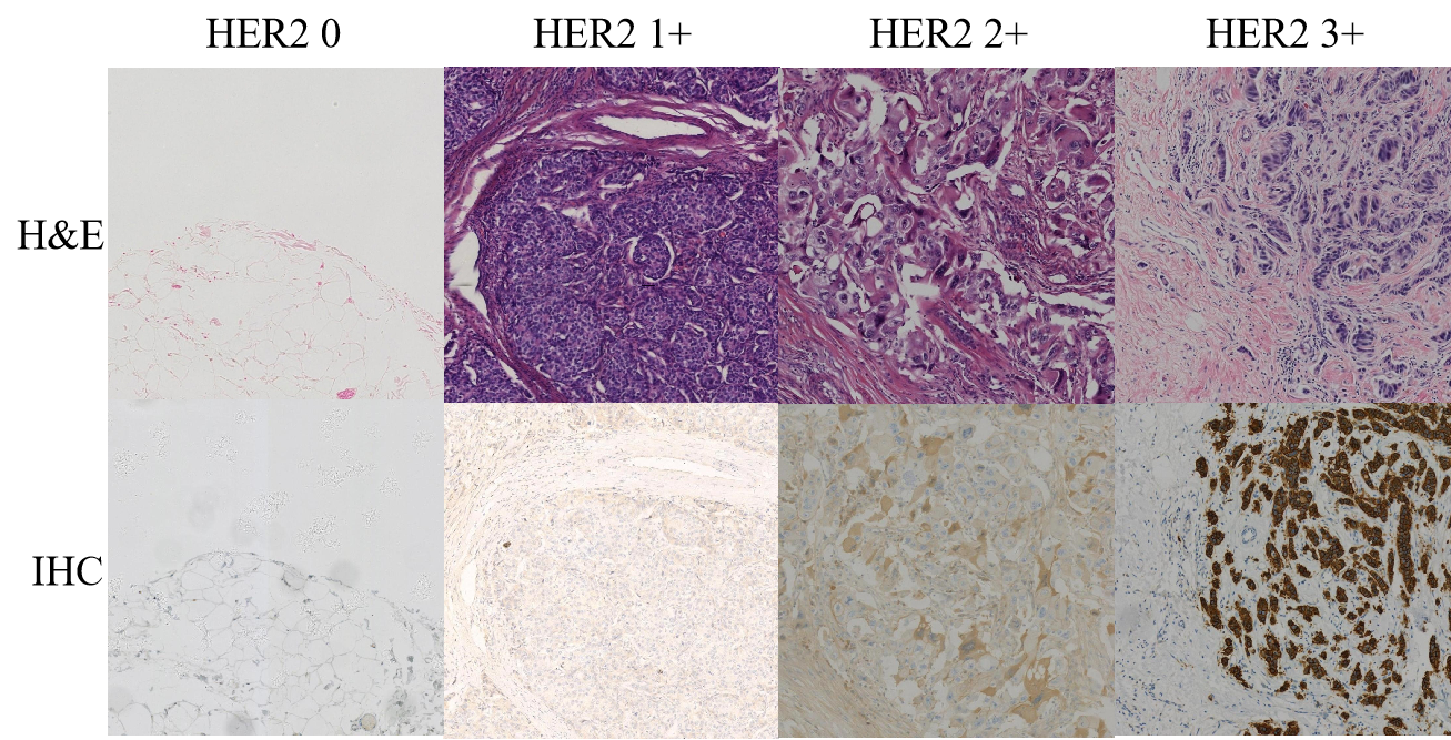
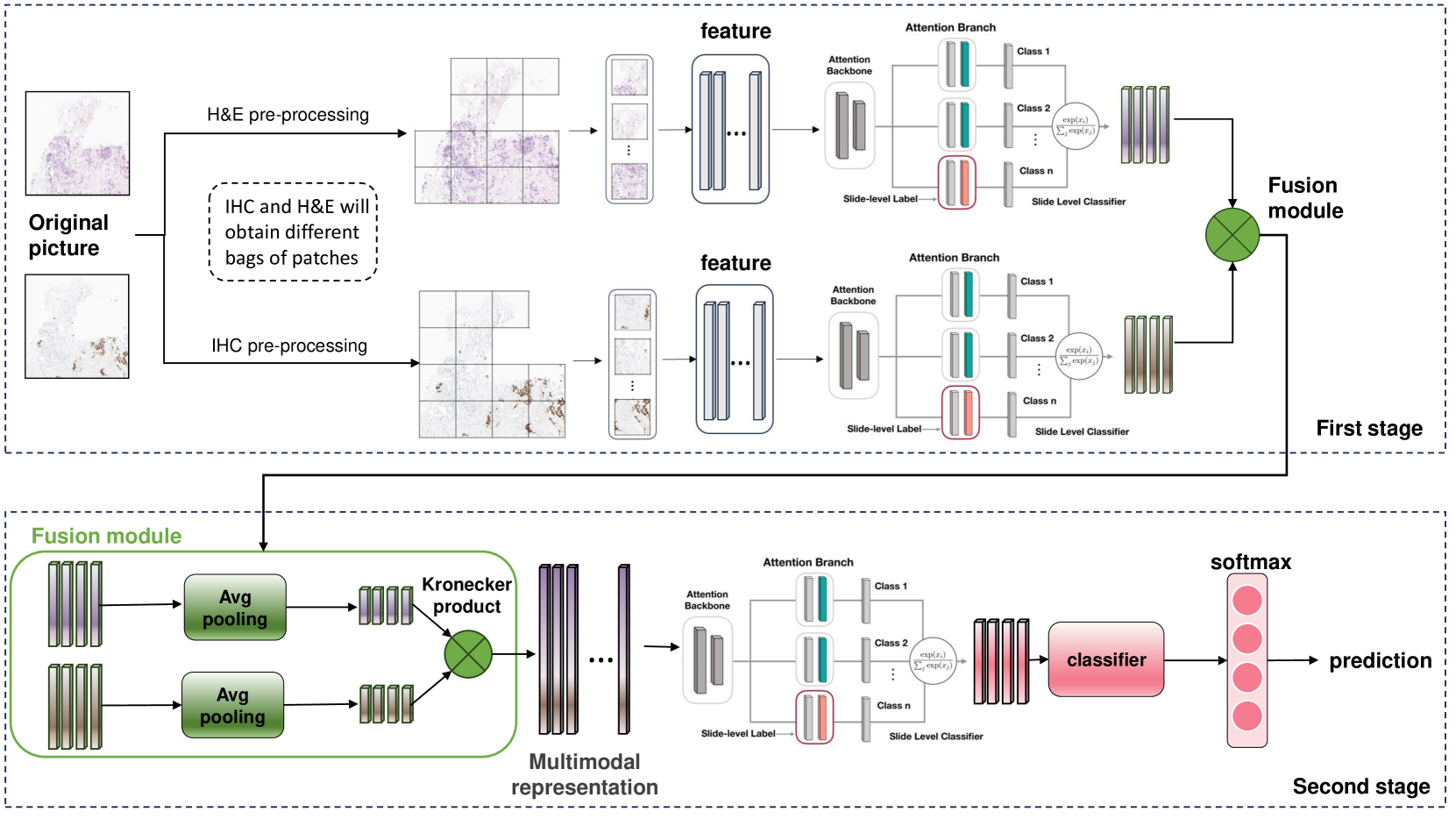
Immunohistochemistry (IHC) plays a crucial role in pathology as it detects the over-expression of protein in tissue samples. However, there are still fewer machine learning model studies on IHC's impact on accurate cancer grading. We discovered that IHC and H&E possess distinct advantages and disadvantages while possessing certain complementary qualities. Building on this observation, we developed a two-stage multi-modal bilinear model with a feature pooling module. This model aims to maximize the potential of both IHC and HE's feature representation, resulting in improved performance compared to their individual use. Our experiments demonstrate that incorporating IHC data into machine learning models, alongside H&E stained images, leads to superior predictive results for cancer grading. The proposed framework achieves an impressive ACC higher of 0.953 on the public dataset BCI.
Read more5/15/2024

0
HER2 and FISH Status Prediction in Breast Biopsy H&E-Stained Images Using Deep Learning
Ardhendu Sekhar, Vrinda Goel, Garima Jain, Abhijeet Patil, Ravi Kant Gupta, Amit Sethi
The current standard for detecting human epidermal growth factor receptor 2 (HER2) status in breast cancer patients relies on HER2 amplification, identified through fluorescence in situ hybridization (FISH) or immunohistochemistry (IHC). However, hematoxylin and eosin (H&E) tumor stains are more widely available, and accurately predicting HER2 status using H&E could reduce costs and expedite treatment selection. Deep Learning algorithms for H&E have shown effectiveness in predicting various cancer features and clinical outcomes, including moderate success in HER2 status prediction. In this work, we employed a customized weak supervision classification technique combined with MoCo-v2 contrastive learning to predict HER2 status. We trained our pipeline on 182 publicly available H&E Whole Slide Images (WSIs) from The Cancer Genome Atlas (TCGA), for which annotations by the pathology team at Yale School of Medicine are publicly available. Our pipeline achieved an Area Under the Curve (AUC) of 0.85 across four different test folds. Additionally, we tested our model on 44 H&E slides from the TCGA-BRCA dataset, which had an HER2 score of 2+ and included corresponding HER2 status and FISH test results. These cases are considered equivocal for IHC, requiring an expensive FISH test on their IHC slides for disambiguation. Our pipeline demonstrated an AUC of 0.81 on these challenging H&E slides. Reducing the need for FISH test can have significant implications in cancer treatment equity for underserved populations.
Read more8/29/2024