AWGUNET: Attention-Aided Wavelet Guided U-Net for Nuclei Segmentation in Histopathology Images

0
Sign in to get full access
Overview
- This paper introduces AWGUNet, a deep learning model for nuclei segmentation in histopathology images.
- The model combines a U-Net architecture with attention mechanisms and wavelet-based feature extraction to improve segmentation performance.
- Experiments on multiple histopathology datasets demonstrate the effectiveness of the proposed approach compared to previous methods.
Plain English Explanation
In medical imaging, being able to accurately identify and segment individual cell nuclei in histology images is an important task. This allows doctors and researchers to better understand the structure and composition of biological tissues, which can provide valuable insights for disease diagnosis and treatment.
AWGUNet is a new deep learning model designed to tackle the challenge of nuclei segmentation. It builds upon the popular U-Net architecture, which has shown strong performance for various medical image segmentation tasks.
The key innovations in AWGUNet are the addition of attention mechanisms and wavelet-based feature extraction. Attention helps the model focus on the most relevant visual features for identifying nuclei, while the wavelet approach allows it to capture information at multiple scales, from coarse to fine details.
By combining these techniques, the researchers were able to develop a model that outperformed previous state-of-the-art methods on several histopathology datasets. This suggests AWGUNet could be a valuable tool for automating and improving the accuracy of nuclei segmentation in digital pathology workflows.
Nuclei instance segmentation in cryosectioned H&E-stained histological images and deep learning-based brain image segmentation are related tasks that also leverage deep learning for medical image analysis.
Technical Explanation
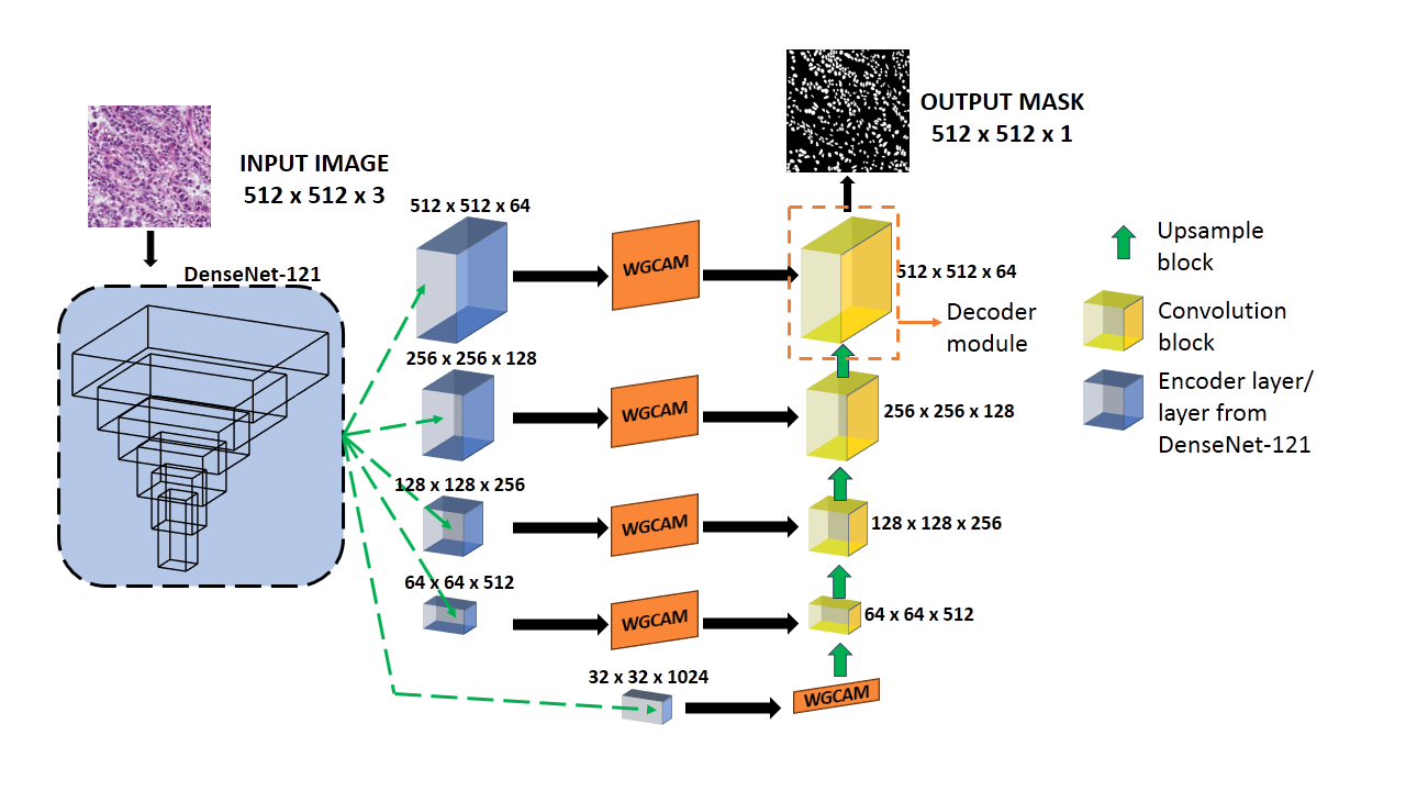
The AWGUNet model consists of an encoder-decoder U-Net architecture with several key components:
-
Wavelet Guided Feature Extraction: The input image is first passed through a wavelet decomposition module, which extracts features at multiple scales. This allows the model to capture both coarse-level and fine-grained visual cues relevant for nuclei segmentation.
-
Attention Mechanism: The encoder features are then passed through an attention module, which learns to selectively emphasize the most informative features for the segmentation task. This helps the model focus on the most relevant parts of the image.
-
Decoder with Skip Connections: The encoded features are passed to the decoder, which progressively upsamples the features to produce the final segmentation map. Skip connections from the encoder help preserve fine spatial details.
The researchers trained and evaluated AWGUNet on several publicly available histopathology datasets, including the TNBC and Pannuke datasets. They compared the performance of AWGUNet to other state-of-the-art nuclei segmentation models and showed that their approach achieved superior results in terms of segmentation accuracy metrics.
Critical Analysis
The paper provides a thorough evaluation of the AWGUNet model and demonstrates its effectiveness for nuclei segmentation in histopathology images. However, a few potential limitations and areas for future work are worth noting:
-
Dataset Bias: The model was trained and evaluated on a limited number of publicly available datasets, which may not capture the full diversity of histological samples encountered in real-world clinical settings. Additional validation on more diverse datasets would help assess the model's broader applicability.
-
Computational Complexity: The attention mechanism and wavelet decomposition components add some computational overhead compared to a standard U-Net. The trade-off between model complexity and inference speed may be an important consideration for deployments in resource-constrained environments.
-
Interpretability: As with many deep learning models, the internal workings of AWGUNet are not easily interpretable. Developing methods to better explain the model's decision-making process could improve trust and facilitate its adoption in clinical settings.
-
Generalization to other Tasks: While the paper focuses on nuclei segmentation, the core ideas of wavelet-guided feature extraction and attention-based processing could potentially be applied to other medical image analysis tasks, such as tumor segmentation or tissue classification. Exploring these extensions could broaden the impact of the proposed approach.
Conclusion
The AWGUNet model presented in this paper represents a promising advance in the field of histopathology image analysis. By combining the strengths of U-Net, attention mechanisms, and wavelet-based feature extraction, the authors have developed a highly effective nuclei segmentation model that outperforms previous state-of-the-art approaches.
While the model shows strong performance on the evaluated datasets, further research is needed to address potential limitations and explore the broader applicability of the proposed techniques. As the field of computational pathology continues to evolve, innovations like AWGUNet will play an increasingly important role in automating and enhancing the analysis of digital pathology images, ultimately supporting more accurate disease diagnosis and better-informed clinical decision-making.
This summary was produced with help from an AI and may contain inaccuracies - check out the links to read the original source documents!
Related Papers
0
AWGUNET: Attention-Aided Wavelet Guided U-Net for Nuclei Segmentation in Histopathology Images
Ayush Roy, Payel Pramanik, Dmitrii Kaplun, Sergei Antonov, Ram Sarkar
Accurate nuclei segmentation in histopathological images is crucial for cancer diagnosis. Automating this process offers valuable support to clinical experts, as manual annotation is time-consuming and prone to human errors. However, automating nuclei segmentation presents challenges due to uncertain cell boundaries, intricate staining, and diverse structures. In this paper, we present a segmentation approach that combines the U-Net architecture with a DenseNet-121 backbone, harnessing the strengths of both to capture comprehensive contextual and spatial information. Our model introduces the Wavelet-guided channel attention module to enhance cell boundary delineation, along with a learnable weighted global attention module for channel-specific attention. The decoder module, composed of an upsample block and convolution block, further refines segmentation in handling staining patterns. The experimental results conducted on two publicly accessible histopathology datasets, namely Monuseg and TNBC, underscore the superiority of our proposed model, demonstrating its potential to advance histopathological image analysis and cancer diagnosis. The code is made available at: https://github.com/AyushRoy2001/AWGUNET.
Read more6/13/2024

0
GRU-Net for breast histopathology image segmentation
Ayush Roy, Payel Pramanik, Sohom Ghosal, Daria Valenkova, Dmitrii Kaplun, Ram Sarkar
Breast cancer is a major global health concern. Pathologists face challenges in analyzing complex features from pathological images, which is a time-consuming and labor-intensive task. Therefore, efficient computer-based diagnostic tools are needed for early detection and treatment planning. This paper presents a modified version of MultiResU-Net for histopathology image segmentation, which is selected as the backbone for its ability to analyze and segment complex features at multiple scales and ensure effective feature flow via skip connections. The modified version also utilizes the Gaussian distribution-based Attention Module (GdAM) to incorporate histopathology-relevant text information in a Gaussian distribution. The sampled features from the Gaussian text feature-guided distribution highlight specific spatial regions based on prior knowledge. Finally, using the Controlled Dense Residual Block (CDRB) on skip connections of MultiResU-Net, the information is transferred from the encoder layers to the decoder layers in a controlled manner using a scaling parameter derived from the extracted spatial features. We validate our approach on two diverse breast cancer histopathology image datasets: TNBC and MonuSeg, demonstrating superior segmentation performance compared to state-of-the-art methods. The code for our proposed model is available on https://github.com/AyushRoy2001/GRU-Net.
Read more8/2/2024
0
Hybrid Multihead Attentive Unet-3D for Brain Tumor Segmentation
Muhammad Ansab Butt, Absaar Ul Jabbar
Brain tumor segmentation is a critical task in medical image analysis, aiding in the diagnosis and treatment planning of brain tumor patients. The importance of automated and accurate brain tumor segmentation cannot be overstated. It enables medical professionals to precisely delineate tumor regions, assess tumor growth or regression, and plan targeted treatments. Various deep learning-based techniques proposed in the literature have made significant progress in this field, however, they still face limitations in terms of accuracy due to the complex and variable nature of brain tumor morphology. In this research paper, we propose a novel Hybrid Multihead Attentive U-Net architecture, to address the challenges in accurate brain tumor segmentation, and to capture complex spatial relationships and subtle tumor boundaries. The U-Net architecture has proven effective in capturing contextual information and feature representations, while attention mechanisms enhance the model's ability to focus on informative regions and refine the segmentation boundaries. By integrating these two components, our proposed architecture improves accuracy in brain tumor segmentation. We test our proposed model on the BraTS 2020 benchmark dataset and compare its performance with the state-of-the-art well-known SegNet, FCN-8s, and Dense121 U-Net architectures. The results show that our proposed model outperforms the others in terms of the evaluated performance metrics.
Read more5/24/2024
🧠
0
Channel Boosted CNN-Transformer-based Multi-Level and Multi-Scale Nuclei Segmentation
Zunaira Rauf, Abdul Rehman Khan, Asifullah Khan
Accurate nuclei segmentation is an essential foundation for various applications in computational pathology, including cancer diagnosis and treatment planning. Even slight variations in nuclei representations can significantly impact these downstream tasks. However, achieving accurate segmentation remains challenging due to factors like clustered nuclei, high intra-class variability in size and shape, resemblance to other cells, and color or contrast variations between nuclei and background. Despite the extensive utilization of Convolutional Neural Networks (CNNs) in medical image segmentation, they may have trouble capturing long-range dependencies crucial for accurate nuclei delineation. Transformers address this limitation but might miss essential low-level features. To overcome these limitations, we utilized CNN-Transformer-based techniques for nuclei segmentation in H&E stained histology images. In this work, we proposed two CNN-Transformer architectures, Nuclei Hybrid Vision Transformer (NucleiHVT) and Channel Boosted Nuclei Hybrid Vision Transformer (CB-NucleiHVT), that leverage the strengths of both CNNs and Transformers to effectively learn nuclei boundaries in multi-organ histology images. The first architecture, NucleiHVT is inspired by the UNet architecture and incorporates the dual attention mechanism to capture both multi-level and multi-scale context effectively. The CB-NucleiHVT network, on the other hand, utilizes the concept of channel boosting to learn diverse feature spaces, enhancing the model's ability to distinguish subtle variations in nuclei characteristics. Detailed evaluation of two medical image segmentation datasets shows that the proposed architectures outperform existing CNN-based, Transformer-based, and hybrid methods. The proposed networks demonstrated effective results both in terms of quantitative metrics, and qualitative visual assessment.
Read more7/30/2024