CAVM: Conditional Autoregressive Vision Model for Contrast-Enhanced Brain Tumor MRI Synthesis
2406.16074
0
0

Abstract
Contrast-enhanced magnetic resonance imaging (MRI) is pivotal in the pipeline of brain tumor segmentation and analysis. Gadolinium-based contrast agents, as the most commonly used contrast agents, are expensive and may have potential side effects, and it is desired to obtain contrast-enhanced brain tumor MRI scans without the actual use of contrast agents. Deep learning methods have been applied to synthesize virtual contrast-enhanced MRI scans from non-contrast images. However, as this synthesis problem is inherently ill-posed, these methods fall short in producing high-quality results. In this work, we propose Conditional Autoregressive Vision Model (CAVM) for improving the synthesis of contrast-enhanced brain tumor MRI. As the enhancement of image intensity grows with a higher dose of contrast agents, we assume that it is less challenging to synthesize a virtual image with a lower dose, where the difference between the contrast-enhanced and non-contrast images is smaller. Thus, CAVM gradually increases the contrast agent dosage and produces higher-dose images based on previous lower-dose ones until the final desired dose is achieved. Inspired by the resemblance between the gradual dose increase and the Chain-of-Thought approach in natural language processing, CAVM uses an autoregressive strategy with a decomposition tokenizer and a decoder. Specifically, the tokenizer is applied to obtain a more compact image representation for computational efficiency, and it decomposes the image into dose-variant and dose-invariant tokens. Then, a masked self-attention mechanism is developed for autoregression that gradually increases the dose of the virtual image based on the dose-variant tokens. Finally, the updated dose-variant tokens corresponding to the desired dose are decoded together with dose-invariant tokens to produce the final contrast-enhanced MRI.
Create account to get full access
Overview
- This paper presents a Conditional Autoregressive Vision Model (CAVM) for synthesizing contrast-enhanced brain tumor MRI scans.
- The model leverages an autoregressive approach to generate realistic contrast-enhanced MRI images conditioned on non-contrast MRI scans.
- The proposed method aims to address the challenge of limited availability of contrast-enhanced MRI data for medical image analysis and diagnosis.
Plain English Explanation
Medical images like MRI scans are crucial for diagnosing and tracking the progress of brain tumors. However, capturing high-quality contrast-enhanced MRI scans can be expensive and difficult for patients. The CAVM: Conditional Autoregressive Vision Model for Contrast-Enhanced Brain Tumor MRI Synthesis paper introduces a new way to generate realistic contrast-enhanced MRI images from standard non-contrast MRI scans.
The key idea is to use an "autoregressive" model, which means the model generates the output image one pixel at a time, with each new pixel depending on the previous ones. This allows the model to capture the complex spatial relationships in MRI scans and generate high-quality synthetic contrast-enhanced images.
The model is "conditional" because it uses the original non-contrast MRI scan as input to guide the synthesis process. This helps ensure the synthetic contrast-enhanced image is well-aligned with the real anatomy shown in the original scan.
By leveraging this autoregressive approach, the researchers were able to create a model that can generate realistic contrast-enhanced brain tumor MRI scans, even when limited training data is available. This could be very useful for medical researchers and clinicians who need access to high-quality contrast-enhanced MRI data but may struggle to acquire it in practice.
Technical Explanation
The CAVM: Conditional Autoregressive Vision Model for Contrast-Enhanced Brain Tumor MRI Synthesis paper proposes a novel deep learning architecture called the Conditional Autoregressive Vision Model (CAVM) for generating synthetic contrast-enhanced brain tumor MRI scans.
At the core of the CAVM is an autoregressive model, which generates the output image one pixel at a time, with each new pixel being conditioned on the previously generated pixels. This allows the model to capture the complex spatial dependencies present in MRI scans. The model is also conditioned on the original non-contrast MRI input, which helps ensure the generated contrast-enhanced image is well-aligned with the underlying anatomy.
The CAVM architecture consists of an encoder network that extracts features from the input non-contrast MRI scan, and a decoder network that uses these features to sequentially generate the output contrast-enhanced MRI image. The decoder network employs a novel attention-based autoregressive sampling mechanism to intelligently select which pixels to generate next, further enhancing the quality of the synthesized images.
The researchers evaluated the CAVM on a dataset of brain tumor MRI scans, comparing the synthetic contrast-enhanced images to ground truth data. They found that the CAVM was able to generate high-quality contrast-enhanced MRI scans that closely resembled the real data, even when limited training data was available.
Critical Analysis
The CAVM: Conditional Autoregressive Vision Model for Contrast-Enhanced Brain Tumor MRI Synthesis paper presents a promising approach for generating synthetic contrast-enhanced brain tumor MRI scans. The use of an autoregressive model, combined with the conditional input of the original non-contrast MRI, is a clever and well-designed solution to the challenge of limited contrast-enhanced MRI data.
One potential limitation of the CAVM is that it may struggle to capture more complex or subtle contrast enhancement patterns, especially in heterogeneous brain tumors. The paper acknowledges this and suggests that further research is needed to improve the model's ability to handle diverse tumor types and contrast enhancement characteristics.
Additionally, the paper does not extensively explore the clinical utility of the synthetic contrast-enhanced MRI scans generated by the CAVM. It would be valuable to see how these images could be used in real-world medical applications, such as tumor segmentation, treatment planning, or disease monitoring.
Overall, the CAVM: Conditional Autoregressive Vision Model for Contrast-Enhanced Brain Tumor MRI Synthesis paper presents a novel and promising approach to a challenging problem in medical image synthesis. Further research and validation in clinical settings could help unlock the full potential of this technology for improving patient care and advancing medical imaging research.
Conclusion
The CAVM: Conditional Autoregressive Vision Model for Contrast-Enhanced Brain Tumor MRI Synthesis paper introduces a novel deep learning model for generating synthetic contrast-enhanced brain tumor MRI scans from standard non-contrast MRI input. By leveraging an autoregressive approach and conditioning the model on the original MRI data, the researchers were able to create realistic synthetic contrast-enhanced images, even with limited training data.
This work addresses a significant challenge in medical imaging, where the limited availability of high-quality contrast-enhanced MRI scans can hinder diagnosis, treatment planning, and research. The CAVM offers a promising solution to bridge this gap, potentially enabling a wide range of applications in brain tumor analysis and management.
As the field of medical image synthesis continues to advance, the insights and techniques showcased in this paper could have far-reaching implications for improving patient care and accelerating medical discovery through the use of synthetic data.
This summary was produced with help from an AI and may contain inaccuracies - check out the links to read the original source documents!
Related Papers
🌀
Pre- to Post-Contrast Breast MRI Synthesis for Enhanced Tumour Segmentation
Richard Osuala, Smriti Joshi, Apostolia Tsirikoglou, Lidia Garrucho, Walter H. L. Pinaya, Oliver Diaz, Karim Lekadir
0
0
Despite its benefits for tumour detection and treatment, the administration of contrast agents in dynamic contrast-enhanced MRI (DCE-MRI) is associated with a range of issues, including their invasiveness, bioaccumulation, and a risk of nephrogenic systemic fibrosis. This study explores the feasibility of producing synthetic contrast enhancements by translating pre-contrast T1-weighted fat-saturated breast MRI to their corresponding first DCE-MRI sequence leveraging the capabilities of a generative adversarial network (GAN). Additionally, we introduce a Scaled Aggregate Measure (SAMe) designed for quantitatively evaluating the quality of synthetic data in a principled manner and serving as a basis for selecting the optimal generative model. We assess the generated DCE-MRI data using quantitative image quality metrics and apply them to the downstream task of 3D breast tumour segmentation. Our results highlight the potential of post-contrast DCE-MRI synthesis in enhancing the robustness of breast tumour segmentation models via data augmentation. Our code is available at https://github.com/RichardObi/pre_post_synthesis.
6/3/2024
📈
Synthetic Brain Images: Bridging the Gap in Brain Mapping With Generative Adversarial Model
Drici Mourad, Kazeem Oluwakemi Oseni
0
0
Magnetic Resonance Imaging (MRI) is a vital modality for gaining precise anatomical information, and it plays a significant role in medical imaging for diagnosis and therapy planning. Image synthesis problems have seen a revolution in recent years due to the introduction of deep learning techniques, specifically Generative Adversarial Networks (GANs). This work investigates the use of Deep Convolutional Generative Adversarial Networks (DCGAN) for producing high-fidelity and realistic MRI image slices. The suggested approach uses a dataset with a variety of brain MRI scans to train a DCGAN architecture. While the discriminator network discerns between created and real slices, the generator network learns to synthesise realistic MRI image slices. The generator refines its capacity to generate slices that closely mimic real MRI data through an adversarial training approach. The outcomes demonstrate that the DCGAN promise for a range of uses in medical imaging research, since they show that it can effectively produce MRI image slices if we train them for a consequent number of epochs. This work adds to the expanding corpus of research on the application of deep learning techniques for medical image synthesis. The slices that are could be produced possess the capability to enhance datasets, provide data augmentation in the training of deep learning models, as well as a number of functions are made available to make MRI data cleaning easier, and a three ready to use and clean dataset on the major anatomical plans.
4/16/2024

Towards Learning Contrast Kinetics with Multi-Condition Latent Diffusion Models
Richard Osuala, Daniel Lang, Preeti Verma, Smriti Joshi, Apostolia Tsirikoglou, Grzegorz Skorupko, Kaisar Kushibar, Lidia Garrucho, Walter H. L. Pinaya, Oliver Diaz, Julia Schnabel, Karim Lekadir
0
0
Contrast agents in dynamic contrast enhanced magnetic resonance imaging allow to localize tumors and observe their contrast kinetics, which is essential for cancer characterization and respective treatment decision-making. However, contrast agent administration is not only associated with adverse health risks, but also restricted for patients during pregnancy, and for those with kidney malfunction, or other adverse reactions. With contrast uptake as key biomarker for lesion malignancy, cancer recurrence risk, and treatment response, it becomes pivotal to reduce the dependency on intravenous contrast agent administration. To this end, we propose a multi-conditional latent diffusion model capable of acquisition time-conditioned image synthesis of DCE-MRI temporal sequences. To evaluate medical image synthesis, we additionally propose and validate the Fr'echet radiomics distance as an image quality measure based on biomarker variability between synthetic and real imaging data. Our results demonstrate our method's ability to generate realistic multi-sequence fat-saturated breast DCE-MRI and uncover the emerging potential of deep learning based contrast kinetics simulation. We publicly share our accessible codebase at https://github.com/RichardObi/ccnet and provide a user-friendly library for Fr'echet radiomics distance calculation at https://pypi.org/project/frd-score.
5/2/2024
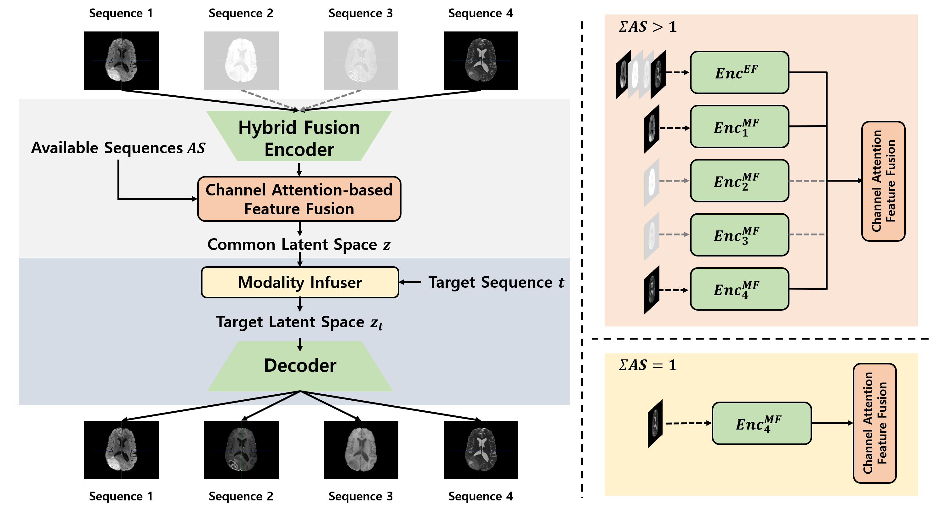
A Unified Framework for Synthesizing Multisequence Brain MRI via Hybrid Fusion
Jihoon Cho, Jonghye Woo, Jinah Park
0
0
Multisequence Magnetic Resonance Imaging (MRI) provides a reliable diagnosis in clinical applications through complementary information within sequences. However, in practice, the absence of certain MR sequences is a common problem that can lead to inconsistent analysis results. In this work, we propose a novel unified framework for synthesizing multisequence MR images, called Hybrid Fusion GAN (HF-GAN). We introduce a hybrid fusion encoder designed to ensure the disentangled extraction of complementary and modality-specific information, along with a channel attention-based feature fusion module that integrates the features into a common latent space handling the complexity from combinations of accessible MR sequences. Common feature representations are transformed into a target latent space via the modality infuser to synthesize missing MR sequences. We have performed experiments on multisequence brain MRI datasets from healthy individuals and patients diagnosed with brain tumors. Experimental results show that our method outperforms state-of-the-art methods in both quantitative and qualitative comparisons. In addition, a detailed analysis of our framework demonstrates the superiority of our designed modules and their effectiveness for use in data imputation tasks.
6/24/2024