CT-AGRG: Automated Abnormality-Guided Report Generation from 3D Chest CT Volumes

0

Sign in to get full access
Overview
- Automated generation of radiology reports from 3D chest CT scans
- Leverages AI to identify and describe abnormalities in medical images
- Aims to streamline the reporting process and improve consistency
Plain English Explanation
This research paper presents a system called CT-AGRG that can automatically generate radiology reports from 3D chest CT scans. The key innovation is that the system is designed to identify and describe any abnormalities it detects in the medical images, rather than just providing a generic summary.
The goal is to streamline the radiology reporting process and improve the consistency of the reports. Currently, radiologists have to manually review the images and dictate their findings, which can be time-consuming and prone to variation. By automating this task with AI, the hope is that reports can be generated more efficiently and with greater standardization.
The system works by first using deep learning models to analyze the 3D CT scans and detect any abnormalities, such as tumors or signs of disease. It then generates natural language descriptions of these findings, incorporating medical terminology as appropriate. The reports can be customized based on the specific needs of the healthcare organization or clinicians.
Overall, this research represents an important step towards improving the radiology reporting workflow through the use of advanced AI techniques. By automating the detection and description of abnormalities, it has the potential to save time, reduce errors, and provide more consistent and informative reports to support patient care.
Technical Explanation
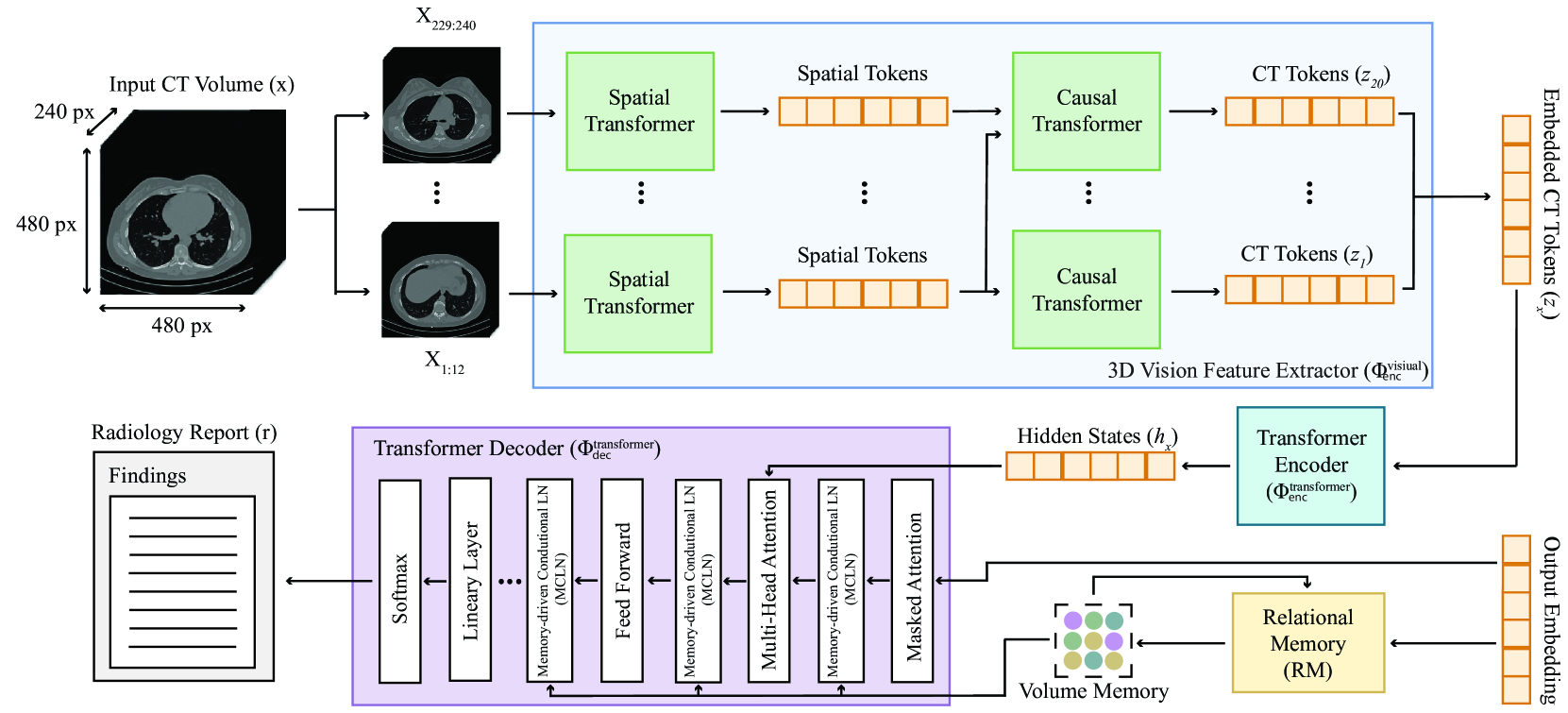
The CT-AGRG system leverages a multi-stage deep learning architecture to generate radiology reports from 3D chest CT volumes. The first stage involves a 3D convolutional neural network (CNN) that is trained to detect and segment various anatomical structures and abnormalities within the CT scans.
These detected findings are then passed to a natural language generation (NLG) module, which is responsible for converting the identified abnormalities into coherent, grammatically correct textual descriptions. The NLG component utilizes a transformer-based language model that has been fine-tuned on a large corpus of existing radiology reports.
The system also incorporates an "abnormality-guided" approach, where the NLG module prioritizes the generation of descriptions for the most significant detected abnormalities. This helps ensure that the most clinically relevant information is included in the final report.
The researchers evaluated their system on a dataset of over 30,000 chest CT scans and corresponding radiology reports. They found that the automatically generated reports achieved high scores on various natural language processing metrics, suggesting that the system is able to produce high-quality, informative reports that are comparable to those created by human radiologists.
Critical Analysis
The CT-AGRG system represents an impressive step forward in the field of automated radiology report generation. By leveraging advanced deep learning and natural language processing techniques, the researchers have developed a system that can efficiently analyze medical images and translate the findings into coherent, clinically relevant reports.
One potential limitation of the system is its reliance on a pre-defined set of abnormalities that the AI has been trained to detect. While this "abnormality-guided" approach helps ensure the most important information is included, it may miss or underemphasize less common or atypical findings that could still be clinically significant.
Additionally, the system's performance was evaluated primarily on standard natural language processing metrics, such as BLEU and ROUGE scores. While these metrics provide a useful benchmark, they may not fully capture the clinical accuracy and usefulness of the generated reports from the perspective of radiologists and clinicians.
Further research could explore ways to make the system more adaptable and open-ended, allowing it to detect a wider range of abnormalities and generate more personalized, context-aware reports. Integrating the system with electronic health records and soliciting feedback from end-users could also help refine the system and ensure it meets the needs of the healthcare professionals who would be relying on it.
Conclusion
The CT-AGRG system represents a significant advancement in the field of automated radiology report generation. By leveraging state-of-the-art deep learning and natural language processing techniques, the researchers have developed a system that can efficiently analyze 3D chest CT scans and generate informative, clinically relevant reports.
This technology has the potential to streamline the radiology reporting process, improve consistency, and free up radiologists to focus on more complex tasks. As the system continues to be refined and integrated into clinical workflows, it could have a meaningful impact on patient care by providing timely, standardized radiology reports to support diagnostic and treatment decisions.
Overall, this research highlights the growing potential of AI-powered systems to augment and enhance the work of healthcare professionals, ultimately improving the quality and efficiency of medical care.
This summary was produced with help from an AI and may contain inaccuracies - check out the links to read the original source documents!
Related Papers
0
CT-AGRG: Automated Abnormality-Guided Report Generation from 3D Chest CT Volumes
Theo Di Piazza
The rapid increase of computed tomography (CT) scans and their time-consuming manual analysis have created an urgent need for robust automated analysis techniques in clinical settings. These aim to assist radiologists and help them managing their growing workload. Existing methods typically generate entire reports directly from 3D CT images, without explicitly focusing on observed abnormalities. This unguided approach often results in repetitive content or incomplete reports, failing to prioritize anomaly-specific descriptions. We propose a new anomaly-guided report generation model, which first predicts abnormalities and then generates targeted descriptions for each. Evaluation on a public dataset demonstrates significant improvements in report quality and clinical relevance. We extend our work by conducting an ablation study to demonstrate its effectiveness.
Read more9/5/2024
0
CT2Rep: Automated Radiology Report Generation for 3D Medical Imaging
Ibrahim Ethem Hamamci, Sezgin Er, Bjoern Menze
Medical imaging plays a crucial role in diagnosis, with radiology reports serving as vital documentation. Automating report generation has emerged as a critical need to alleviate the workload of radiologists. While machine learning has facilitated report generation for 2D medical imaging, extending this to 3D has been unexplored due to computational complexity and data scarcity. We introduce the first method to generate radiology reports for 3D medical imaging, specifically targeting chest CT volumes. Given the absence of comparable methods, we establish a baseline using an advanced 3D vision encoder in medical imaging to demonstrate our method's effectiveness, which leverages a novel auto-regressive causal transformer. Furthermore, recognizing the benefits of leveraging information from previous visits, we augment CT2Rep with a cross-attention-based multi-modal fusion module and hierarchical memory, enabling the incorporation of longitudinal multimodal data. Access our code at https://github.com/ibrahimethemhamamci/CT2Rep
Read more7/8/2024

0
Automatically Generating Narrative-Style Radiology Reports from Volumetric CT Images; a Proof of Concept
Marijn Borghouts
The world faces a shortage of radiologists, leading to longer treatment times and increased stress, negatively impacting patient safety and workforce morale. Integrating artificial intelligence to interpret radiographic images and generate descriptive reports offers a promising solution. However, limited research exists on generating natural language descriptions for volumetric medical images. This study introduces a deep learning-based proof of concept model to accurately identify abnormalities in volumetric CT data and generate narrative-style reports. Various encoder-decoder models were assessed for their efficacy in clinically relevant and surrogate tasks. Clinically relevant tasks involved identifying and describing pulmonary nodules and pleural effusions, while surrogate tasks involved recognizing and describing artificial abnormalities such as mirroring, rotation, and lung lobe occlusion. The results show high accuracy in detecting combinations of artificial abnormalities, with the best model achieving a classification accuracy of 0.97 on an independent dataset with a homogeneously distributed 11-class problem. Furthermore, the best model consistently generated coherent radiology reports in natural language, with a next-word prediction accuracy of 0.84. Additionally, 65% of these reports were factually accurate regarding the identified artificial abnormalities. Unfortunately, these models did not replicate this success for clinically relevant tasks. Overall, this study provides a working proof of concept model for a challenge yet to be fully addressed by the scientific community. Given the success on surrogate tasks, the leap to clinically relevant tasks seems feasible. Acquiring a significantly larger high-quality dataset appears to be the most promising path forward, alongside more computational resources for end-to-end model training.
Read more6/19/2024

0
AutoRG-Brain: Grounded Report Generation for Brain MRI
Jiayu Lei, Xiaoman Zhang, Chaoyi Wu, Lisong Dai, Ya Zhang, Yanyong Zhang, Yanfeng Wang, Weidi Xie, Yuehua Li
Radiologists are tasked with interpreting a large number of images in a daily base, with the responsibility of generating corresponding reports. This demanding workload elevates the risk of human error, potentially leading to treatment delays, increased healthcare costs, revenue loss, and operational inefficiencies. To address these challenges, we initiate a series of work on grounded Automatic Report Generation (AutoRG), starting from the brain MRI interpretation system, which supports the delineation of brain structures, the localization of anomalies, and the generation of well-organized findings. We make contributions from the following aspects, first, on dataset construction, we release a comprehensive dataset encompassing segmentation masks of anomaly regions and manually authored reports, termed as RadGenome-Brain MRI. This data resource is intended to catalyze ongoing research and development in the field of AI-assisted report generation systems. Second, on system design, we propose AutoRG-Brain, the first brain MRI report generation system with pixel-level grounded visual clues. Third, for evaluation, we conduct quantitative assessments and human evaluations of brain structure segmentation, anomaly localization, and report generation tasks to provide evidence of its reliability and accuracy. This system has been integrated into real clinical scenarios, where radiologists were instructed to write reports based on our generated findings and anomaly segmentation masks. The results demonstrate that our system enhances the report-writing skills of junior doctors, aligning their performance more closely with senior doctors, thereby boosting overall productivity.
Read more7/31/2024