Deconvolving Complex Neuronal Networks into Interpretable Task-Specific Connectomes
2407.00201
0
0
Abstract
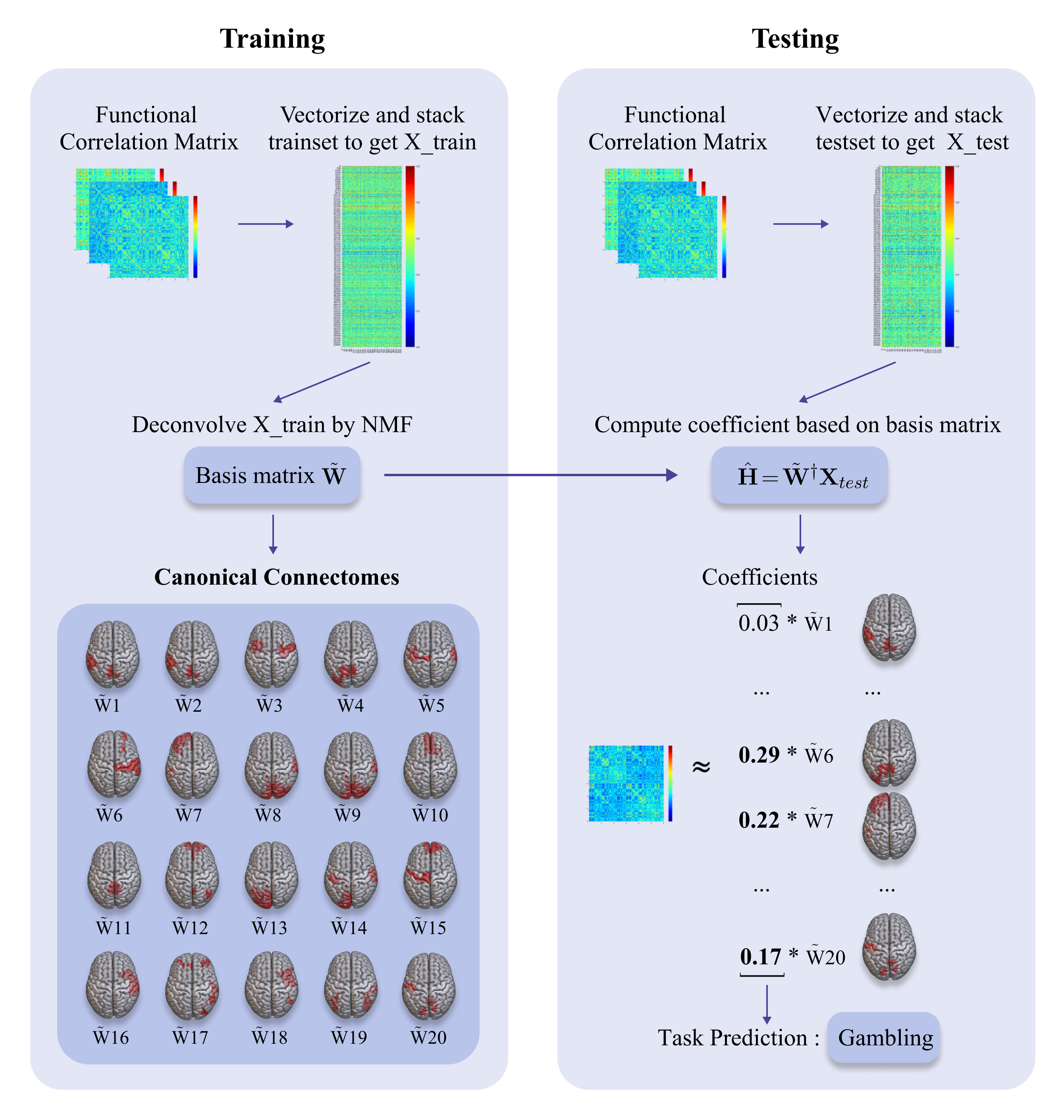
Task-specific functional MRI (fMRI) images provide excellent modalities for studying the neuronal basis of cognitive processes. We use fMRI data to formulate and solve the problem of deconvolving task-specific aggregate neuronal networks into a set of basic building blocks called canonical networks, to use these networks for functional characterization, and to characterize the physiological basis of these responses by mapping them to regions of the brain. Our results show excellent task-specificity of canonical networks, i.e., the expression of a small number of canonical networks can be used to accurately predict tasks; generalizability across cohorts, i.e., canonical networks are conserved across diverse populations, studies, and acquisition protocols; and that canonical networks have strong anatomical and physiological basis. From a methods perspective, the problem of identifying these canonical networks poses challenges rooted in the high dimensionality, small sample size, acquisition variability, and noise. Our deconvolution technique is based on non-negative matrix factorization (NMF) that identifies canonical networks as factors of a suitably constructed matrix. We demonstrate that our method scales to large datasets, yields stable and accurate factors, and is robust to noise.
Create account to get full access
Overview
- This research paper presents a novel approach for deconvolving complex neuronal networks into interpretable task-specific connectomes.
- The method involves extracting task-specific neural circuit representations from complex neural network models.
- The goal is to improve the interpretability and explainability of deep learning models used for neuroimaging analysis and brain-computer interfaces.
Plain English Explanation
The human brain is an incredibly complex and intricate system, with billions of neurons and trillions of connections between them. When researchers use deep learning models to analyze brain imaging data or develop brain-computer interfaces, these models can become quite complex and difficult to understand.
This research paper introduces a new technique to "deconvolve" or break down these complex neural network models into more interpretable and task-specific "connectomes" - maps of the key connections and pathways in the brain that are relevant for a particular task or function.
By extracting these task-specific representations from the complex neural networks, the researchers aim to make the models more transparent and easier for humans to understand. This could lead to better insights into how the brain works and ultimately, more effective brain-computer interfaces and neuroimaging tools.
The key innovation is the ability to distill the essential features of a complex neural network model down to the core connections and pathways that are most relevant for a given task. This allows researchers to move beyond "black box" models and develop a deeper, more interpretable understanding of the neural mechanisms underlying various cognitive and behavioral functions.
Technical Explanation
The researchers developed a novel method for "deconvolving" complex neural network models into more interpretable task-specific connectomes. This builds on previous work on explainable AI for brain networks.
The approach involves training a neural network model to perform a specific task, such as classifying brain activity patterns. The model learns a complex representation of the underlying neural circuits. The researchers then use a specialized deconvolution algorithm to extract the key connections and pathways that are most relevant for the task at hand.
This allows them to generate an interpretable, task-specific "connectome" that highlights the essential neural mechanisms driving the model's performance. The technique is related to other work on discovering robust biomarkers for neurological disorders from functional brain imaging data.
The deconvolution process leverages recent advances in graph neural networks and differentiable pooling operators. It complements other work on generating brain imaging data from graph representations and using vision-to-language models to enhance visual reconstruction of brain activity.
By extracting these task-specific connectomes, the researchers aim to provide neuroscientists and clinicians with a more interpretable and transparent view into the neural underpinnings of cognitive functions and brain disorders. This aligns with broader efforts towards explainable automated neuroanatomy.
Critical Analysis
The proposed deconvolution method represents an important step forward in making complex neural network models used for neuroimaging and brain-computer interfaces more interpretable and explainable. By distilling the essential neural circuits driving task performance, the technique provides valuable insights that can complement traditional neuroscientific approaches.
However, the paper does acknowledge some limitations. The deconvolution process relies on the fidelity of the original neural network model, and errors or biases in that model could be propagated to the extracted connectomes. Additionally, the task-specificity of the connectomes means they may not capture the full complexity and flexibility of the brain's neural mechanisms.
Further research is needed to fully validate the approach across a wider range of neuroimaging tasks and applications. Careful comparison to ground truth neuroscientific data and exploration of edge cases will be important to assess the reliability and generalizability of the technique.
Overall, this work represents a significant advancement in the field of explainable AI for neuroscience and brain-computer interfaces. By making complex neural network models more interpretable, it has the potential to accelerate scientific discovery and lead to more effective and personalized clinical applications.
Conclusion
This research paper presents a novel method for deconvolving complex neural network models used for neuroimaging and brain-computer interfaces into more interpretable task-specific connectomes. By extracting the essential neural circuits driving model performance, the technique provides valuable insights into the underlying neural mechanisms.
The ability to generate these explainable, task-specific representations of brain function has important implications for neuroscience research, clinical applications, and the development of advanced brain-computer interfaces. While the approach has some limitations, it represents a significant step forward in making complex deep learning models more transparent and accessible to researchers and clinicians.
As the field of AI-powered neurotechnology continues to advance, techniques like this one will be crucial for building trust, generating new scientific insights, and ultimately, improving outcomes for individuals with neurological and psychiatric disorders.
This summary was produced with help from an AI and may contain inaccuracies - check out the links to read the original source documents!
Related Papers
🏷️
Contrastive Graph Pooling for Explainable Classification of Brain Networks
Jiaxing Xu, Qingtian Bian, Xinhang Li, Aihu Zhang, Yiping Ke, Miao Qiao, Wei Zhang, Wei Khang Jeremy Sim, Bal'azs Guly'as
0
0
Functional magnetic resonance imaging (fMRI) is a commonly used technique to measure neural activation. Its application has been particularly important in identifying underlying neurodegenerative conditions such as Parkinson's, Alzheimer's, and Autism. Recent analysis of fMRI data models the brain as a graph and extracts features by graph neural networks (GNNs). However, the unique characteristics of fMRI data require a special design of GNN. Tailoring GNN to generate effective and domain-explainable features remains challenging. In this paper, we propose a contrastive dual-attention block and a differentiable graph pooling method called ContrastPool to better utilize GNN for brain networks, meeting fMRI-specific requirements. We apply our method to 5 resting-state fMRI brain network datasets of 3 diseases and demonstrate its superiority over state-of-the-art baselines. Our case study confirms that the patterns extracted by our method match the domain knowledge in neuroscience literature, and disclose direct and interesting insights. Our contributions underscore the potential of ContrastPool for advancing the understanding of brain networks and neurodegenerative conditions. The source code is available at https://github.com/AngusMonroe/ContrastPool.
4/15/2024

Discovering robust biomarkers of neurological disorders from functional MRI using graph neural networks: A Review
Yi Hao Chan, Deepank Girish, Sukrit Gupta, Jing Xia, Chockalingam Kasi, Yinan He, Conghao Wang, Jagath C. Rajapakse
0
0
Graph neural networks (GNN) have emerged as a popular tool for modelling functional magnetic resonance imaging (fMRI) datasets. Many recent studies have reported significant improvements in disorder classification performance via more sophisticated GNN designs and highlighted salient features that could be potential biomarkers of the disorder. In this review, we provide an overview of how GNN and model explainability techniques have been applied on fMRI datasets for disorder prediction tasks, with a particular emphasis on the robustness of biomarkers produced for neurodegenerative diseases and neuropsychiatric disorders. We found that while most studies have performant models, salient features highlighted in these studies vary greatly across studies on the same disorder and little has been done to evaluate their robustness. To address these issues, we suggest establishing new standards that are based on objective evaluation metrics to determine the robustness of these potential biomarkers. We further highlight gaps in the existing literature and put together a prediction-attribution-evaluation framework that could set the foundations for future research on improving the robustness of potential biomarkers discovered via GNNs.
5/2/2024
🛸
Brain Imaging-to-Graph Generation using Adversarial Hierarchical Diffusion Models for MCI Causality Analysis
Qiankun Zuo, Hao Tian, Chi-Man Pun, Hongfei Wang, Yudong Zhang, Jin Hong
0
0
Effective connectivity can describe the causal patterns among brain regions. These patterns have the potential to reveal the pathological mechanism and promote early diagnosis and effective drug development for cognitive disease. However, the current methods utilize software toolkits to extract empirical features from brain imaging to estimate effective connectivity. These methods heavily rely on manual parameter settings and may result in large errors during effective connectivity estimation. In this paper, a novel brain imaging-to-graph generation (BIGG) framework is proposed to map functional magnetic resonance imaging (fMRI) into effective connectivity for mild cognitive impairment (MCI) analysis. To be specific, the proposed BIGG framework is based on the diffusion denoising probabilistic models (DDPM), where each denoising step is modeled as a generative adversarial network (GAN) to progressively translate the noise and conditional fMRI to effective connectivity. The hierarchical transformers in the generator are designed to estimate the noise at multiple scales. Each scale concentrates on both spatial and temporal information between brain regions, enabling good quality in noise removal and better inference of causal relations. Meanwhile, the transformer-based discriminator constrains the generator to further capture global and local patterns for improving high-quality and diversity generation. By introducing the diffusive factor, the denoising inference with a large sampling step size is more efficient and can maintain high-quality results for effective connectivity generation. Evaluations of the ADNI dataset demonstrate the feasibility and efficacy of the proposed model. The proposed model not only achieves superior prediction performance compared with other competing methods but also predicts MCI-related causal connections that are consistent with clinical studies.
6/4/2024
🖼️
Neuro-Vision to Language: Image Reconstruction and Interaction via Non-invasive Brain Recordings
Guobin Shen, Dongcheng Zhao, Xiang He, Linghao Feng, Yiting Dong, Jihang Wang, Qian Zhang, Yi Zeng
0
0
Decoding non-invasive brain recordings is pivotal for advancing our understanding of human cognition but faces challenges due to individual differences and complex neural signal representations. Traditional methods often require customized models and extensive trials, lacking interpretability in visual reconstruction tasks. Our framework integrates 3D brain structures with visual semantics using a Vision Transformer 3D. This unified feature extractor efficiently aligns fMRI features with multiple levels of visual embeddings, eliminating the need for subject-specific models and allowing extraction from single-trial data. The extractor consolidates multi-level visual features into one network, simplifying integration with Large Language Models (LLMs). Additionally, we have enhanced the fMRI dataset with diverse fMRI-image-related textual data to support multimodal large model development. Integrating with LLMs enhances decoding capabilities, enabling tasks such as brain captioning, complex reasoning, concept localization, and visual reconstruction. Our approach demonstrates superior performance across these tasks, precisely identifying language-based concepts within brain signals, enhancing interpretability, and providing deeper insights into neural processes. These advances significantly broaden the applicability of non-invasive brain decoding in neuroscience and human-computer interaction, setting the stage for advanced brain-computer interfaces and cognitive models.
5/24/2024