Towards Explainable Automated Neuroanatomy
2404.05814
0
0
Abstract
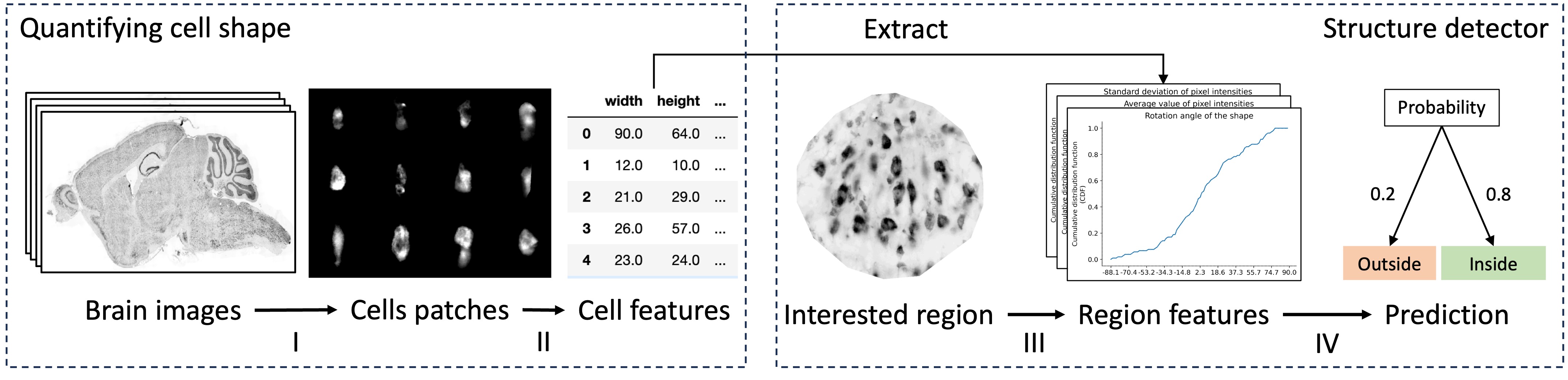
We present a novel method for quantifying the microscopic structure of brain tissue. It is based on the automated recognition of interpretable features obtained by analyzing the shapes of cells. This contrasts with prevailing methods of brain anatomical analysis in two ways. First, contemporary methods use gray-scale values derived from smoothed version of the anatomical images, which dissipated valuable information from the texture of the images. Second, contemporary analysis uses the output of black-box Convolutional Neural Networks, while our system makes decisions based on interpretable features obtained by analyzing the shapes of individual cells. An important benefit of this open-box approach is that the anatomist can understand and correct the decisions made by the computer. Our proposed system can accurately localize and identify existing brain structures. This can be used to align and coregistar brains and will facilitate connectomic studies for reverse engineering of brain circuitry.
Create account to get full access
Overview
- Explores the use of machine learning techniques to automate the analysis of brain anatomy
- Focuses on developing explainable models that can provide insights into the underlying neuroanatomical structures
- Aims to enhance the interpretability and transparency of automated neuroanatomy methods
Plain English Explanation
This research paper investigates the use of advanced machine learning algorithms to automatically analyze brain anatomy. The goal is to develop models that can not only accurately identify and segment different brain structures, but also provide explanations for their predictions. This would make the automated neuroanatomy process more transparent and easier for researchers and clinicians to understand.
The key idea is to move beyond "black box" machine learning models, which can be highly accurate but difficult to interpret, and instead create "explainable AI" models that can explain their reasoning. This could help unlock new insights into the complex structure and function of the brain, and potentially lead to better-informed clinical decisions.
Technical Explanation
The researchers propose a novel approach that combines advanced computer vision techniques with innovative methods for "explainable machine learning". They start by developing a deep learning model that can accurately segment and classify different brain regions based on magnetic resonance imaging (MRI) data.
To make this model more interpretable, the researchers integrate "attention mechanisms" that highlight the specific image features the model is focusing on to make its predictions. They also explore "concept-based explanations", where the model's decisions are explained in terms of higher-level neuroanatomical concepts that are more intuitive for human users.
Additionally, the researchers investigate quantitative methods for describing the shapes and textures of brain cells, known as "neuronal morphology". These shape features are then used as inputs to the explainable machine learning models, potentially providing new insights into the relationship between cellular structure and brain function.
Critical Analysis
The researchers acknowledge that developing truly "explainable" machine learning models for neuroanatomy is a significant challenge. While the proposed techniques show promise, there are still limitations in terms of the level of detail and sophistication of the explanations provided.
Additionally, the reliance on MRI data, which has relatively coarse spatial resolution, may limit the ability to capture fine-grained neuroanatomical features. The researchers suggest that integrating higher-resolution imaging modalities, such as microscopy, could provide more detailed insights.
Furthermore, the generalizability of the proposed methods to diverse brain imaging datasets and clinical applications remains to be thoroughly evaluated. Continued research and validation in real-world settings will be crucial to assess the practical utility of this approach.
Conclusion
This research represents an important step towards developing more transparent and interpretable machine learning tools for automated neuroanatomy. By combining advanced computer vision, explainable AI, and quantitative morphology analysis, the researchers aim to unlock new insights into the complex structure and function of the human brain.
While challenges remain, the potential benefits of this approach are significant. Improving the interpretability of automated neuroanatomy could lead to better-informed clinical decisions, more efficient research workflows, and a deeper understanding of the underlying biological mechanisms that shape the brain.
This summary was produced with help from an AI and may contain inaccuracies - check out the links to read the original source documents!
Related Papers
✨
Computational limits to the legibility of the imaged human brain
James K Ruffle, Robert J Gray, Samia Mohinta, Guilherme Pombo, Chaitanya Kaul, Harpreet Hyare, Geraint Rees, Parashkev Nachev
0
0
Our knowledge of the organisation of the human brain at the population-level is yet to translate into power to predict functional differences at the individual-level, limiting clinical applications, and casting doubt on the generalisability of inferred mechanisms. It remains unknown whether the difficulty arises from the absence of individuating biological patterns within the brain, or from limited power to access them with the models and compute at our disposal. Here we comprehensively investigate the resolvability of such patterns with data and compute at unprecedented scale. Across 23 810 unique participants from UK Biobank, we systematically evaluate the predictability of 25 individual biological characteristics, from all available combinations of structural and functional neuroimaging data. Over 4526 GPU hours of computation, we train, optimize, and evaluate out-of-sample 700 individual predictive models, including fully-connected feed-forward neural networks of demographic, psychological, serological, chronic disease, and functional connectivity characteristics, and both uni- and multi-modal 3D convolutional neural network models of macro- and micro-structural brain imaging. We find a marked discrepancy between the high predictability of sex (balanced accuracy 99.7%), age (mean absolute error 2.048 years, R2 0.859), and weight (mean absolute error 2.609Kg, R2 0.625), for which we set new state-of-the-art performance, and the surprisingly low predictability of other characteristics. Neither structural nor functional imaging predicted psychology better than the coincidence of chronic disease (p<0.05). Serology predicted chronic disease (p<0.05) and was best predicted by it (p<0.001), followed by structural neuroimaging (p<0.05). Our findings suggest either more informative imaging or more powerful models are needed to decipher individual level characteristics from the human brain.
4/4/2024
🔎
A self-supervised text-vision framework for automated brain abnormality detection
David A. Wood, Emily Guilhem, Sina Kafiabadi, Ayisha Al Busaidi, Kishan Dissanayake, Ahmed Hammam, Nina Mansoor, Matthew Townend, Siddharth Agarwal, Yiran Wei, Asif Mazumder, Gareth J. Barker, Peter Sasieni, Sebastien Ourselin, James H. Cole, Thomas C. Booth
0
0
Artificial neural networks trained on large, expert-labelled datasets are considered state-of-the-art for a range of medical image recognition tasks. However, categorically labelled datasets are time-consuming to generate and constrain classification to a pre-defined, fixed set of classes. For neuroradiological applications in particular, this represents a barrier to clinical adoption. To address these challenges, we present a self-supervised text-vision framework that learns to detect clinically relevant abnormalities in brain MRI scans by directly leveraging the rich information contained in accompanying free-text neuroradiology reports. Our training approach consisted of two-steps. First, a dedicated neuroradiological language model - NeuroBERT - was trained to generate fixed-dimensional vector representations of neuroradiology reports (N = 50,523) via domain-specific self-supervised learning tasks. Next, convolutional neural networks (one per MRI sequence) learnt to map individual brain scans to their corresponding text vector representations by optimising a mean square error loss. Once trained, our text-vision framework can be used to detect abnormalities in unreported brain MRI examinations by scoring scans against suitable query sentences (e.g., 'there is an acute stroke', 'there is hydrocephalus' etc.), enabling a range of classification-based applications including automated triage. Potentially, our framework could also serve as a clinical decision support tool, not only by suggesting findings to radiologists and detecting errors in provisional reports, but also by retrieving and displaying examples of pathologies from historical examinations that could be relevant to the current case based on textual descriptors.
6/13/2024
👁️
An explainable three dimension framework to uncover learning patterns: A unified look in variable sulci recognition
Michail Mamalakis, Heloise de Vareilles, Atheer AI-Manea, Samantha C. Mitchell, Ingrid Arartz, Lynn Egeland Morch-Johnsen, Jane Garrison, Jon Simons, Pietro Lio, John Suckling, Graham Murray
0
0
Detecting the significant features of the learning process of an artificial intelligence framework in the entire training and validation dataset can be determined as 'global' explanations. Studies in the literature lack of accurate, low-complexity, and three-dimensional (3D) global explanations which are crucial in neuroimaging, a field with a complex representational space that demands more than basic two-dimensional interpretations. To fill this gap, we developed a novel explainable artificial intelligence (XAI) 3D-Framework that provides robust, faithful, and low-complexity global explanations. We evaluated our framework on various 3D deep learning networks trained, validated, and tested on a well-annotated cohort of 596 subjects from the TOP-OSLO study. The focus was on the presence and absence of the paracingulate sulcus, a variable feature of brain morphology correlated with psychotic conditions. Our proposed 3D-Framework outperforms traditional XAI methods in terms of faithfulness for global explanations. As a result, we were able to use these robust explanations to uncover new patterns that not only enhance the credibility and reliability of the training process but also reveal promising new biomarkers and significantly related sub-regions. For the first time, our developed 3D-Framework proposes a way for the scientific community to utilize global explanations to discover novel patterns in this specific neuroscientific application and beyond. This study can helps improve the trustworthiness of AI training processes and push the boundaries of our understanding by revealing new patterns in neuroscience and beyond.
6/11/2024
BrainFounder: Towards Brain Foundation Models for Neuroimage Analysis
Joseph Cox, Peng Liu, Skylar E. Stolte, Yunchao Yang, Kang Liu, Kyle B. See, Huiwen Ju, Ruogu Fang
0
0
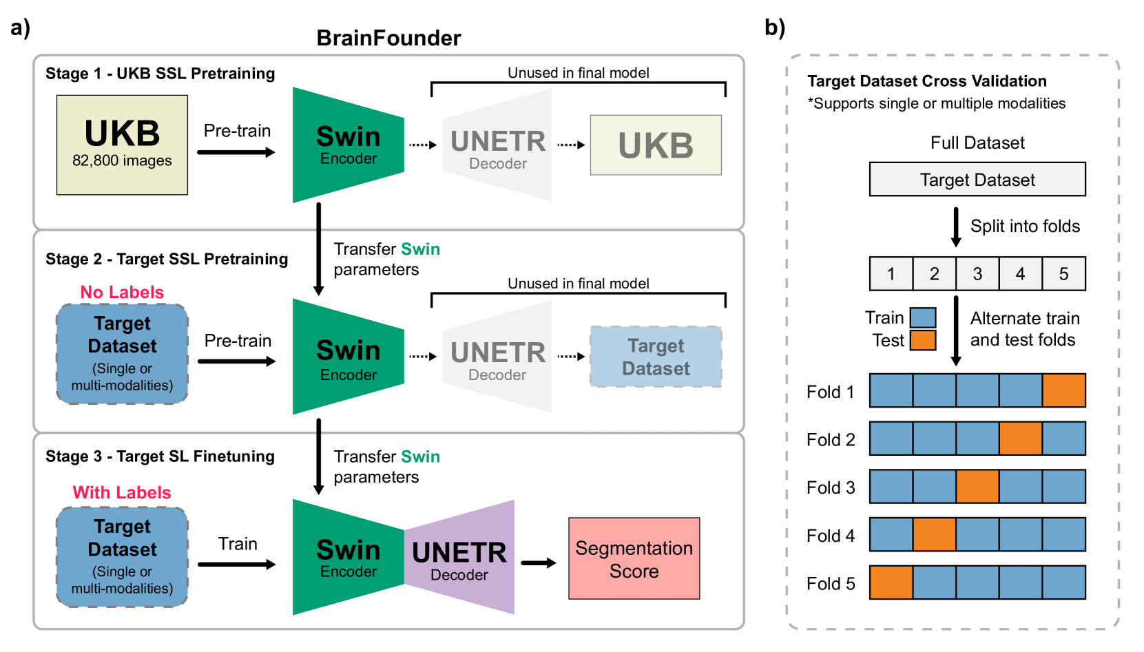
The burgeoning field of brain health research increasingly leverages artificial intelligence (AI) to interpret and analyze neurological data. This study introduces a novel approach towards the creation of medical foundation models by integrating a large-scale multi-modal magnetic resonance imaging (MRI) dataset derived from 41,400 participants in its own. Our method involves a novel two-stage pretraining approach using vision transformers. The first stage is dedicated to encoding anatomical structures in generally healthy brains, identifying key features such as shapes and sizes of different brain regions. The second stage concentrates on spatial information, encompassing aspects like location and the relative positioning of brain structures. We rigorously evaluate our model, BrainFounder, using the Brain Tumor Segmentation (BraTS) challenge and Anatomical Tracings of Lesions After Stroke v2.0 (ATLAS v2.0) datasets. BrainFounder demonstrates a significant performance gain, surpassing the achievements of the previous winning solutions using fully supervised learning. Our findings underscore the impact of scaling up both the complexity of the model and the volume of unlabeled training data derived from generally healthy brains, which enhances the accuracy and predictive capabilities of the model in complex neuroimaging tasks with MRI. The implications of this research provide transformative insights and practical applications in healthcare and make substantial steps towards the creation of foundation models for Medical AI. Our pretrained models and training code can be found at https://github.com/lab-smile/GatorBrain.
6/18/2024