Deep learning-based auto-segmentation of paraganglioma for growth monitoring

0
🤿
Sign in to get full access
Overview
- Paragangliomas are rare neuroendocrine tumors that typically form along major blood vessels and nerve pathways in the head and neck region.
- Accurately measuring the volume of these tumors is crucial for monitoring and modeling their growth over time.
- However, existing measurement tools are time-consuming and suffer from tumor-shape assumptions and observer-to-observer variation.
- Improved measurement techniques could enable growth model studies and provide valuable insights into tumor development, helping prevent severe symptoms in patients.
Plain English Explanation
Paragangliomas are a type of rare tumor that typically grow along the body's major blood vessels and nerves, often in the head and neck area. Accurately measuring the size of these tumors over time is very important for keeping track of how they are developing and developing models to predict their growth. However, the tools currently used to do these measurements are time-consuming and make assumptions about the shape of the tumor, and different doctors may measure the same tumor differently.
Improved measurement techniques could allow for more detailed studies of how these tumors grow over time, which could provide valuable insights. This could help doctors know which patients really need treatment to prevent severe symptoms, and avoid unnecessarily treating patients who don't need it. The paper proposes using a deep learning model to automatically measure the volume of these tumors, and shows that this method performs as well as manual measurement by experts.
Technical Explanation
The paper presents an automated tumor volume measurement method based on a deep learning segmentation model using no-new-UNnet (nnUNet). The authors assess the performance of the model by comparing its outputs to manual delineations by a senior otorhinolaryngologist, as well as analyzing the variation in manual delineations by multiple observers.
The results indicate that the automatic method performs at least equal to manual delineation. The authors then use the created model and a proposed linking procedure to track tumor volume over time and show how additional volume measurements affect the fit of known growth functions.
Critical Analysis
The paper acknowledges the limitations of the study, including the relatively small dataset and the need for further validation on a larger, more diverse set of paraganglioma cases. Additionally, the authors note that the linking procedure used to track tumors over time requires manual intervention and could benefit from further automation.
While the automated volume measurement method shows promising results, some concerns remain about its robustness and generalizability. Applying deep learning segmentation to small tumor volumes can be challenging, and the authors do not provide a detailed analysis of potential failure modes or edge cases.
Further research and validation on a larger and more diverse patient population would be needed to fully assess the clinical utility of this approach and its potential impact on patient care and tumor growth modeling.
Conclusion
This paper presents an automated method for measuring the volume of paraganglioma tumors using deep learning. The proposed approach performs comparably to manual delineation by experts and could enable more detailed studies of tumor growth over time, potentially leading to improved patient care and treatment decisions.
While the results are encouraging, the authors acknowledge the need for further validation and refinement of the method. Continued research in this area could yield valuable insights into the development of these rare but clinically significant tumors.
This summary was produced with help from an AI and may contain inaccuracies - check out the links to read the original source documents!
Related Papers
🤿
0
Deep learning-based auto-segmentation of paraganglioma for growth monitoring
E. M. C. Sijben, J. C. Jansen, M. de Ridder, P. A. N. Bosman, T. Alderliesten
Volume measurement of a paraganglioma (a rare neuroendocrine tumor that typically forms along major blood vessels and nerve pathways in the head and neck region) is crucial for monitoring and modeling tumor growth in the long term. However, in clinical practice, using available tools to do these measurements is time-consuming and suffers from tumor-shape assumptions and observer-to-observer variation. Growth modeling could play a significant role in solving a decades-old dilemma (stemming from uncertainty regarding how the tumor will develop over time). By giving paraganglioma patients treatment, severe symptoms can be prevented. However, treating patients who do not actually need it, comes at the cost of unnecessary possible side effects and complications. Improved measurement techniques could enable growth model studies with a large amount of tumor volume data, possibly giving valuable insights into how these tumors develop over time. Therefore, we propose an automated tumor volume measurement method based on a deep learning segmentation model using no-new-UNnet (nnUNet). We assess the performance of the model based on visual inspection by a senior otorhinolaryngologist and several quantitative metrics by comparing model outputs with manual delineations, including a comparison with variation in manual delineation by multiple observers. Our findings indicate that the automatic method performs (at least) equal to manual delineation. Finally, using the created model, and a linking procedure that we propose to track the tumor over time, we show how additional volume measurements affect the fit of known growth functions.
Read more4/12/2024
🤿
0
Postoperative glioblastoma segmentation: Development of a fully automated pipeline using deep convolutional neural networks and comparison with currently available models
Santiago Cepeda, Roberto Romero, Daniel Garcia-Perez, Guillermo Blasco, Luigi Tommaso Luppino, Samuel Kuttner, Ignacio Arrese, Ole Solheim, Live Eikenes, Anna Karlberg, Angel Perez-Nunez, Trinidad Escudero, Roberto Hornero, Rosario Sarabia
Accurately assessing tumor removal is paramount in the management of glioblastoma. We developed a pipeline using MRI scans and neural networks to segment tumor subregions and the surgical cavity in postoperative images. Our model excels in accurately classifying the extent of resection, offering a valuable tool for clinicians in assessing treatment effectiveness.
Read more4/19/2024
0
Function Class Learning with Genetic Programming: Towards Explainable Meta Learning for Tumor Growth Functionals
E. M. C. Sijben, J. C. Jansen, P. A. N. Bosman, T. Alderliesten
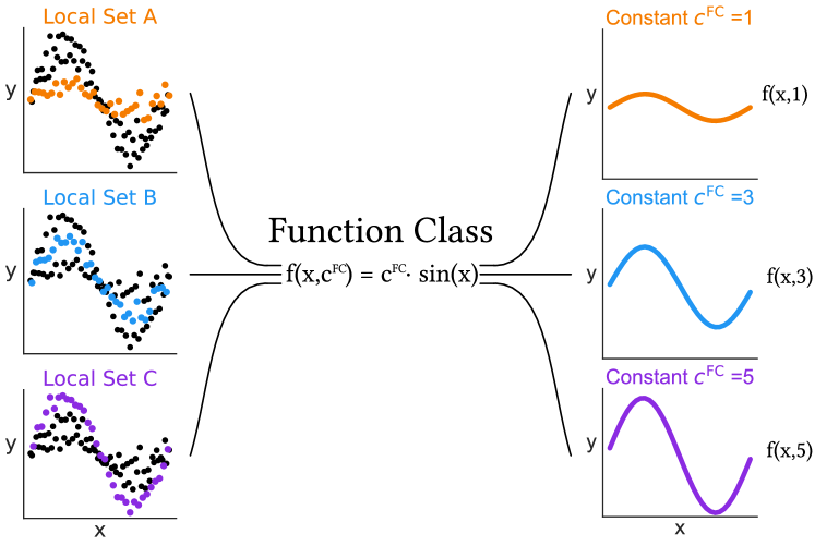
Paragangliomas are rare, primarily slow-growing tumors for which the underlying growth pattern is unknown. Therefore, determining the best care for a patient is hard. Currently, if no significant tumor growth is observed, treatment is often delayed, as treatment itself is not without risk. However, by doing so, the risk of (irreversible) adverse effects due to tumor growth may increase. Being able to predict the growth accurately could assist in determining whether a patient will need treatment during their lifetime and, if so, the timing of this treatment. The aim of this work is to learn the general underlying growth pattern of paragangliomas from multiple tumor growth data sets, in which each data set contains a tumor's volume over time. To do so, we propose a novel approach based on genetic programming to learn a function class, i.e., a parameterized function that can be fit anew for each tumor. We do so in a unique, multi-modal, multi-objective fashion to find multiple potentially interesting function classes in a single run. We evaluate our approach on a synthetic and a real-world data set. By analyzing the resulting function classes, we can effectively explain the general patterns in the data.
Read more4/10/2024
🤿
0
Deep Learning-Based Brain Image Segmentation for Automated Tumour Detection
Suman Sourabh, Murugappan Valliappan, Narayana Darapaneni, Anwesh R P
Introduction: The present study on the development and evaluation of an automated brain tumor segmentation technique based on deep learning using the 3D U-Net model. Objectives: The objective is to leverage state-of-the-art convolutional neural networks (CNNs) on a large dataset of brain MRI scans for segmentation. Methods: The proposed methodology applies pre-processing techniques for enhanced performance and generalizability. Results: Extensive validation on an independent dataset confirms the model's robustness and potential for integration into clinical workflows. The study emphasizes the importance of data pre-processing and explores various hyperparameters to optimize the model's performance. The 3D U-Net, has given IoUs for training and validation dataset have been 0.8181 and 0.66 respectively. Conclusion: Ultimately, this comprehensive framework showcases the efficacy of deep learning in automating brain tumour detection, offering valuable support in clinical practice.
Read more4/10/2024