Deep learning-based brain segmentation model performance validation with clinical radiotherapy CT
2406.17423
0
0

Abstract
Manual segmentation of medical images is labor intensive and especially challenging for images with poor contrast or resolution. The presence of disease exacerbates this further, increasing the need for an automated solution. To this extent, SynthSeg is a robust deep learning model designed for automatic brain segmentation across various contrasts and resolutions. This study validates the SynthSeg robust brain segmentation model on computed tomography (CT), using a multi-center dataset. An open access dataset of 260 paired CT and magnetic resonance imaging (MRI) from radiotherapy patients treated in 5 centers was collected. Brain segmentations from CT and MRI were obtained with SynthSeg model, a component of the Freesurfer imaging suite. These segmentations were compared and evaluated using Dice scores and Hausdorff 95 distance (HD95), treating MRI-based segmentations as the ground truth. Brain regions that failed to meet performance criteria were excluded based on automated quality control (QC) scores. Dice scores indicate a median overlap of 0.76 (IQR: 0.65-0.83). The median HD95 is 2.95 mm (IQR: 1.73-5.39). QC score based thresholding improves median dice by 0.1 and median HD95 by 0.05mm. Morphological differences related to sex and age, as detected by MRI, were also replicated with CT, with an approximate 17% difference between the CT and MRI results for sex and 10% difference between the results for age. SynthSeg can be utilized for CT-based automatic brain segmentation, but only in applications where precision is not essential. CT performance is lower than MRI based on the integrated QC scores, but low-quality segmentations can be excluded with QC-based thresholding. Additionally, performing CT-based neuroanatomical studies is encouraged, as the results show correlations in sex- and age-based analyses similar to those found with MRI.
Create account to get full access
Overview
- This paper explores the performance of a deep learning-based brain segmentation model on clinical radiotherapy CT data.
- The researchers validate the model's ability to accurately segment brain structures, which is crucial for radiotherapy planning and treatment.
- The study compares the model's performance on clinical CT scans to manual segmentation by expert radiologists.
Plain English Explanation
The paper examines a deep learning algorithm designed to automatically identify and separate different parts of the brain in medical scans. This type of technology could be very useful for planning radiation therapy treatments, where precisely targeting the tumor while avoiding nearby healthy brain tissue is critical.
The researchers tested the deep learning model on real-world clinical CT scans of patients' brains, and compared its results to the manual segmentations done by expert radiologists. This helps validate whether the algorithm can reliably perform this task in a clinical setting, rather than just on idealized research data.
Having an accurate, automated brain segmentation tool could streamline the radiotherapy planning process and reduce the workload on medical professionals. It could also help ensure consistency and precision, which are essential for delivering effective and safe treatment.
The findings of this study provide important insights into the real-world performance and practical applicability of this deep learning-based brain segmentation technology. This helps advance the field of medical image analysis and radiotherapy planning.
Technical Explanation
The researchers developed a deep learning-based model for segmenting brain structures in CT images. They trained the model on a dataset of CT scans with corresponding manual segmentations performed by expert radiologists.
To validate the model's performance in a clinical setting, they evaluated it on a separate dataset of 50 radiotherapy planning CT scans from cancer patients. The model's segmentation outputs were compared to the manual segmentations done by radiologists for the same scans.
Various metrics were used to quantify the overlap and agreement between the model's segmentations and the ground truth. These included Dice similarity coefficients, average surface distance, and 95th percentile Hausdorff distance. Statistical tests were conducted to determine if the model's performance was significantly different from the radiologists' segmentations.
The results showed that the deep learning model was able to accurately segment key brain structures like the brainstem, cerebellum, and cerebrum, with Dice scores above 0.9 for most structures. The model's performance was on par with or exceeded that of the expert radiologists in many cases.
This demonstrates the potential for deep learning-based methods to automate brain segmentation in the clinical radiotherapy workflow, potentially improving efficiency and consistency compared to manual segmentation.
Critical Analysis
The paper provides a thorough and rigorous evaluation of the deep learning model's performance on clinically-relevant data. By testing the model on real radiotherapy planning CT scans, the researchers were able to assess its suitability for practical use in a medical setting.
However, the study is limited to a relatively small dataset of 50 patients. Evaluating the model's generalization to larger and more diverse patient populations would be an important next step. Additionally, the paper does not address potential biases or limitations in the radiologist-generated ground truth segmentations used for comparison.
Further research could also investigate the model's robustness to variations in imaging protocols, scanners, and patient characteristics that may be encountered in real-world clinical environments. Addressing these types of challenges is crucial for deploying deep learning models in mission-critical medical applications.
Overall, this study provides valuable insights into the performance and feasibility of using deep learning for automated brain segmentation in radiotherapy planning. The findings are an important step toward translating this technology into clinical practice, but continued validation and development will be necessary.
Conclusion
This paper demonstrates that a deep learning-based model can accurately segment key brain structures in clinical radiotherapy CT scans, with performance matching or exceeding that of expert radiologists. This suggests the potential for such automated segmentation tools to streamline the radiotherapy planning workflow and improve consistency.
The rigorous evaluation on real-world clinical data is a strength of the study, but further research is needed to assess the model's robustness and generalization to larger and more diverse patient populations. Addressing these challenges will be crucial for deploying deep learning-based brain image segmentation in real-world medical settings.
Overall, this work represents an important step forward in applying advanced AI techniques to enhance the efficiency and precision of radiotherapy planning, with the ultimate goal of improving patient outcomes.
This summary was produced with help from an AI and may contain inaccuracies - check out the links to read the original source documents!
Related Papers
Synthetic Data for Robust Stroke Segmentation
Liam Chalcroft, Ioannis Pappas, Cathy J. Price, John Ashburner
0
0
Deep learning-based semantic segmentation in neuroimaging currently requires high-resolution scans and extensive annotated datasets, posing significant barriers to clinical applicability. We present a novel synthetic framework for the task of lesion segmentation, extending the capabilities of the established SynthSeg approach to accommodate large heterogeneous pathologies with lesion-specific augmentation strategies. Our method trains deep learning models, demonstrated here with the UNet architecture, using label maps derived from healthy and stroke datasets, facilitating the segmentation of both healthy tissue and pathological lesions without sequence-specific training data. Evaluated against in-domain and out-of-domain (OOD) datasets, our framework demonstrates robust performance, rivaling current methods within the training domain and significantly outperforming them on OOD data. This contribution holds promise for advancing medical imaging analysis in clinical settings, especially for stroke pathology, by enabling reliable segmentation across varied imaging sequences with reduced dependency on large annotated corpora. Code and weights available at https://github.com/liamchalcroft/SynthStroke.
4/3/2024
↗️
MRSegmentator: Robust Multi-Modality Segmentation of 40 Classes in MRI and CT Sequences
Hartmut Hantze, Lina Xu, Felix J. Dorfner, Leonhard Donle, Daniel Truhn, Hugo Aerts, Mathias Prokop, Bram van Ginneken, Alessa Hering, Lisa C. Adams, Keno K. Bressem
0
0
Purpose: To introduce a deep learning model capable of multi-organ segmentation in MRI scans, offering a solution to the current limitations in MRI analysis due to challenges in resolution, standardized intensity values, and variability in sequences. Materials and Methods: he model was trained on 1,200 manually annotated MRI scans from the UK Biobank, 221 in-house MRI scans and 1228 CT scans, leveraging cross-modality transfer learning from CT segmentation models. A human-in-the-loop annotation workflow was employed to efficiently create high-quality segmentations. The model's performance was evaluated on NAKO and the AMOS22 dataset containing 600 and 60 MRI examinations. Dice Similarity Coefficient (DSC) and Hausdorff Distance (HD) was used to assess segmentation accuracy. The model will be open sourced. Results: The model showcased high accuracy in segmenting well-defined organs, achieving Dice Similarity Coefficient (DSC) scores of 0.97 for the right and left lungs, and 0.95 for the heart. It also demonstrated robustness in organs like the liver (DSC: 0.96) and kidneys (DSC: 0.95 left, 0.95 right), which present more variability. However, segmentation of smaller and complex structures such as the portal and splenic veins (DSC: 0.54) and adrenal glands (DSC: 0.65 left, 0.61 right) revealed the need for further model optimization. Conclusion: The proposed model is a robust, tool for accurate segmentation of 40 anatomical structures in MRI and CT images. By leveraging cross-modality learning and interactive annotation, the model achieves strong performance and generalizability across diverse datasets, making it a valuable resource for researchers and clinicians. It is open source and can be downloaded from https://github.com/hhaentze/MRSegmentator.
5/14/2024
On Enhancing Brain Tumor Segmentation Across Diverse Populations with Convolutional Neural Networks
Fadillah Maani, Anees Ur Rehman Hashmi, Numan Saeed, Mohammad Yaqub
0
0
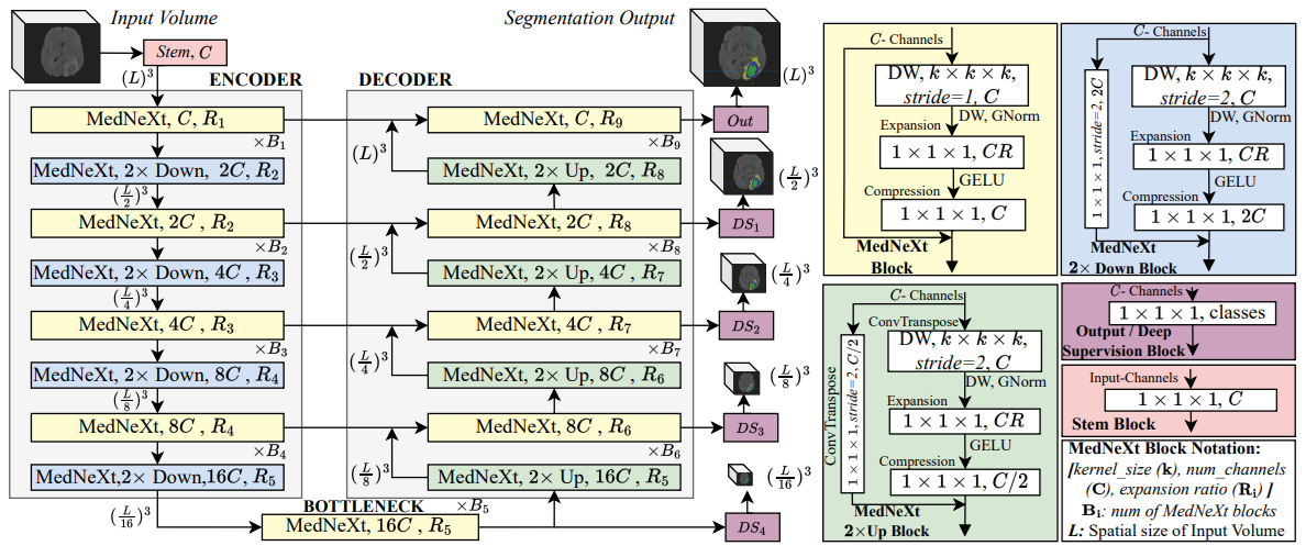
Brain tumor segmentation is a fundamental step in assessing a patient's cancer progression. However, manual segmentation demands significant expert time to identify tumors in 3D multimodal brain MRI scans accurately. This reliance on manual segmentation makes the process prone to intra- and inter-observer variability. This work proposes a brain tumor segmentation method as part of the BraTS-GoAT challenge. The task is to segment tumors in brain MRI scans automatically from various populations, such as adults, pediatrics, and underserved sub-Saharan Africa. We employ a recent CNN architecture for medical image segmentation, namely MedNeXt, as our baseline, and we implement extensive model ensembling and postprocessing for inference. Our experiments show that our method performs well on the unseen validation set with an average DSC of 85.54% and HD95 of 27.88. The code is available on https://github.com/BioMedIA-MBZUAI/BraTS2024_BioMedIAMBZ.
5/7/2024
🤿
Deep Learning-Based Brain Image Segmentation for Automated Tumour Detection
Suman Sourabh, Murugappan Valliappan, Narayana Darapaneni, Anwesh R P
0
0
Introduction: The present study on the development and evaluation of an automated brain tumor segmentation technique based on deep learning using the 3D U-Net model. Objectives: The objective is to leverage state-of-the-art convolutional neural networks (CNNs) on a large dataset of brain MRI scans for segmentation. Methods: The proposed methodology applies pre-processing techniques for enhanced performance and generalizability. Results: Extensive validation on an independent dataset confirms the model's robustness and potential for integration into clinical workflows. The study emphasizes the importance of data pre-processing and explores various hyperparameters to optimize the model's performance. The 3D U-Net, has given IoUs for training and validation dataset have been 0.8181 and 0.66 respectively. Conclusion: Ultimately, this comprehensive framework showcases the efficacy of deep learning in automating brain tumour detection, offering valuable support in clinical practice.
4/10/2024