A Deep Learning-Driven Pipeline for Differentiating Hypertrophic Cardiomyopathy from Cardiac Amyloidosis Using 2D Multi-View Echocardiography

0
🤿
Sign in to get full access
Overview
- This paper introduces a novel multi-view deep learning approach to differentiate between two heart conditions: hypertrophic cardiomyopathy (HCM) and cardiac amyloidosis (CA).
- HCM and CA exhibit similar echocardiographic characteristics, making them challenging to diagnose.
- The method classifies 2D echocardiography data into five distinct views, extracts features from each view, and combines them to classify the disease.
- The approach was evaluated on a dataset of 212 HCM patients, 30 CA patients, and 200 individuals with normal cardiac function, achieving high precision, recall, and micro-F1 score.
Plain English Explanation
The paper describes a new technique to help doctors better identify two heart conditions, hypertrophic cardiomyopathy (HCM) and cardiac amyloidosis (CA). These conditions can both lead to heart failure if left untreated, but they can be tricky to tell apart because they have similar patterns on echocardiograms (ultrasound images of the heart).
The new approach works by first dividing the echocardiogram into five different views of the heart. It then analyzes each view separately to extract important features, and combines this information to classify whether the patient has HCM, CA, or a normal, healthy heart. This multi-view analysis helps the system make more accurate diagnoses compared to looking at the echocardiogram as a whole.
The researchers tested this approach on a dataset of 442 patients, including 212 with HCM, 30 with CA, and 200 with normal heart function. The method was able to correctly identify the heart condition with a high degree of precision and accuracy, demonstrating its potential to aid doctors in making faster and more reliable diagnoses.
Technical Explanation
The paper presents a novel multi-view deep learning approach for differentiating between hypertrophic cardiomyopathy (HCM) and cardiac amyloidosis (CA) using 2D echocardiography.
The method begins by classifying the 2D echocardiography data into five distinct views: apical 4-chamber, parasternal long axis of the left ventricle, parasternal short axis at the levels of the mitral valve, papillary muscle, and apex. It then extracts features from each view separately and combines these five feature sets to perform disease classification.
The researchers evaluated their approach on a dataset of 212 patients diagnosed with HCM, 30 patients diagnosed with CA, and 200 individuals with normal cardiac function. This multi-view analysis achieved a precision of 0.905, a recall of 0.905, and a micro-F1 score of 0.904, demonstrating its effectiveness in accurately identifying HCM and CA.
The multi-view approach is crucial because HCM and CA exhibit similar echocardiographic characteristics, often leading to diagnostic challenges. By extracting features from multiple distinct views of the heart, the system is better able to capture the nuanced differences between these two conditions and make more reliable diagnoses.
Critical Analysis
The paper provides a comprehensive evaluation of the proposed multi-view deep learning approach, including comparisons to other state-of-the-art methods. However, the authors acknowledge several limitations and areas for future research.
One limitation is the relatively small sample size, particularly for the cardiac amyloidosis (CA) class, which had only 30 patients. Larger and more diverse datasets would be needed to further validate the generalizability of the approach.
Additionally, the paper does not address the potential challenges of deploying such a system in a clinical setting, such as the need for robust data preprocessing, model interpretability, and integration with existing hospital workflows. Explainable AI techniques could be explored to provide clinicians with more transparent and trustworthy diagnoses.
Despite these limitations, the multi-view deep learning approach demonstrated promising results in differentiating between HCM and CA, which could have significant implications for improving cardiovascular disease diagnosis and management. Further research and validation in larger, more diverse patient cohorts would be valuable to assess the broader applicability of this technique.
Conclusion
This paper presents a novel multi-view deep learning approach for differentiating between hypertrophic cardiomyopathy (HCM) and cardiac amyloidosis (CA) using 2D echocardiography. By extracting features from multiple distinct views of the heart and combining them for disease classification, the method achieved high precision, recall, and micro-F1 score, demonstrating its potential to aid clinicians in making accurate diagnoses.
The ability to reliably distinguish between these two conditions is crucial, as they exhibit similar echocardiographic characteristics but require different treatment approaches. The multi-view analysis proposed in this paper represents an important step forward in improving cardiovascular disease diagnosis and management, and further research in larger, more diverse patient populations could lead to even greater advancements in this field.
This summary was produced with help from an AI and may contain inaccuracies - check out the links to read the original source documents!
Related Papers
🤿
0
A Deep Learning-Driven Pipeline for Differentiating Hypertrophic Cardiomyopathy from Cardiac Amyloidosis Using 2D Multi-View Echocardiography
Bo Peng, Xiaofeng Li, Xinyu Li, Zhenghan Wang, Hui Deng, Xiaoxian Luo, Lixue Yin, Hongmei Zhang
Hypertrophic cardiomyopathy (HCM) and cardiac amyloidosis (CA) are both heart conditions that can progress to heart failure if untreated. They exhibit similar echocardiographic characteristics, often leading to diagnostic challenges. This paper introduces a novel multi-view deep learning approach that utilizes 2D echocardiography for differentiating between HCM and CA. The method begins by classifying 2D echocardiography data into five distinct echocardiographic views: apical 4-chamber, parasternal long axis of left ventricle, parasternal short axis at levels of the mitral valve, papillary muscle, and apex. It then extracts features of each view separately and combines five features for disease classification. A total of 212 patients diagnosed with HCM, and 30 patients diagnosed with CA, along with 200 individuals with normal cardiac function(Normal), were enrolled in this study from 2018 to 2022. This approach achieved a precision, recall of 0.905, and micro-F1 score of 0.904, demonstrating its effectiveness in accurately identifying HCM and CA using a multi-view analysis.
Read more4/26/2024

0
Multimodal Fusion of Echocardiography and Electronic Health Records for the Detection of Cardiac Amyloidosis
Zishun Feng, Joseph A. Sivak, Ashok K. Krishnamurthy
Cardiac amyloidosis, a rare and highly morbid condition, presents significant challenges for detection through echocardiography. Recently, there has been a surge in proposing machine-learning algorithms to identify cardiac amyloidosis, with the majority being imaging-based deep-learning approaches that require extensive data. In this study, we introduce a novel transformer-based multimodal fusion algorithm that leverages information from both imaging and electronic health records. Specifically, our approach utilizes echocardiography videos from both the parasternal long-axis (PLAX) view and the apical 4-chamber (A4C) view along with patients' demographic data, laboratory tests, and cardiac metrics to predict the probability of cardiac amyloidosis. We evaluated our method using 5-fold cross-validation on a dataset comprising 41 patients and achieved an Area Under the Receiver Operating Characteristic curve (AUROC) of 0.94. The experimental results demonstrate that our approach can achieve competitive results with a significantly smaller dataset compared to prior imaging-based methods that required data from thousands of patients. This underscores the potential of leveraging multimodal data to enhance diagnostic accuracy in the identification of complex cardiac conditions such as cardiac amyloidosis.
Read more6/10/2024
👁️
0
Automatic Cardiac Pathology Recognition in Echocardiography Images Using Higher Order Dynamic Mode Decomposition and a Vision Transformer for Small Datasets
Andr'es Bell-Navas, Nourelhouda Groun, Mar'ia Villalba-Orero, Enrique Lara-Pezzi, Jes'us Garicano-Mena, Soledad Le Clainche
Heart diseases are the main international cause of human defunction. According to the WHO, nearly 18 million people decease each year because of heart diseases. Also considering the increase of medical data, much pressure is put on the health industry to develop systems for early and accurate heart disease recognition. In this work, an automatic cardiac pathology recognition system based on a novel deep learning framework is proposed, which analyses in real-time echocardiography video sequences. The system works in two stages. The first one transforms the data included in a database of echocardiography sequences into a machine-learning-compatible collection of annotated images which can be used in the training stage of any kind of machine learning-based framework, and more specifically with deep learning. This includes the use of the Higher Order Dynamic Mode Decomposition (HODMD) algorithm, for the first time to the authors' knowledge, for both data augmentation and feature extraction in the medical field. The second stage is focused on building and training a Vision Transformer (ViT), barely explored in the related literature. The ViT is adapted for an effective training from scratch, even with small datasets. The designed neural network analyses images from an echocardiography sequence to predict the heart state. The results obtained show the superiority of the proposed system and the efficacy of the HODMD algorithm, even outperforming pretrained Convolutional Neural Networks (CNNs), which are so far the method of choice in the literature.
Read more5/1/2024
0
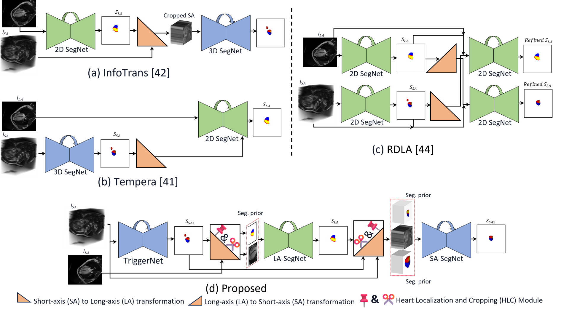
Multi-view Cardiac Image Segmentation via Trans-Dimensional Priors
Abbas Khan, Muhammad Asad, Martin Benning, Caroline Roney, Gregory Slabaugh
We propose a novel multi-stage trans-dimensional architecture for multi-view cardiac image segmentation. Our method exploits the relationship between long-axis (2D) and short-axis (3D) magnetic resonance (MR) images to perform a sequential 3D-to-2D-to-3D segmentation, segmenting the long-axis and short-axis images. In the first stage, 3D segmentation is performed using the short-axis image, and the prediction is transformed to the long-axis view and used as a segmentation prior in the next stage. In the second step, the heart region is localized and cropped around the segmentation prior using a Heart Localization and Cropping (HLC) module, focusing the subsequent model on the heart region of the image, where a 2D segmentation is performed. Similarly, we transform the long-axis prediction to the short-axis view, localize and crop the heart region and again perform a 3D segmentation to refine the initial short-axis segmentation. We evaluate our proposed method on the Multi-Disease, Multi-View & Multi-Center Right Ventricular Segmentation in Cardiac MRI (M&Ms-2) dataset, where our method outperforms state-of-the-art methods in segmenting cardiac regions of interest in both short-axis and long-axis images. The pre-trained models, source code, and implementation details will be publicly available.
Read more4/26/2024