A Deep Look Into -- Automated Lung X-Ray Abnormality Detection System

0
🤿
Sign in to get full access
Overview
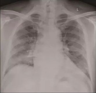
- Automated system to detect abnormalities in lung X-ray images
- Aims to distinguish normal X-ray images from those showing signs of infection
- Highlights areas of concern for further analysis by experts
- Motivated by the need for faster, more efficient disease detection during pandemics
Plain English Explanation
The research paper presents an automated lung X-ray abnormality detection system. This system can identify whether an X-ray image shows signs of infection or if it is normal. It also highlights the specific areas in the X-ray that the system believes may be abnormal, to help experts analyze the images more efficiently.
The researchers were motivated by the recent pandemic, which has shown the need for faster and more accessible methods of detecting infectious diseases. X-rays can be a useful tool for this, and an automated system can make the process quicker and less reliant on expert analysis alone.
The paper compares the performance of three existing machine learning models - DenseNet, MobileNet, and VGG16 - on the task of classifying X-ray images. However, the researchers found that these models tended to "overfit," meaning they performed well on the training data but struggled with new, unseen X-ray images. This was likely due to the black-and-white nature of the X-ray images.
To address this, the researchers developed a new model called V-BreathNet, which achieved over 96% accuracy in detecting abnormalities in the X-ray images. This suggests that not all state-of-the-art machine learning models are well-suited for working with black-and-white medical images, and that specialized approaches may be needed.
Technical Explanation
The researchers evaluated the performance of three pre-existing convolutional neural network (CNN) models - DenseNet, MobileNet, and VGG16 - on the task of classifying lung X-ray images as either normal or showing signs of infection.
They found that these models achieved high accuracy rates on the training data, but suffered from overfitting, meaning they performed poorly on new, unseen X-ray images. The researchers hypothesized that this was due to the black-and-white nature of the X-ray images, which may have limited the ability of these general-purpose models to learn relevant features.
To address this, the researchers developed a new model called V-BreathNet, which was specifically designed for working with black-and-white medical images. V-BreathNet achieved over 96% accuracy in detecting abnormalities in the X-ray images, outperforming the pre-existing models.
Critical Analysis
The researchers acknowledge several limitations of their study. First, the dataset used was relatively small, which may have contributed to the overfitting observed in the pre-existing models. Expanding the dataset with a wider range of X-ray images could help improve the models' generalization capabilities.
Additionally, the paper does not provide a detailed analysis of the types of abnormalities the V-BreathNet model was able to detect, or how its performance compares to human experts in identifying specific lung conditions. Further research could explore these aspects to better understand the model's clinical utility.
It would also be valuable to investigate how the V-BreathNet model might perform on other types of medical imaging data, such as lung sound analysis or blood test parameters for COVID-19 detection. This could help determine the broader applicability of the specialized approach used in developing V-BreathNet.
Conclusion
This research presents a promising automated system for detecting abnormalities in lung X-ray images. The development of the V-BreathNet model, which was specifically designed for working with black-and-white medical images, demonstrates the importance of tailoring machine learning approaches to the unique characteristics of the data.
The high accuracy achieved by V-BreathNet suggests that this type of specialized system could be a valuable tool for healthcare providers, potentially helping to speed up the process of identifying and diagnosing lung conditions, especially during times of increased demand, such as pandemics. However, further research is needed to fully understand the model's capabilities and limitations in a clinical setting.
This summary was produced with help from an AI and may contain inaccuracies - check out the links to read the original source documents!
Related Papers
🤿
0
A Deep Look Into -- Automated Lung X-Ray Abnormality Detection System
Nagullas KS, Vivekanand. V, Narayana Darapaneni, Anwesh R P
Introduction: Automated Lung X-Ray Abnormality Detection System is the application which distinguish the normal x-ray images from infected x-ray images and highlight area considered for prediction, with the recent pandemic a need to have a non-conventional method and faster detecting diseases, for which X ray serves the purpose. Obectives: As of current situation any viral disease that is infectious is potential pandemic, so there is need for cheap and early detection system. Methods: This research will help to eases the work of expert to do further analysis. Accuracy of three different preexisting models such as DenseNet, MobileNet and VGG16 were high but models over-fitted primarily due to black and white images. Results: This led to building up new method such as as V-BreathNet which gave more than 96% percent accuracy. Conclusion: Thus, it can be stated that not all state-of art CNN models can be used on B/W images. In conclusion not all state-of-art CNN models can be used on B/W images.
Read more4/9/2024
0
Automatic Detection of COVID-19 from Chest X-ray Images Using Deep Learning Model
Alloy Das, Rohit Agarwal, Rituparna Singh, Arindam Chowdhury, Debashis Nandi
The infectious disease caused by novel corona virus (2019-nCoV) has been widely spreading since last year and has shaken the entire world. It has caused an unprecedented effect on daily life, global economy and public health. Hence this disease detection has life-saving importance for both patients as well as doctors. Due to limited test kits, it is also a daunting task to test every patient with severe respiratory problems using conventional techniques (RT-PCR). Thus implementing an automatic diagnosis system is urgently required to overcome the scarcity problem of Covid-19 test kits at hospital, health care systems. The diagnostic approach is mainly classified into two categories-laboratory based and Chest radiography approach. In this paper, a novel approach for computerized corona virus (2019-nCoV) detection from lung x-ray images is presented. Here, we propose models using deep learning to show the effectiveness of diagnostic systems. In the experimental result, we evaluate proposed models on publicly available data-set which exhibit satisfactory performance and promising results compared with other previous existing methods.
Read more8/28/2024
🧠
0
A design of Convolutional Neural Network model for the Diagnosis of the COVID-19
Xinyuan Song
With the spread of COVID-19 around the globe over the past year, the usage of artificial intelligence (AI) algorithms and image processing methods to analyze the X-ray images of patients' chest with COVID-19 has become essential. The COVID-19 virus recognition in the lung area of a patient is one of the basic and essential needs of clicical centers and hospitals. Most research in this field has been devoted to papers on the basis of deep learning methods utilizing CNNs (Convolutional Neural Network), which mainly deal with the screening of sick and healthy people.In this study, a new structure of a 19-layer CNN has been recommended for accurately recognition of the COVID-19 from the X-ray pictures of chest. The offered CNN is developed to serve as a precise diagnosis system for a three class (viral pneumonia, Normal, COVID) and a four classclassification (Lung opacity, Normal, COVID-19, and pneumonia). A comparison is conducted among the outcomes of the offered procedure and some popular pretrained networks, including Inception, Alexnet, ResNet50, Squeezenet, and VGG19 and based on Specificity, Accuracy, Precision, Sensitivity, Confusion Matrix, and F1-score. The experimental results of the offered CNN method specify its dominance over the existing published procedures. This method can be a useful tool for clinicians in deciding properly about COVID-19.
Read more4/17/2024
🔎
0
CoVid-19 Detection leveraging Vision Transformers and Explainable AI
Pangoth Santhosh Kumar, Kundrapu Supriya, Mallikharjuna Rao K, Taraka Satya Krishna Teja Malisetti
Lung disease is a common health problem in many parts of the world. It is a significant risk to people health and quality of life all across the globe since it is responsible for five of the top thirty leading causes of death. Among them are COVID 19, pneumonia, and tuberculosis, to name just a few. It is critical to diagnose lung diseases in their early stages. Several different models including machine learning and image processing have been developed for this purpose. The earlier a condition is diagnosed, the better the patient chances of making a full recovery and surviving into the long term. Thanks to deep learning algorithms, there is significant promise for the autonomous, rapid, and accurate identification of lung diseases based on medical imaging. Several different deep learning strategies, including convolutional neural networks (CNN), vanilla neural networks, visual geometry group based networks (VGG), and capsule networks , are used for the goal of making lung disease forecasts. The standard CNN has a poor performance when dealing with rotated, tilted, or other aberrant picture orientations. As a result of this, within the scope of this study, we have suggested a vision transformer based approach end to end framework for the diagnosis of lung disorders. In the architecture, data augmentation, training of the suggested models, and evaluation of the models are all included. For the purpose of detecting lung diseases such as pneumonia, Covid 19, lung opacity, and others, a specialised Compact Convolution Transformers (CCT) model have been tested and evaluated on datasets such as the Covid 19 Radiography Database. The model has achieved a better accuracy for both its training and validation purposes on the Covid 19 Radiography Database.
Read more5/7/2024