DP-MDM: Detail-Preserving MR Reconstruction via Multiple Diffusion Models
2405.05763
0
0
🔍
Abstract
Detail features of magnetic resonance images play a cru-cial role in accurate medical diagnosis and treatment, as they capture subtle changes that pose challenges for doc-tors when performing precise judgments. However, the widely utilized naive diffusion model has limitations, as it fails to accurately capture more intricate details. To en-hance the quality of MRI reconstruction, we propose a comprehensive detail-preserving reconstruction method using multiple diffusion models to extract structure and detail features in k-space domain instead of image do-main. Moreover, virtual binary modal masks are utilized to refine the range of values in k-space data through highly adaptive center windows, which allows the model to focus its attention more efficiently. Last but not least, an inverted pyramid structure is employed, where the top-down image information gradually decreases, ena-bling a cascade representation. The framework effective-ly represents multi-scale sampled data, taking into ac-count the sparsity of the inverted pyramid architecture, and utilizes cascade training data distribution to repre-sent multi-scale data. Through a step-by-step refinement approach, the method refines the approximation of de-tails. Finally, the proposed method was evaluated by con-ducting experiments on clinical and public datasets. The results demonstrate that the proposed method outper-forms other methods.
Create account to get full access
Overview
- Magnetic resonance imaging (MRI) plays a crucial role in accurate medical diagnosis and treatment, but the widely used naive diffusion model has limitations in capturing intricate details.
- To enhance the quality of MRI reconstruction, the researchers propose a comprehensive detail-preserving reconstruction method using multiple diffusion models.
- The method extracts structure and detail features in the k-space domain, utilizes virtual binary modal masks to refine the range of values, and employs an inverted pyramid structure to represent multi-scale sampled data.
Plain English Explanation
Doctors rely on MRI scans to make precise diagnoses and determine the best treatments for their patients. These scans can detect subtle changes in the body that are challenging for doctors to interpret accurately. The standard technique for reconstructing MRI images, known as the naive diffusion model, has limitations in capturing the fine details needed for effective medical decision-making.
To address this, the researchers developed a new method that uses multiple diffusion models to extract structural and detailed features from the k-space data (the raw data collected by the MRI machine) rather than the final image. This allows the model to focus on preserving the intricate details that are crucial for diagnosis.
The method also utilizes virtual binary modal masks to refine the range of values in the k-space data, helping the model concentrate on the most important information. Finally, it employs an inverted pyramid structure that gradually decreases the top-down image information, enabling a step-by-step refinement of the details.
Technical Explanation
The proposed method uses multiple diffusion models to extract structure and detail features from the k-space domain, rather than the image domain. This allows the model to capture more intricate details that are crucial for accurate medical diagnosis and treatment.
The researchers also utilize virtual binary modal masks to refine the range of values in the k-space data. These highly adaptive center windows enable the model to focus its attention more efficiently on the most relevant information.
Furthermore, the framework employs an inverted pyramid structure, where the top-down image information gradually decreases. This enables a cascade representation of multi-scale sampled data, taking into account the sparsity of the inverted pyramid architecture and utilizing cascade training data distribution to represent multi-scale data.
Through this step-by-step refinement approach, the proposed method effectively represents and refines the approximation of details in the MRI reconstruction process.
Critical Analysis
The researchers acknowledge that the proposed method has some limitations, as it may not be able to capture all the intricate details in certain complex cases. Additionally, the computational complexity of the method could be a concern, especially for real-time clinical applications.
While the results demonstrate the method's superiority over other techniques, it would be valuable to see how the method performs on a wider range of MRI data, including various pathologies and imaging modalities. Further research could also explore ways to optimize the computational efficiency of the framework without compromising its detail-preserving capabilities.
Conclusion
The proposed detail-preserving reconstruction method using multiple diffusion models represents a significant advancement in the field of MRI reconstruction. By focusing on preserving the crucial details in the k-space domain, rather than the image domain, the method can significantly improve the accuracy of medical diagnoses and the effectiveness of treatments.
The innovative use of virtual binary modal masks and the inverted pyramid structure are key components that enable the method to refine the details in a step-by-step manner, resulting in higher-quality MRI reconstructions. While the method has some limitations, the researchers have made an important contribution to the field and have opened up new avenues for further research and development in this area.
This summary was produced with help from an AI and may contain inaccuracies - check out the links to read the original source documents!
Related Papers

Rethinking Diffusion Model for Multi-Contrast MRI Super-Resolution
Guangyuan Li, Chen Rao, Juncheng Mo, Zhanjie Zhang, Wei Xing, Lei Zhao
0
0
Recently, diffusion models (DM) have been applied in magnetic resonance imaging (MRI) super-resolution (SR) reconstruction, exhibiting impressive performance, especially with regard to detailed reconstruction. However, the current DM-based SR reconstruction methods still face the following issues: (1) They require a large number of iterations to reconstruct the final image, which is inefficient and consumes a significant amount of computational resources. (2) The results reconstructed by these methods are often misaligned with the real high-resolution images, leading to remarkable distortion in the reconstructed MR images. To address the aforementioned issues, we propose an efficient diffusion model for multi-contrast MRI SR, named as DiffMSR. Specifically, we apply DM in a highly compact low-dimensional latent space to generate prior knowledge with high-frequency detail information. The highly compact latent space ensures that DM requires only a few simple iterations to produce accurate prior knowledge. In addition, we design the Prior-Guide Large Window Transformer (PLWformer) as the decoder for DM, which can extend the receptive field while fully utilizing the prior knowledge generated by DM to ensure that the reconstructed MR image remains undistorted. Extensive experiments on public and clinical datasets demonstrate that our DiffMSR outperforms state-of-the-art methods.
4/9/2024
🤔
A Survey of Emerging Applications of Diffusion Probabilistic Models in MRI
Yuheng Fan, Hanxi Liao, Shiqi Huang, Yimin Luo, Huazhu Fu, Haikun Qi
0
0
Diffusion probabilistic models (DPMs) which employ explicit likelihood characterization and a gradual sampling process to synthesize data, have gained increasing research interest. Despite their huge computational burdens due to the large number of steps involved during sampling, DPMs are widely appreciated in various medical imaging tasks for their high-quality and diversity of generation. Magnetic resonance imaging (MRI) is an important medical imaging modality with excellent soft tissue contrast and superb spatial resolution, which possesses unique opportunities for DPMs. Although there is a recent surge of studies exploring DPMs in MRI, a survey paper of DPMs specifically designed for MRI applications is still lacking. This review article aims to help researchers in the MRI community to grasp the advances of DPMs in different applications. We first introduce the theory of two dominant kinds of DPMs, categorized according to whether the diffusion time step is discrete or continuous, and then provide a comprehensive review of emerging DPMs in MRI, including reconstruction, image generation, image translation, segmentation, anomaly detection, and further research topics. Finally, we discuss the general limitations as well as limitations specific to the MRI tasks of DPMs and point out potential areas that are worth further exploration.
5/9/2024
📈
MSDiff: Multi-Scale Diffusion Model for Ultra-Sparse View CT Reconstruction
Pinhuang Tan, Mengxiao Geng, Jingya Lu, Liu Shi, Bin Huang, Qiegen Liu
0
0
Computed Tomography (CT) technology reduces radiation haz-ards to the human body through sparse sampling, but fewer sampling angles pose challenges for image reconstruction. Score-based generative models are widely used in sparse-view CT re-construction, performance diminishes significantly with a sharp reduction in projection angles. Therefore, we propose an ultra-sparse view CT reconstruction method utilizing multi-scale dif-fusion models (MSDiff), designed to concentrate on the global distribution of information and facilitate the reconstruction of sparse views with local image characteristics. Specifically, the proposed model ingeniously integrates information from both comprehensive sampling and selectively sparse sampling tech-niques. Through precise adjustments in diffusion model, it is capable of extracting diverse noise distribution, furthering the understanding of the overall structure of images, and aiding the fully sampled model in recovering image information more effec-tively. By leveraging the inherent correlations within the projec-tion data, we have designed an equidistant mask, enabling the model to focus its attention more effectively. Experimental re-sults demonstrated that the multi-scale model approach signifi-cantly improved the quality of image reconstruction under ultra-sparse angles, with good generalization across various datasets.
5/10/2024
Discrepancy-based Diffusion Models for Lesion Detection in Brain MRI
Keqiang Fan, Xiaohao Cai, Mahesan Niranjan
0
0
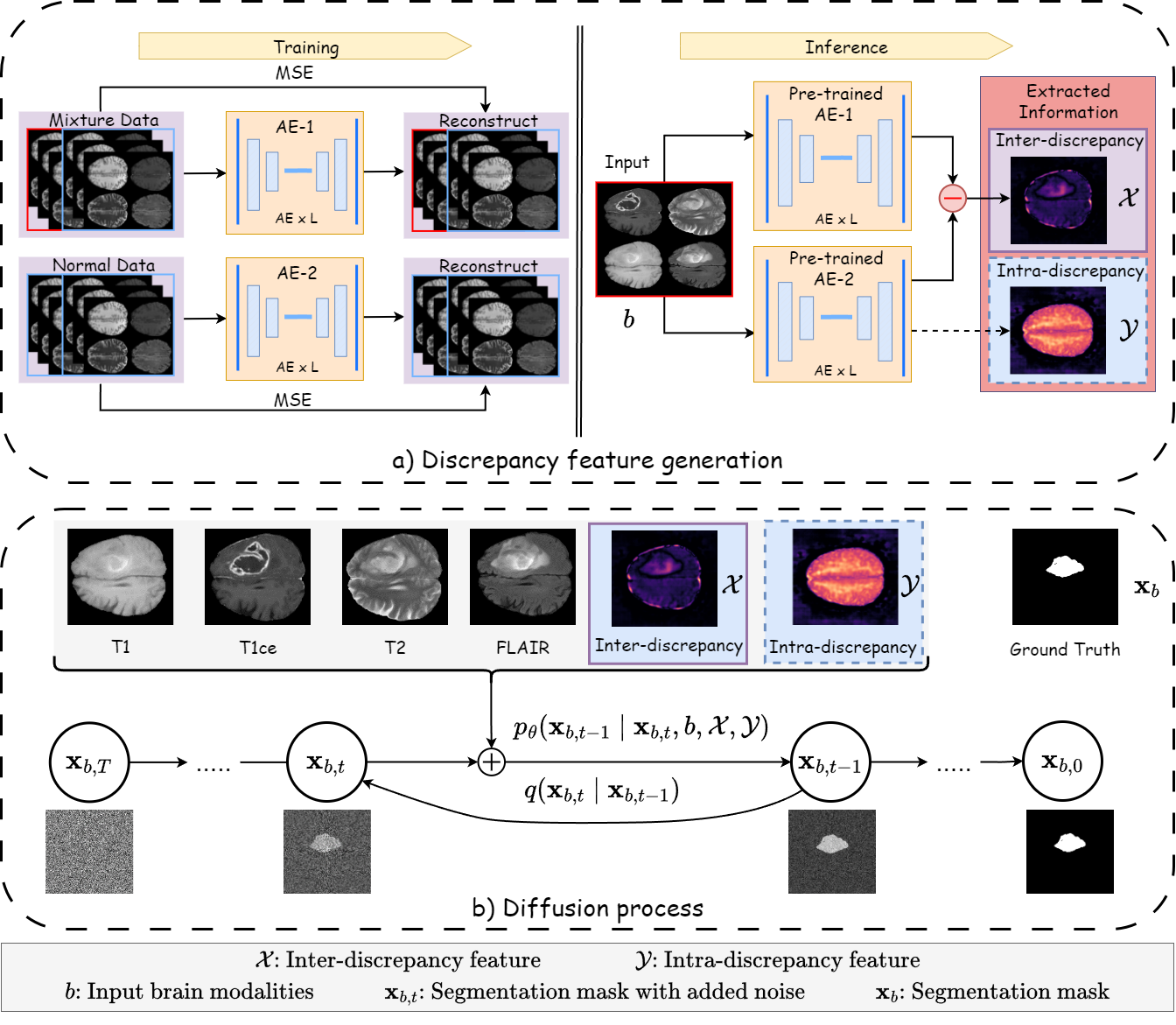
Diffusion probabilistic models (DPMs) have exhibited significant effectiveness in computer vision tasks, particularly in image generation. However, their notable performance heavily relies on labelled datasets, which limits their application in medical images due to the associated high-cost annotations. Current DPM-related methods for lesion detection in medical imaging, which can be categorized into two distinct approaches, primarily rely on image-level annotations. The first approach, based on anomaly detection, involves learning reference healthy brain representations and identifying anomalies based on the difference in inference results. In contrast, the second approach, resembling a segmentation task, employs only the original brain multi-modalities as prior information for generating pixel-level annotations. In this paper, our proposed model - discrepancy distribution medical diffusion (DDMD) - for lesion detection in brain MRI introduces a novel framework by incorporating distinctive discrepancy features, deviating from the conventional direct reliance on image-level annotations or the original brain modalities. In our method, the inconsistency in image-level annotations is translated into distribution discrepancies among heterogeneous samples while preserving information within homogeneous samples. This property retains pixel-wise uncertainty and facilitates an implicit ensemble of segmentation, ultimately enhancing the overall detection performance. Thorough experiments conducted on the BRATS2020 benchmark dataset containing multimodal MRI scans for brain tumour detection demonstrate the great performance of our approach in comparison to state-of-the-art methods.
5/9/2024