Discrepancy-based Diffusion Models for Lesion Detection in Brain MRI
2405.04974
0
0
Abstract
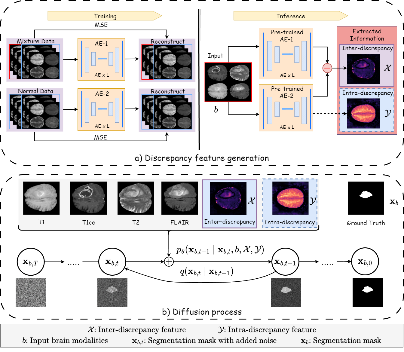
Diffusion probabilistic models (DPMs) have exhibited significant effectiveness in computer vision tasks, particularly in image generation. However, their notable performance heavily relies on labelled datasets, which limits their application in medical images due to the associated high-cost annotations. Current DPM-related methods for lesion detection in medical imaging, which can be categorized into two distinct approaches, primarily rely on image-level annotations. The first approach, based on anomaly detection, involves learning reference healthy brain representations and identifying anomalies based on the difference in inference results. In contrast, the second approach, resembling a segmentation task, employs only the original brain multi-modalities as prior information for generating pixel-level annotations. In this paper, our proposed model - discrepancy distribution medical diffusion (DDMD) - for lesion detection in brain MRI introduces a novel framework by incorporating distinctive discrepancy features, deviating from the conventional direct reliance on image-level annotations or the original brain modalities. In our method, the inconsistency in image-level annotations is translated into distribution discrepancies among heterogeneous samples while preserving information within homogeneous samples. This property retains pixel-wise uncertainty and facilitates an implicit ensemble of segmentation, ultimately enhancing the overall detection performance. Thorough experiments conducted on the BRATS2020 benchmark dataset containing multimodal MRI scans for brain tumour detection demonstrate the great performance of our approach in comparison to state-of-the-art methods.
Create account to get full access
Overview
- This paper presents a novel diffusion-based approach for detecting lesions in brain MRI scans.
- The key idea is to use a discrepancy-based diffusion model, which learns to identify differences between a healthy brain and a brain with lesions.
- The model is trained on pairs of healthy and lesioned brain scans, and can then be used to detect lesions in new images.
- The authors demonstrate the effectiveness of their approach on several brain MRI datasets, showing improved lesion detection compared to other state-of-the-art methods.
Plain English Explanation
The paper describes a new way to detect abnormal growths or injuries (called "lesions") in brain MRI scans using a special type of machine learning model called a "diffusion model". Diffusion models work by learning the differences between healthy brain scans and brain scans with lesions.
The researchers train their model on pairs of healthy and lesioned brain scans. Once trained, the model can then be used to analyze a new brain scan and identify any potential lesions. This is helpful for doctors who need to quickly and accurately detect brain abnormalities in medical images.
Compared to other lesion detection methods, the authors show that their diffusion-based approach performs better at finding lesions in the brain MRI data they tested. This suggests it could be a useful tool for improving medical diagnosis and treatment planning for patients with brain injuries or diseases.
Technical Explanation
The paper introduces a discrepancy-based diffusion model for detecting lesions in brain MRI scans. Diffusion models are a type of generative AI model that can learn complex data distributions, such as the appearance of healthy and lesioned brain anatomy.
The key idea is to train the diffusion model on pairs of healthy and lesioned brain scans. This allows the model to learn the "discrepancies" or differences between normal and abnormal brain structures. At inference time, the model can then be used to identify lesions in new brain MRI images by detecting these discrepancies.
The authors evaluate their approach on several brain MRI datasets, including the ISLES and BRATS challenges. They show that their discrepancy-based diffusion model outperforms other state-of-the-art lesion detection methods in terms of segmentation accuracy and lesion localization.
Critical Analysis
The paper presents a promising new approach for brain lesion detection, but there are a few potential limitations worth considering:
-
The model relies on having access to paired healthy and lesioned brain scans for training, which may not always be available in practice. Cross-modal domain adaptation techniques could help address this issue.
-
The evaluation is primarily focused on segmentation accuracy, but lesion detection in real-world clinical settings may require additional considerations like false positive rates and interpretability.
-
The paper does not provide much insight into the types of lesions the model is best suited for detecting. Further analysis of the model's performance on different lesion characteristics (e.g., size, location, etiology) would be helpful.
-
While the authors demonstrate state-of-the-art results, there may be room for improvement by combining their discrepancy-based approach with other advanced techniques like DiffSeg for more robust lesion segmentation.
Overall, this research represents an exciting step forward in the use of diffusion models for medical image analysis, and the authors' insights could inspire further innovations in this area.
Conclusion
This paper presents a novel diffusion-based approach for detecting lesions in brain MRI scans. By learning to identify discrepancies between healthy and lesioned brain anatomy, the model can accurately locate abnormalities in new images.
The authors demonstrate the effectiveness of their method on several benchmark datasets, showing improved lesion detection performance compared to other state-of-the-art techniques. While the approach has some limitations, it represents a promising step towards more accurate and efficient computer-aided diagnosis of brain diseases and injuries.
Looking ahead, further research could explore ways to make the model more robust and generalizable, with the ultimate goal of developing AI tools that can assist clinicians in providing better care for patients with neurological conditions.
This summary was produced with help from an AI and may contain inaccuracies - check out the links to read the original source documents!
Related Papers
✅
Conditional Diffusion Models for Semantic 3D Brain MRI Synthesis
Zolnamar Dorjsembe, Hsing-Kuo Pao, Sodtavilan Odonchimed, Furen Xiao
0
0
Artificial intelligence (AI) in healthcare, especially in medical imaging, faces challenges due to data scarcity and privacy concerns. Addressing these, we introduce Med-DDPM, a diffusion model designed for 3D semantic brain MRI synthesis. This model effectively tackles data scarcity and privacy issues by integrating semantic conditioning. This involves the channel-wise concatenation of a conditioning image to the model input, enabling control in image generation. Med-DDPM demonstrates superior stability and performance compared to existing 3D brain imaging synthesis methods. It generates diverse, anatomically coherent images with high visual fidelity. In terms of dice score accuracy in the tumor segmentation task, Med-DDPM achieves 0.6207, close to the 0.6531 accuracy of real images, and outperforms baseline models. Combined with real images, it further increases segmentation accuracy to 0.6675, showing the potential of our proposed method for data augmentation. This model represents the first use of a diffusion model in 3D semantic brain MRI synthesis, producing high-quality images. Its semantic conditioning feature also shows potential for image anonymization in biomedical imaging, addressing data and privacy issues. We provide the code and model weights for Med-DDPM on our GitHub repository (https://github.com/mobaidoctor/med-ddpm/) to support reproducibility.
4/22/2024
🤔
A Survey of Emerging Applications of Diffusion Probabilistic Models in MRI
Yuheng Fan, Hanxi Liao, Shiqi Huang, Yimin Luo, Huazhu Fu, Haikun Qi
0
0
Diffusion probabilistic models (DPMs) which employ explicit likelihood characterization and a gradual sampling process to synthesize data, have gained increasing research interest. Despite their huge computational burdens due to the large number of steps involved during sampling, DPMs are widely appreciated in various medical imaging tasks for their high-quality and diversity of generation. Magnetic resonance imaging (MRI) is an important medical imaging modality with excellent soft tissue contrast and superb spatial resolution, which possesses unique opportunities for DPMs. Although there is a recent surge of studies exploring DPMs in MRI, a survey paper of DPMs specifically designed for MRI applications is still lacking. This review article aims to help researchers in the MRI community to grasp the advances of DPMs in different applications. We first introduce the theory of two dominant kinds of DPMs, categorized according to whether the diffusion time step is discrete or continuous, and then provide a comprehensive review of emerging DPMs in MRI, including reconstruction, image generation, image translation, segmentation, anomaly detection, and further research topics. Finally, we discuss the general limitations as well as limitations specific to the MRI tasks of DPMs and point out potential areas that are worth further exploration.
5/9/2024
📉
Re-DiffiNet: Modeling discrepancies in tumor segmentation using diffusion models
Tianyi Ren, Abhishek Sharma, Juampablo Heras Rivera, Harshitha Rebala, Ethan Honey, Agamdeep Chopra, Jacob Ruzevick, Mehmet Kurt
0
0
Identification of tumor margins is essential for surgical decision-making for glioblastoma patients and provides reliable assistance for neurosurgeons. Despite improvements in deep learning architectures for tumor segmentation over the years, creating a fully autonomous system suitable for clinical floors remains a formidable challenge because the model predictions have not yet reached the desired level of accuracy and generalizability for clinical applications. Generative modeling techniques have seen significant improvements in recent times. Specifically, Generative Adversarial Networks (GANs) and Denoising-diffusion-based models (DDPMs) have been used to generate higher-quality images with fewer artifacts and finer attributes. In this work, we introduce a framework called Re-Diffinet for modeling the discrepancy between the outputs of a segmentation model like U-Net and the ground truth, using DDPMs. By explicitly modeling the discrepancy, the results show an average improvement of 0.55% in the Dice score and 16.28% in HD95 from cross-validation over 5-folds, compared to the state-of-the-art U-Net segmentation model.
4/11/2024
🧠
Cross-Modal Domain Adaptation in Brain Disease Diagnosis: Maximum Mean Discrepancy-based Convolutional Neural Networks
Xuran Zhu
0
0
Brain disorders are a major challenge to global health, causing millions of deaths each year. Accurate diagnosis of these diseases relies heavily on advanced medical imaging techniques such as Magnetic Resonance Imaging (MRI) and Computed Tomography (CT). However, the scarcity of annotated data poses a significant challenge in deploying machine learning models for medical diagnosis. To address this limitation, deep learning techniques have shown considerable promise. Domain adaptation techniques enhance a model's ability to generalize across imaging modalities by transferring knowledge from one domain (e.g., CT images) to another (e.g., MRI images). Such cross-modality adaptation is essential to improve the ability of models to consistently generalize across different imaging modalities. This study collected relevant resources from the Kaggle website and employed the Maximum Mean Difference (MMD) method - a popular domain adaptation method - to reduce the differences between imaging domains. By combining MMD with Convolutional Neural Networks (CNNs), the accuracy and utility of the model is obviously enhanced. The excellent experimental results highlight the great potential of data-driven domain adaptation techniques to improve diagnostic accuracy and efficiency, especially in resource-limited environments. By bridging the gap between different imaging modalities, the study aims to provide clinicians with more reliable diagnostic tools.
5/7/2024