Efficient and Accurate Pneumonia Detection Using a Novel Multi-Scale Transformer Approach

0
Sign in to get full access
Overview
- Presents a novel multi-scale transformer approach for efficient and accurate pneumonia detection
- Demonstrates superior performance compared to existing state-of-the-art models
- Leverages a multi-scale architecture to capture features at different granularities
Plain English Explanation
The paper introduces a new transformer-based approach for detecting pneumonia in medical images. Pneumonia is a serious lung infection that can be challenging to diagnose, but early detection is crucial for effective treatment.
The key innovation is the use of a multi-scale architecture, which means the model looks at the images at different levels of detail. This allows it to capture both the high-level structural features and the fine-grained textural details that are indicative of pneumonia.
By combining these multi-scale representations, the model is able to make more accurate and reliable pneumonia diagnoses compared to existing techniques, including convolutional neural networks and other transformer-based approaches.
The authors demonstrate the effectiveness of their method on standard pneumonia detection benchmarks, showing that it outperforms the current state-of-the-art models in terms of both accuracy and efficiency.
Technical Explanation
The paper proposes a novel multi-scale transformer architecture for pneumonia detection. The key components are:
-
Multi-Scale Encoder: The input image is passed through a series of transformer blocks that operate at different spatial resolutions. This allows the model to capture features at multiple scales, from coarse-grained structural information to fine-grained textural details.
-
Attention Fusion: The features extracted at each scale are then fused using a attention-based mechanism, which learns to adaptively combine the multi-scale representations based on the specific characteristics of the input image.
-
Classification Head: The fused multi-scale features are passed through a final classification layer to predict whether the input image shows signs of pneumonia.
The authors conduct extensive experiments on publicly available pneumonia detection datasets, comparing their approach to state-of-the-art convolutional neural network and transformer-based models. Their results demonstrate that the multi-scale transformer outperforms these baselines in terms of both classification accuracy and inference efficiency.
Critical Analysis
The paper presents a compelling and well-designed approach for pneumonia detection, with a strong theoretical foundation and robust experimental evaluation. However, there are a few potential limitations and areas for further research:
-
Dataset Bias: The authors use standard benchmark datasets for pneumonia detection, which may not fully capture the diversity of real-world medical imaging data. Further testing on more diverse and representative datasets would help validate the generalizability of the approach.
-
Interpretability: While the multi-scale transformer architecture is effective, it may be less interpretable than simpler models. Incorporating explainability techniques could help clinicians understand the model's decision-making process and build trust in the system.
-
Real-World Deployment: The paper focuses on the technical performance of the model, but does not address the practical challenges of deploying such a system in a clinical setting. Further research is needed to explore the operational and regulatory considerations for integrating this technology into real-world medical workflows.
Conclusion
The proposed multi-scale transformer approach represents a significant advancement in the field of automated pneumonia detection. By leveraging a multi-scale architecture, the model is able to achieve state-of-the-art performance in both accuracy and efficiency, with the potential to enhance disease diagnosis and improve patient outcomes.
While there are some areas for further refinement and real-world validation, this research demonstrates the power of transformer-based approaches in the medical imaging domain, and suggests promising avenues for continued innovation and development in this important field.
This summary was produced with help from an AI and may contain inaccuracies - check out the links to read the original source documents!
Related Papers
0
Efficient and Accurate Pneumonia Detection Using a Novel Multi-Scale Transformer Approach
Alireza Saber, Pouria Parhami, Alimihammad Siahkarzadeh, Amirreza Fateh
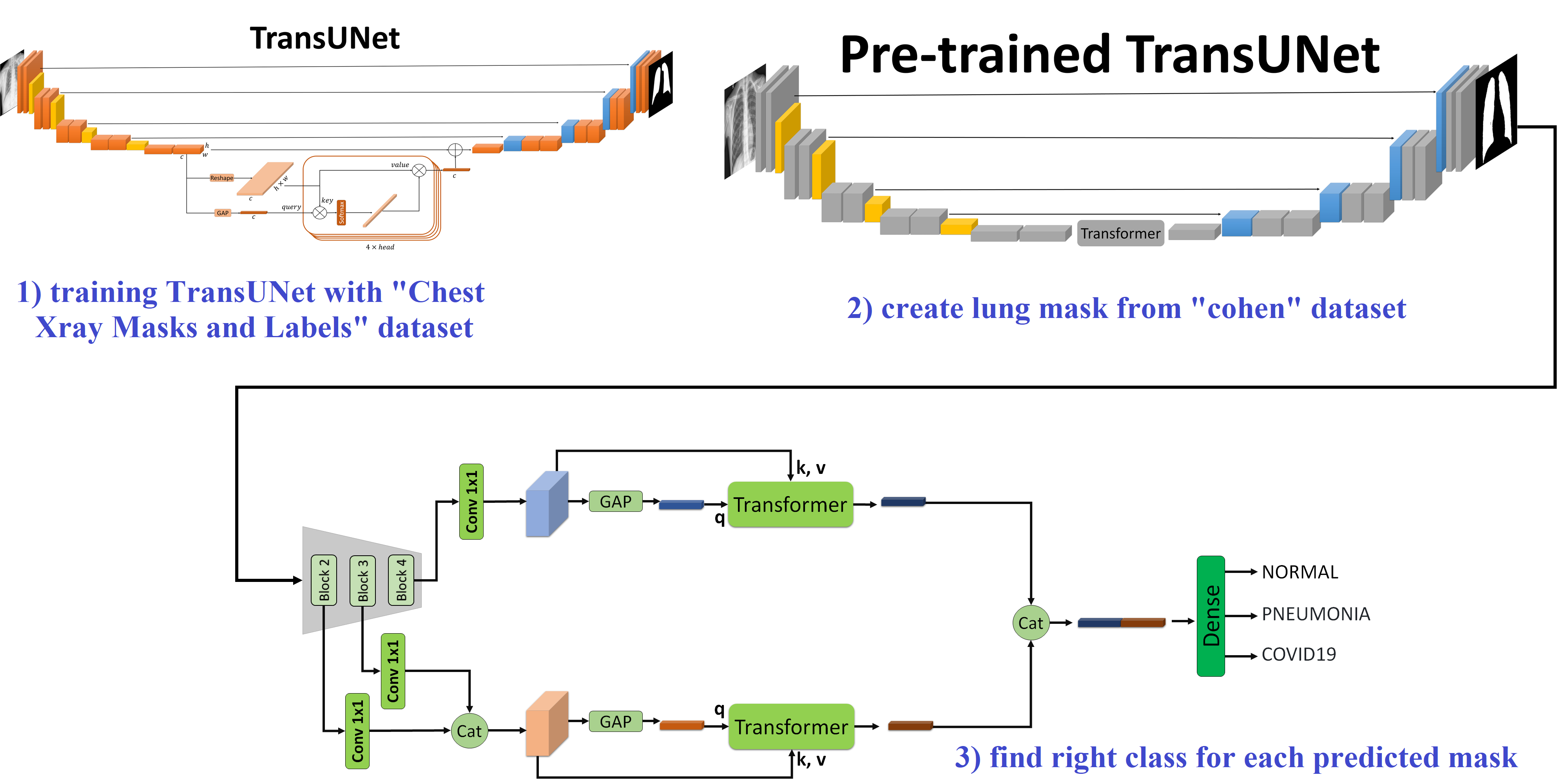
Pneumonia, a severe respiratory disease, poses significant diagnostic challenges, especially in underdeveloped regions. Traditional diagnostic methods, such as chest X-rays, suffer from variability in interpretation among radiologists, necessitating reliable automated tools. In this study, we propose a novel approach combining deep learning and transformer-based attention mechanisms to enhance pneumonia detection from chest X-rays. Our method begins with lung segmentation using a TransUNet model that integrates our specialized transformer module, which has fewer parameters compared to common transformers while maintaining performance. This model is trained on the Chest Xray Masks and Labels dataset and then applied to the Kermany and Cohen datasets to isolate lung regions, enhancing subsequent classification tasks. For classification, we employ pre-trained ResNet models (ResNet-50 and ResNet-101) to extract multi-scale feature maps, processed through our modified transformer module. By employing our specialized transformer, we attain superior results with significantly fewer parameters compared to common transformer models. Our approach achieves high accuracy rates of 92.79% on the Kermany dataset and 95.11% on the Cohen dataset, ensuring robust and efficient performance suitable for resource-constrained environments. https://github.com/amirrezafateh/Multi-Scale-Transformer-Pneumonia
Read more8/9/2024
0
FA-Net: A Fuzzy Attention-aided Deep Neural Network for Pneumonia Detection in Chest X-Rays
Ayush Roy, Anurag Bhattacharjee, Diego Oliva, Oscar Ramos-Soto, Francisco J. Alvarez-Padilla, Ram Sarkar
Pneumonia is a respiratory infection caused by bacteria, fungi, or viruses. It affects many people, particularly those in developing or underdeveloped nations with high pollution levels, unhygienic living conditions, overcrowding, and insufficient medical infrastructure. Pneumonia can cause pleural effusion, where fluids fill the lungs, leading to respiratory difficulty. Early diagnosis is crucial to ensure effective treatment and increase survival rates. Chest X-ray imaging is the most commonly used method for diagnosing pneumonia. However, visual examination of chest X-rays can be difficult and subjective. In this study, we have developed a computer-aided diagnosis system for automatic pneumonia detection using chest X-ray images. We have used DenseNet-121 and ResNet50 as the backbone for the binary class (pneumonia and normal) and multi-class (bacterial pneumonia, viral pneumonia, and normal) classification tasks, respectively. We have also implemented a channel-specific spatial attention mechanism, called Fuzzy Channel Selective Spatial Attention Module (FCSSAM), to highlight the specific spatial regions of relevant channels while removing the irrelevant channels of the extracted features by the backbone. We evaluated the proposed approach on a publicly available chest X-ray dataset, using binary and multi-class classification setups. Our proposed method achieves accuracy rates of 97.15% and 79.79% for the binary and multi-class classification setups, respectively. The results of our proposed method are superior to state-of-the-art (SOTA) methods. The code of the proposed model will be available at: https://github.com/AyushRoy2001/FA-Net.
Read more6/24/2024
🤿
0
Pneumonia Diagnosis through pixels -- A Deep Learning Model for detection and classification
Amit Karanth Gurpur, Janani S, Ajeetha B, Brintha Therese A, Rajeswaran Rangasami
Manual identification and classification of pneumonia and COVID-19 infection is a cumbersome process that, if delayed can cause irreversible damage to the patient. We have compiled CT scan images from various sources, namely, from the China Consortium of Chest CT Image Investigation (CC-CCII), the Negin Radiology located at Sari in Iran, an open access COVID-19 repository from Havard dataverse, and Sri Ramachandra University, Chennai, India. The images were preprocessed using various methods such as normalization, sharpening, median filter application, binarizing, and cropping to ensure uniformity while training the models. We present an ensemble classification approach using deep learning and machine learning methods to classify patients with the said diseases. Our ensemble model uses pre-trained networks such as ResNet-18 and ResNet-50 for classification and MobileNetV2 for feature extraction. The features from MobileNetV2 are used by the gradient-boosting classifier for the classification of patients. Using ResNet-18, ResNet-50, and the MobileNetV2 aided gradient boosting classifier, we propose an ensemble model with an accuracy of 98 percent on unseen data.
Read more4/22/2024
🔎
0
CoVid-19 Detection leveraging Vision Transformers and Explainable AI
Pangoth Santhosh Kumar, Kundrapu Supriya, Mallikharjuna Rao K, Taraka Satya Krishna Teja Malisetti
Lung disease is a common health problem in many parts of the world. It is a significant risk to people health and quality of life all across the globe since it is responsible for five of the top thirty leading causes of death. Among them are COVID 19, pneumonia, and tuberculosis, to name just a few. It is critical to diagnose lung diseases in their early stages. Several different models including machine learning and image processing have been developed for this purpose. The earlier a condition is diagnosed, the better the patient chances of making a full recovery and surviving into the long term. Thanks to deep learning algorithms, there is significant promise for the autonomous, rapid, and accurate identification of lung diseases based on medical imaging. Several different deep learning strategies, including convolutional neural networks (CNN), vanilla neural networks, visual geometry group based networks (VGG), and capsule networks , are used for the goal of making lung disease forecasts. The standard CNN has a poor performance when dealing with rotated, tilted, or other aberrant picture orientations. As a result of this, within the scope of this study, we have suggested a vision transformer based approach end to end framework for the diagnosis of lung disorders. In the architecture, data augmentation, training of the suggested models, and evaluation of the models are all included. For the purpose of detecting lung diseases such as pneumonia, Covid 19, lung opacity, and others, a specialised Compact Convolution Transformers (CCT) model have been tested and evaluated on datasets such as the Covid 19 Radiography Database. The model has achieved a better accuracy for both its training and validation purposes on the Covid 19 Radiography Database.
Read more5/7/2024