An efficient method to automate tooth identification and 3D bounding box extraction from Cone Beam CT Images

0

Sign in to get full access
Overview
• This paper presents an efficient method to automate tooth identification and 3D bounding box extraction from Cone Beam CT (CBCT) images. • The proposed approach leverages deep learning techniques to accurately locate and segment individual teeth in CBCT scans. • The method also extracts 3D bounding boxes around each tooth, providing a comprehensive solution for dental imaging analysis.
Plain English Explanation
Cone Beam CT (CBCT) scans are a type of 3D medical imaging used in dentistry to get detailed views of a patient's teeth and jaw. Analyzing these scans can be important for various dental procedures and treatments, but manually identifying and measuring each individual tooth is a tedious and time-consuming process.
The researchers in this paper developed an automated system to make this task much easier. Their method uses deep learning, a type of artificial intelligence, to automatically detect and outline each tooth in a CBCT scan. The system can not only identify where each tooth is located, but also draw a 3D box around it to measure its size and position.
This automated tooth detection and 3D measurement could save dentists a significant amount of time and effort when working with CBCT scans. It could also help improve the accuracy and consistency of dental analyses compared to manual methods. Overall, the proposed approach represents an efficient way to get valuable information from these 3D dental images.
Technical Explanation
The key components of the proposed method include:
-
A deep learning-based tooth detection model that localizes and segments individual teeth in CBCT scans. This leverages a convolutional neural network architecture to identify the precise outlines of each tooth.
-
A post-processing pipeline that groups the segmented tooth regions into individual 3D bounding boxes. This involves steps like connecting adjacent tooth regions and fitting 3D cuboids to each tooth.
-
An evaluation on a dataset of CBCT scans, demonstrating the method's ability to accurately detect and measure teeth compared to manual ground truth annotations.
The authors show that their automated approach achieves high precision and recall in tooth identification, outperforming previous state-of-the-art methods. The 3D bounding box extraction also enables downstream applications like orthodontic treatment planning and dental prosthetic design.
Critical Analysis
The paper provides a comprehensive solution for automating a key task in dental imaging analysis. However, a few potential limitations and areas for future research are worth noting:
-
The training and evaluation was conducted on a relatively small dataset of CBCT scans. Expanding the dataset size and diversity could further improve the model's robustness.
-
The approach assumes that teeth are fully visible and unobstructed in the CBCT scans. Handling partially occluded or impacted teeth may require additional techniques.
-
While the 3D bounding boxes are useful, extracting more detailed 3D tooth models could enable even more sophisticated dental applications.
Overall, this work demonstrates a strong technical advancement in automating a common dental imaging analysis task. Further research in this direction could lead to significant time and cost savings for dentists, as well as more consistent and reliable dental treatments for patients.
Conclusion
This paper presents an efficient deep learning-based method to automate the identification and 3D measurement of individual teeth in Cone Beam CT scans. By accurately localizing and segmenting each tooth, and then extracting 3D bounding boxes around them, the proposed approach can significantly streamline the analysis of these 3D dental images.
The authors show that their method outperforms previous state-of-the-art techniques, and discuss how the automated tooth detection and measurement capabilities could benefit a range of dental applications, from treatment planning to prosthetic design. While some limitations and areas for future work are noted, this research represents an important step forward in leveraging computer vision and AI for dental imaging analysis.
This summary was produced with help from an AI and may contain inaccuracies - check out the links to read the original source documents!
Related Papers
0
An efficient method to automate tooth identification and 3D bounding box extraction from Cone Beam CT Images
Ignacio Garrido Botella, Ignacio Arranz 'Agueda, Juan Carlos Armenteros Carmona, Oleg Vorontsov, Fernando Bay'on Robledo, Evgeny Solovykh, Obrubov Aleksandr Andreevich, Adri'an Alonso Barriuso
Accurate identification, localization, and segregation of teeth from Cone Beam Computed Tomography (CBCT) images are essential for analyzing dental pathologies. Modeling an individual tooth can be challenging and intricate to accomplish, especially when fillings and other restorations introduce artifacts. This paper proposes a method for automatically detecting, identifying, and extracting teeth from CBCT images. Our approach involves dividing the three-dimensional images into axial slices for image detection. Teeth are pinpointed and labeled using a single-stage object detector. Subsequently, bounding boxes are delineated and identified to create three-dimensional representations of each tooth. The proposed solution has been successfully integrated into the dental analysis tool Dentomo.
Read more7/11/2024
0
A Multi-Stage Framework for 3D Individual Tooth Segmentation in Dental CBCT
Chunshi Wang, Bin Zhao, Shuxue Ding
Cone beam computed tomography (CBCT) is a common way of diagnosing dental related diseases. Accurate segmentation of 3D tooth is of importance for the treatment. Although deep learning based methods have achieved convincing results in medical image processing, they need a large of annotated data for network training, making it very time-consuming in data collection and annotation. Besides, domain shift widely existing in the distribution of data acquired by different devices impacts severely the model generalization. To resolve the problem, we propose a multi-stage framework for 3D tooth segmentation in dental CBCT, which achieves the third place in the Semi-supervised Teeth Segmentation 3D (STS-3D) challenge. The experiments on validation set compared with other semi-supervised segmentation methods further indicate the validity of our approach.
Read more7/16/2024

0
Instance Segmentation and Teeth Classification in Panoramic X-rays
Devichand Budagam, Ayush Kumar, Sayan Ghosh, Anuj Shrivastav, Azamat Zhanatuly Imanbayev, Iskander Rafailovich Akhmetov, Dmitrii Kaplun, Sergey Antonov, Artem Rychenkov, Gleb Cyganov, Aleksandr Sinitca
Teeth segmentation and recognition are critical in various dental applications and dental diagnosis. Automatic and accurate segmentation approaches have been made possible by integrating deep learning models. Although teeth segmentation has been studied in the past, only some techniques were able to effectively classify and segment teeth simultaneously. This article offers a pipeline of two deep learning models, U-Net and YOLOv8, which results in BB-UNet, a new architecture for the classification and segmentation of teeth on panoramic X-rays that is efficient and reliable. We have improved the quality and reliability of teeth segmentation by utilising the YOLOv8 and U-Net capabilities. The proposed networks have been evaluated using the mean average precision (mAP) and dice coefficient for YOLOv8 and BB-UNet, respectively. We have achieved a 3% increase in mAP score for teeth classification compared to existing methods, and a 10-15% increase in dice coefficient for teeth segmentation compared to U-Net across different categories of teeth. A new Dental dataset was created based on UFBA-UESC dataset with Bounding-Box and Polygon annotations of 425 dental panoramic X-rays. The findings of this research pave the way for a wider adoption of object detection models in the field of dental diagnosis.
Read more6/7/2024
0
STS MICCAI 2023 Challenge: Grand challenge on 2D and 3D semi-supervised tooth segmentation
Yaqi Wang, Yifan Zhang, Xiaodiao Chen, Shuai Wang, Dahong Qian, Fan Ye, Feng Xu, Hongyuan Zhang, Qianni Zhang, Chengyu Wu, Yunxiang Li, Weiwei Cui, Shan Luo, Chengkai Wang, Tianhao Li, Yi Liu, Xiang Feng, Huiyu Zhou, Dongyun Liu, Qixuan Wang, Zhouhao Lin, Wei Song, Yuanlin Li, Bing Wang, Chunshi Wang, Qiupu Chen, Mingqian Li
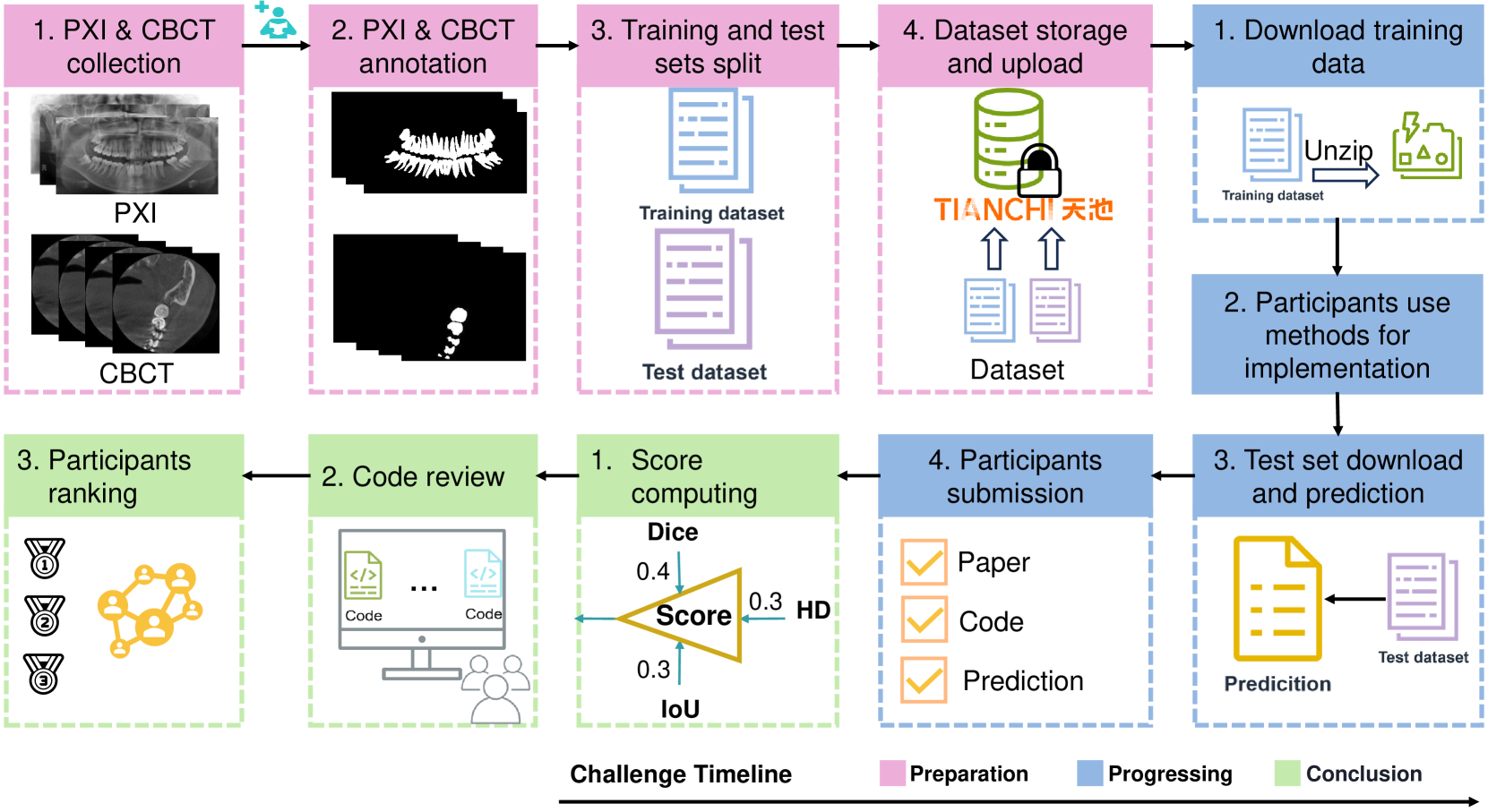
Computer-aided design (CAD) tools are increasingly popular in modern dental practice, particularly for treatment planning or comprehensive prognosis evaluation. In particular, the 2D panoramic X-ray image efficiently detects invisible caries, impacted teeth and supernumerary teeth in children, while the 3D dental cone beam computed tomography (CBCT) is widely used in orthodontics and endodontics due to its low radiation dose. However, there is no open-access 2D public dataset for children's teeth and no open 3D dental CBCT dataset, which limits the development of automatic algorithms for segmenting teeth and analyzing diseases. The Semi-supervised Teeth Segmentation (STS) Challenge, a pioneering event in tooth segmentation, was held as a part of the MICCAI 2023 ToothFairy Workshop on the Alibaba Tianchi platform. This challenge aims to investigate effective semi-supervised tooth segmentation algorithms to advance the field of dentistry. In this challenge, we provide two modalities including the 2D panoramic X-ray images and the 3D CBCT tooth volumes. In Task 1, the goal was to segment tooth regions in panoramic X-ray images of both adult and pediatric teeth. Task 2 involved segmenting tooth sections using CBCT volumes. Limited labelled images with mostly unlabelled ones were provided in this challenge prompt using semi-supervised algorithms for training. In the preliminary round, the challenge received registration and result submission by 434 teams, with 64 advancing to the final round. This paper summarizes the diverse methods employed by the top-ranking teams in the STS MICCAI 2023 Challenge.
Read more7/19/2024