An Explainable Non-local Network for COVID-19 Diagnosis

0

Sign in to get full access
Overview
- This paper presents an "Explainable Non-local Network for COVID-19 Diagnosis" that uses 3D attention mechanisms to automatically classify COVID-19 cases from chest CT scans.
- The model is designed to be interpretable, allowing visualization of the key features it uses to make predictions.
- Experiments show the model achieves high accuracy on COVID-19 diagnosis while providing explanations for its decisions.
Plain English Explanation
The researchers developed a machine learning model that can analyze 3D chest CT scans and automatically detect whether a patient has COVID-19. Unlike many AI models, this one is designed to be "explainable" - it can show the specific regions of the CT scan that it is focusing on to make its diagnosis.
This is important because it allows doctors and patients to understand how the model is making its decisions, rather than just treating it as a "black box." The model uses a special 3D attention mechanism to identify the key COVID-19 features in the CT scans, and these attention maps can be visualized to see what the model is looking at.
The researchers tested the model on a dataset of COVID-19 and non-COVID chest CT scans, and found that it achieved high accuracy in correctly diagnosing COVID-19 cases. Importantly, the attention visualizations showed that the model was focusing on clinically relevant regions of the lungs, providing confidence that the model is making decisions in a sensible way.
Overall, this work represents an advance in AI-based COVID-19 diagnosis, as it combines high performance with built-in interpretability - allowing doctors and patients to understand and trust the model's decision-making process.
Technical Explanation
The core of the researchers' approach is a 3D convolutional neural network architecture that takes a 3D chest CT scan as input and outputs a classification of whether the scan shows signs of COVID-19.
A key innovation is the use of a "non-local" attention mechanism, which allows the model to dynamically focus on the most relevant regions of the 3D CT volume when making its prediction. This attention mechanism produces a set of weights that indicate how much each spatial location in the CT scan contributes to the final classification.
These attention weights can then be visualized as a 3D heatmap, allowing the model's decision-making process to be explained and interpreted. The researchers found that the attention maps highlighted regions of the lungs that are clinically associated with COVID-19, lending credibility to the model's predictions.
In experiments, the model achieved state-of-the-art performance on a benchmark COVID-19 CT scan dataset, outperforming other AI-based COVID-19 diagnosis approaches. This demonstrates the value of the explainable non-local network architecture for accurate and transparent COVID-19 detection from medical imaging.
Critical Analysis
The researchers acknowledge several limitations of their work. First, the dataset used for evaluation, while sizeable, may not fully capture the diversity of COVID-19 presentations in the real world. Further testing on larger and more heterogeneous datasets would be needed to assess the model's generalization capabilities.
Additionally, while the attention visualizations provide insight into the model's decision-making, they do not constitute a full explanation of the underlying reasoning. There may be complex, higher-level patterns that the model is picking up on that are not easily interpretable through simple visualization.
It would also be important to study how the model performs compared to human radiologists in real-world clinical settings. The true test of the model's usefulness will be its ability to augment and enhance human expert diagnosis, rather than simply matching or exceeding it in a research setting.
Overall, this work represents a promising step towards explainable AI for medical diagnosis, but additional research is needed to fully validate the model's practical utility and robustness.
Conclusion
The "Explainable Non-local Network for COVID-19 Diagnosis" presents an innovative approach to automating COVID-19 detection from chest CT scans while providing transparency into the model's decision-making process. By leveraging 3D attention mechanisms, the model can highlight the clinically relevant regions of the scans that are driving its predictions.
This work demonstrates the potential for AI-based medical imaging analysis to not only achieve high performance, but to do so in an explainable manner that builds trust and confidence. As the use of AI in healthcare continues to expand, approaches like this non-local network will be crucial for ensuring the technology is adopted safely and responsibly.
While further research is needed to fully validate the model's real-world applicability, this paper represents an important step forward in the field of explainable AI for medical diagnosis.
This summary was produced with help from an AI and may contain inaccuracies - check out the links to read the original source documents!
Related Papers
0
An Explainable Non-local Network for COVID-19 Diagnosis
Jingfu Yang, Peng Huang, Jing Hu, Shu Hu, Siwei Lyu, Xin Wang, Jun Guo, Xi Wu
The CNN has achieved excellent results in the automatic classification of medical images. In this study, we propose a novel deep residual 3D attention non-local network (NL-RAN) to classify CT images included COVID-19, common pneumonia, and normal to perform rapid and explainable COVID-19 diagnosis. We built a deep residual 3D attention non-local network that could achieve end-to-end training. The network is embedded with a nonlocal module to capture global information, while a 3D attention module is embedded to focus on the details of the lesion so that it can directly analyze the 3D lung CT and output the classification results. The output of the attention module can be used as a heat map to increase the interpretability of the model. 4079 3D CT scans were included in this study. Each scan had a unique label (novel coronavirus pneumonia, common pneumonia, and normal). The CT scans cohort was randomly split into a training set of 3263 scans, a validation set of 408 scans, and a testing set of 408 scans. And compare with existing mainstream classification methods, such as CovNet, CBAM, ResNet, etc. Simultaneously compare the visualization results with visualization methods such as CAM. Model performance was evaluated using the Area Under the ROC Curve(AUC), precision, and F1-score. The NL-RAN achieved the AUC of 0.9903, the precision of 0.9473, and the F1-score of 0.9462, surpass all the classification methods compared. The heat map output by the attention module is also clearer than the heat map output by CAM. Our experimental results indicate that our proposed method performs significantly better than existing methods. In addition, the first attention module outputs a heat map containing detailed outline information to increase the interpretability of the model. Our experiments indicate that the inference of our model is fast. It can provide real-time assistance with diagnosis.
Read more8/9/2024
🧠
0
A design of Convolutional Neural Network model for the Diagnosis of the COVID-19
Xinyuan Song
With the spread of COVID-19 around the globe over the past year, the usage of artificial intelligence (AI) algorithms and image processing methods to analyze the X-ray images of patients' chest with COVID-19 has become essential. The COVID-19 virus recognition in the lung area of a patient is one of the basic and essential needs of clicical centers and hospitals. Most research in this field has been devoted to papers on the basis of deep learning methods utilizing CNNs (Convolutional Neural Network), which mainly deal with the screening of sick and healthy people.In this study, a new structure of a 19-layer CNN has been recommended for accurately recognition of the COVID-19 from the X-ray pictures of chest. The offered CNN is developed to serve as a precise diagnosis system for a three class (viral pneumonia, Normal, COVID) and a four classclassification (Lung opacity, Normal, COVID-19, and pneumonia). A comparison is conducted among the outcomes of the offered procedure and some popular pretrained networks, including Inception, Alexnet, ResNet50, Squeezenet, and VGG19 and based on Specificity, Accuracy, Precision, Sensitivity, Confusion Matrix, and F1-score. The experimental results of the offered CNN method specify its dominance over the existing published procedures. This method can be a useful tool for clinicians in deciding properly about COVID-19.
Read more4/17/2024
0
An Improved CovidConvLSTM model for pneumonia-COVID-19 detection and classification
Imane Beghoura, Mustapha Benssalah, Fazia Sbargoud
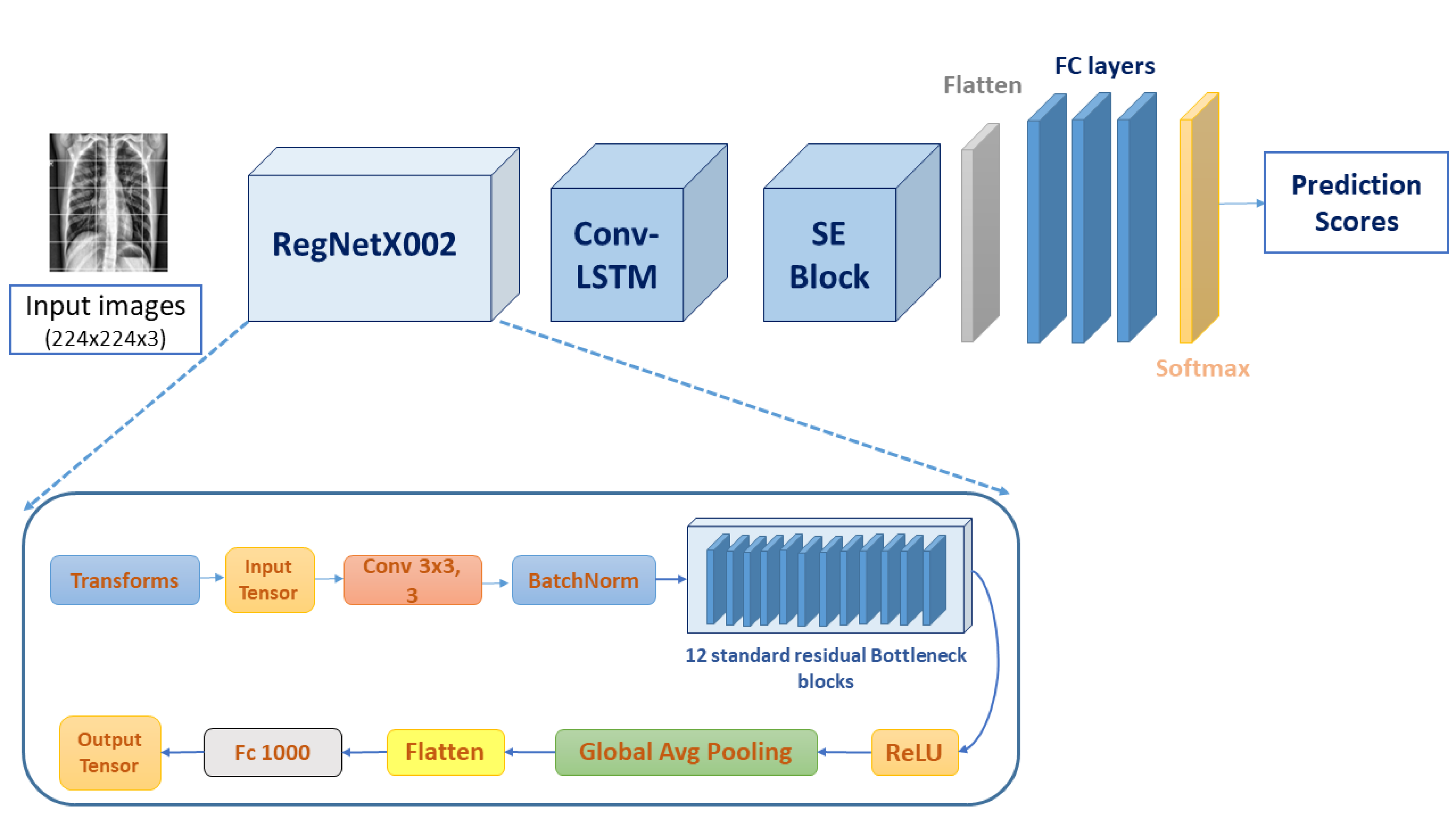
Recently, COVID-19 pandemic has rapidly evolved into a critical global health crisis, profoundly impacting daily life. As a result, CAD systems have gained significant interest for its massive computational capabilities, which facilitate the rapid analysis and interpretation of medical imaging. In particular, Deep Learning (DL )techniques have emerged as critical tools to assist radiologists and pulmonologists in distinguishing COVID-19 patients from other pneumonia types and healthy cases. Unfortunately, existing DL techniques face several challenges such as overfitting, performance degradation, feature irrelevance and redundancy, vanishing gradient problem, and high computational complexity. In this paper we address these challenges by introducing an enhanced Convolutional Neural Network algorithm that combines a bottleneck based model RegNetX002, ConvLstm layer, and Squeeze and Excitation block (SE). Specifically, the RegNetx002 and the ConvLstm layer are used for features map extraction and feature quality enhancement, while the attention mechanism SE block is employed to improve feature representation by highlighting important channel features and suppressing unimportant features. More importantly, The bottleneck module facilitates the extraction of more abstract features while lowering computational costs. Additionally, it incorporates residual connections that helps reducing the vanishing gradient problem. Balanced CPN-CXRPA and imbalanced CXRI-P/C-CXR datasets are used to assess the proposed model. Performance metrics such as accuracy and F1 score are used to evaluate the model efficiency. Using the CPN-CXRPA dataset, our model achieved an accuracy of 98.22%. For the CXRI-P-C-CXR dataset, it achieved 98.78% of both accuracy and F1 score. The experimental results show that this framework outperforms existing models in terms of performance and computational complexity.
Read more8/22/2024
🤿
0
Pneumonia Diagnosis through pixels -- A Deep Learning Model for detection and classification
Amit Karanth Gurpur, Janani S, Ajeetha B, Brintha Therese A, Rajeswaran Rangasami
Manual identification and classification of pneumonia and COVID-19 infection is a cumbersome process that, if delayed can cause irreversible damage to the patient. We have compiled CT scan images from various sources, namely, from the China Consortium of Chest CT Image Investigation (CC-CCII), the Negin Radiology located at Sari in Iran, an open access COVID-19 repository from Havard dataverse, and Sri Ramachandra University, Chennai, India. The images were preprocessed using various methods such as normalization, sharpening, median filter application, binarizing, and cropping to ensure uniformity while training the models. We present an ensemble classification approach using deep learning and machine learning methods to classify patients with the said diseases. Our ensemble model uses pre-trained networks such as ResNet-18 and ResNet-50 for classification and MobileNetV2 for feature extraction. The features from MobileNetV2 are used by the gradient-boosting classifier for the classification of patients. Using ResNet-18, ResNet-50, and the MobileNetV2 aided gradient boosting classifier, we propose an ensemble model with an accuracy of 98 percent on unseen data.
Read more4/22/2024