Explainable vertebral fracture analysis with uncertainty estimation using differentiable rule-based classification

0
🏷️
Sign in to get full access
Overview
- The researchers present a new method for detecting and assessing vertebral fractures in low-dose X-ray images using deep neural networks.
- The method incorporates vertebra detection, keypoint localization, and classification of fracture grade and morphology based on Genant's semi-quantitative criteria.
- The approach provides explainable classifications that align with clinical practices, as well as uncertainty estimates.
- The method outperforms state-of-the-art techniques, achieving a vertebra-level sensitivity of 93% and an end-to-end AUC of 97%.
- The model's reliability is on par with human annotators, as evidenced by a comparison of model uncertainty estimates and intra-reader agreement.
Plain English Explanation
Detecting and assessing vertebral fractures, which are breaks in the bones of the spine, is an important task in medical imaging. The researchers have developed a new deep learning-based method to automate this process using low-dose X-ray images.
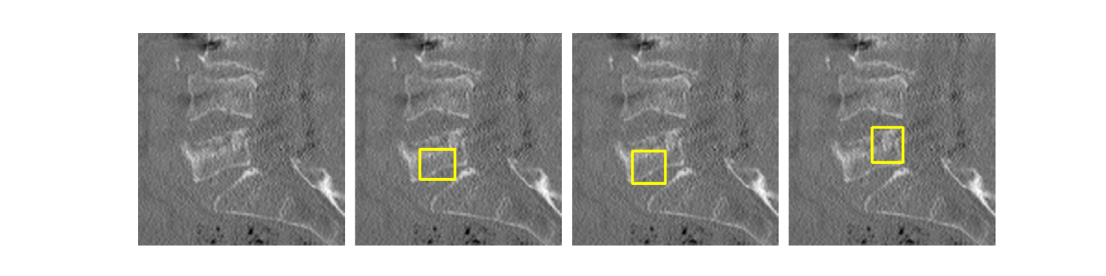
Their approach first identifies the individual vertebrae in the X-ray images and then estimates the specific locations of key points on each vertebra. This information is then used to classify the fracture grade (the severity of the break) and the morphology (the shape) of the vertebra, based on a set of clinical criteria known as Genant's semi-quantitative criteria.
What sets this method apart is that it provides not only the fracture assessment, but also an explanation of how it arrived at that assessment. This aligns with the way clinicians currently evaluate vertebral fractures. Additionally, the method provides an estimate of how confident it is in its assessment, which can help doctors interpret the results.
Importantly, this new technique outperforms previous state-of-the-art methods, achieving very high accuracy in detecting and classifying vertebral fractures. The researchers also show that the model's reliability is comparable to that of human experts who manually assess these X-rays.
Technical Explanation
The key innovation of this work is the incorporation of Genant's semi-quantitative criteria as a differentiable, rule-based means of classifying both vertebra fracture grade and morphology. This allows the deep neural network model to provide explainable classifications that are relatable to current clinical practice, in contrast to previous black-box approaches.
The model first detects the individual vertebrae in the X-ray images using a detection network, then localizes key points on each vertebra using a keypoint estimation network. These vertebra locations and keypoints are then fed into a classification network that assigns a fracture grade (normal, mild, moderate, or severe) and morphology (wedge, biconcave, or crush) based on Genant's criteria.
Importantly, the model also outputs uncertainty estimates for these classifications, providing doctors with a sense of the model's confidence in its assessments. The researchers show that these uncertainty estimates correlate well with inter-reader agreement among human experts, indicating that the model's reliability is on par with human annotators.
Experiments on a challenging dataset demonstrate that this explainable vertebral fracture assessment (XVFA) method surpasses state-of-the-art techniques, achieving a vertebra-level sensitivity of 93% and an end-to-end AUC of 97%. This represents a significant improvement over prior work, including approaches using transfer learning and general-purpose chest X-ray models.
Critical Analysis
The researchers acknowledge several limitations of their study. First, the dataset used for evaluation, while challenging, may not be fully representative of the diversity of vertebral fractures seen in clinical practice. Additionally, the model was trained and evaluated on low-dose radiographs, and its performance on higher-quality images remains to be tested.
Another potential concern is the reliance on Genant's semi-quantitative criteria, which, while widely used, may not capture the full complexity of vertebral fracture assessment. The researchers note that incorporating more nuanced clinical guidelines or leveraging evidential reasoning could further improve the model's performance and clinical relevance.
Overall, the researchers have made a compelling case for the utility of their XVFA method in assisting clinicians with vertebral fracture assessment. By providing explainable classifications and uncertainty estimates, the model has the potential to enhance the interpretability and trustworthiness of automated fracture detection in clinical settings.
Conclusion
The researchers have developed a novel deep learning-based method for explainable vertebral fracture assessment in low-dose radiographs. Their approach, which incorporates vertebra detection, keypoint localization, and classification of fracture grade and morphology, outperforms state-of-the-art techniques while providing clinically relevant explanations and uncertainty estimates.
This work represents an important step towards the integration of AI-powered tools in the clinical workflow for vertebral fracture assessment. By aligning with current practices and providing a measure of model reliability, the XVFA method has the potential to enhance the accuracy and interpretability of automated fracture detection, ultimately improving patient care and outcomes.
This summary was produced with help from an AI and may contain inaccuracies - check out the links to read the original source documents!
Related Papers
🏷️
0
Explainable vertebral fracture analysis with uncertainty estimation using differentiable rule-based classification
Victor W{aa}hlstrand Skarstrom, Lisa Johansson, Jennifer Alv'en, Mattias Lorentzon, Ida Haggstrom
We present a novel method for explainable vertebral fracture assessment (XVFA) in low-dose radiographs using deep neural networks, incorporating vertebra detection and keypoint localization with uncertainty estimates. We incorporate Genant's semi-quantitative criteria as a differentiable rule-based means of classifying both vertebra fracture grade and morphology. Unlike previous work, XVFA provides explainable classifications relatable to current clinical methodology, as well as uncertainty estimations, while at the same time surpassing state-of-the art methods with a vertebra-level sensitivity of 93% and end-to-end AUC of 97% in a challenging setting. Moreover, we compare intra-reader agreement with model uncertainty estimates, with model reliability on par with human annotators.
Read more7/4/2024
0
Enhancing Interpretability of Vertebrae Fracture Grading using Human-interpretable Prototypes
Poulami Sinhamahapatra, Suprosanna Shit, Anjany Sekuboyina, Malek Husseini, David Schinz, Nicolas Lenhart, Joern Menze, Jan Kirschke, Karsten Roscher, Stephan Guennemann
Vertebral fracture grading classifies the severity of vertebral fractures, which is a challenging task in medical imaging and has recently attracted Deep Learning (DL) models. Only a few works attempted to make such models human-interpretable despite the need for transparency and trustworthiness in critical use cases like DL-assisted medical diagnosis. Moreover, such models either rely on post-hoc methods or additional annotations. In this work, we propose a novel interpretable-by-design method, ProtoVerse, to find relevant sub-parts of vertebral fractures (prototypes) that reliably explain the model's decision in a human-understandable way. Specifically, we introduce a novel diversity-promoting loss to mitigate prototype repetitions in small datasets with intricate semantics. We have experimented with the VerSe'19 dataset and outperformed the existing prototype-based method. Further, our model provides superior interpretability against the post-hoc method. Importantly, expert radiologists validated the visual interpretability of our results, showing clinical applicability.
Read more8/1/2024

0
Bone Fracture Classification using Transfer Learning
Shyam Gupta, Dhanisha Sharma
The manual examination of X-ray images for fractures is a time-consuming process that is prone to human error. In this work, we introduce a robust yet simple training loop for the classification of fractures, which significantly outperforms existing methods. Our method achieves superior performance in less than ten epochs and utilizes the latest dataset to deliver the best-performing model for this task. We emphasize the importance of training deep learning models responsibly and efficiently, as well as the critical role of selecting high-quality datasets.
Read more6/26/2024
📈
0
EVA-X: A Foundation Model for General Chest X-ray Analysis with Self-supervised Learning
Jingfeng Yao, Xinggang Wang, Yuehao Song, Huangxuan Zhao, Jun Ma, Yajie Chen, Wenyu Liu, Bo Wang
The diagnosis and treatment of chest diseases play a crucial role in maintaining human health. X-ray examination has become the most common clinical examination means due to its efficiency and cost-effectiveness. Artificial intelligence analysis methods for chest X-ray images are limited by insufficient annotation data and varying levels of annotation, resulting in weak generalization ability and difficulty in clinical dissemination. Here we present EVA-X, an innovative foundational model based on X-ray images with broad applicability to various chest disease detection tasks. EVA-X is the first X-ray image based self-supervised learning method capable of capturing both semantic and geometric information from unlabeled images for universal X-ray image representation. Through extensive experimentation, EVA-X has demonstrated exceptional performance in chest disease analysis and localization, becoming the first model capable of spanning over 20 different chest diseases and achieving leading results in over 11 different detection tasks in the medical field. Additionally, EVA-X significantly reduces the burden of data annotation in the medical AI field, showcasing strong potential in the domain of few-shot learning. The emergence of EVA-X will greatly propel the development and application of foundational medical models, bringing about revolutionary changes in future medical research and clinical practice. Our codes and models are available at: https://github.com/hustvl/EVA-X.
Read more5/9/2024