Guided Context Gating: Learning to leverage salient lesions in retinal fundus images

0
Sign in to get full access
Overview
- This paper proposes a novel deep learning architecture called "Guided Context Gating" (GCG) for the task of retinal lesion detection in fundus images.
- The key idea is to leverage salient lesion information to guide the network's feature extraction and classification process, leading to improved performance on this important medical imaging task.
- The authors demonstrate the effectiveness of GCG on multiple public datasets, showing significant improvements over state-of-the-art methods.
Plain English Explanation
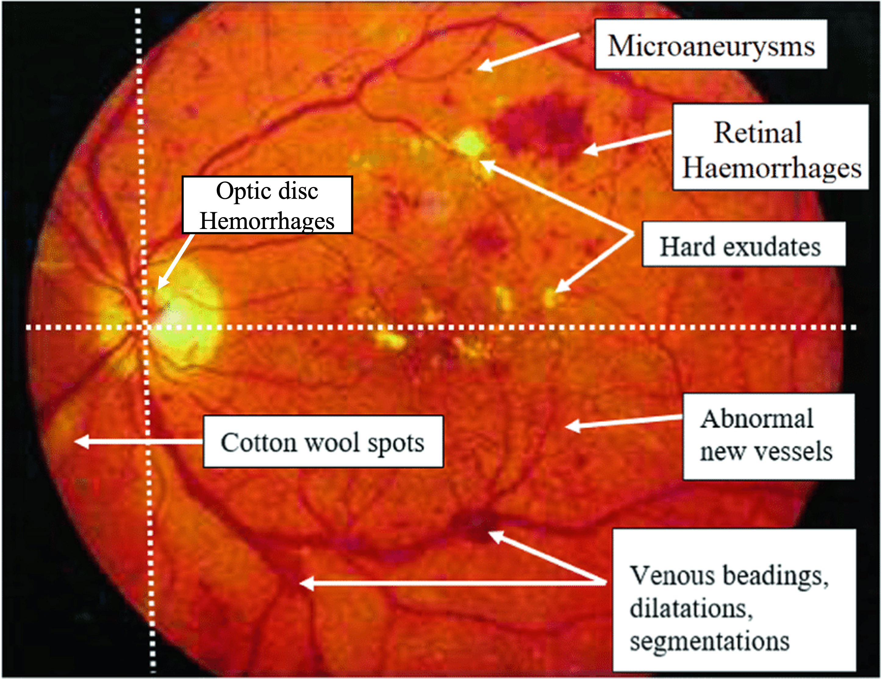
The paper is about a new deep learning model that can help doctors better detect and diagnose eye diseases by analyzing images of the back of the eye (called retinal fundus images). These images contain important information about the health of the eye, including the presence of lesions or abnormalities that can be indicators of diseases like diabetic retinopathy.
The key innovation of this work is a technique called "Guided Context Gating" (GCG). The idea is to have the model first identify the most important or "salient" regions of the eye image that contain lesions or abnormalities. It then uses this information to guide the rest of the model, helping it focus on the relevant parts of the image and make more accurate diagnoses.
This is important because many deep learning models for medical imaging tasks can get distracted by irrelevant parts of the image, leading to mistakes. By explicitly guiding the model to pay attention to the important lesions, the researchers were able to significantly improve the performance of their eye disease detection system.
The authors tested their GCG model on several publicly available datasets of retinal fundus images and showed that it outperformed other state-of-the-art methods. This suggests that their approach could be valuable for developing more accurate and reliable tools to help doctors diagnose and treat eye diseases earlier.
Technical Explanation
The paper introduces a new deep learning architecture called "Guided Context Gating" (GCG) for the task of retinal lesion detection in fundus images. The core idea is to leverage salient lesion information to guide the network's feature extraction and classification process.
Specifically, the GCG model consists of two main components:
- A Saliency Guidance Module that identifies the most important, or salient, regions of the input image that contain lesions or abnormalities.
- A Classification Module that uses the salient region information provided by the Saliency Guidance Module to focus its attention on the relevant parts of the image and make more accurate predictions.
The Saliency Guidance Module is implemented as a small convolutional neural network that outputs a saliency map highlighting the salient regions of the input image. This saliency map is then used to modulate the features extracted by the Classification Module, a process the authors refer to as "guided context gating."
The authors evaluate the GCG model on multiple public retinal fundus image datasets, including Messidor, DRIVE, and REFUGE. They show that GCG outperforms state-of-the-art methods on lesion detection, achieving significant improvements in metrics like area under the ROC curve (AUC-ROC).
Critical Analysis
The key strength of the GCG approach is its ability to explicitly leverage salient lesion information to guide the feature extraction and classification process. This helps the model focus on the most relevant parts of the input image, potentially mitigating issues with shortcut learning or getting distracted by irrelevant image regions.
However, the paper does not provide a deep analysis of the model's failure cases or limitations. It would be valuable to understand scenarios where the GCG model struggles, such as detecting rare or atypical lesions, and how the saliency guidance mechanism might fail in certain situations.
Additionally, the authors could have explored the interpretability of the saliency maps produced by the Saliency Guidance Module. Understanding what visual cues the model is using to identify salient regions could provide insights into the model's decision-making process and help build trust in the system.
Finally, the paper could have discussed the computational overhead and inference time of the GCG model compared to simpler, more lightweight architectures. This information would be important for assessing the practical deployment of the system in real-world clinical settings.
Conclusion
Overall, the Guided Context Gating (GCG) approach presented in this paper is a promising advance in the field of retinal lesion detection. By explicitly leveraging salient lesion information to guide the feature extraction and classification process, the model demonstrates significant performance improvements over state-of-the-art methods on multiple public datasets.
The key innovation of GCG is its ability to focus the model's attention on the most relevant parts of the input image, potentially mitigating issues with shortcut learning and improving the overall reliability and robustness of the system. This work could have important implications for the development of more accurate and trustworthy computer-aided diagnosis tools for eye diseases, ultimately helping clinicians provide better care for their patients.
This summary was produced with help from an AI and may contain inaccuracies - check out the links to read the original source documents!
Related Papers
0
Guided Context Gating: Learning to leverage salient lesions in retinal fundus images
Teja Krishna Cherukuri, Nagur Shareef Shaik, Dong Hye Ye
Effectively representing medical images, especially retinal images, presents a considerable challenge due to variations in appearance, size, and contextual information of pathological signs called lesions. Precise discrimination of these lesions is crucial for diagnosing vision-threatening issues such as diabetic retinopathy. While visual attention-based neural networks have been introduced to learn spatial context and channel correlations from retinal images, they often fall short in capturing localized lesion context. Addressing this limitation, we propose a novel attention mechanism called Guided Context Gating, an unique approach that integrates Context Formulation, Channel Correlation, and Guided Gating to learn global context, spatial correlations, and localized lesion context. Our qualitative evaluation against existing attention mechanisms emphasize the superiority of Guided Context Gating in terms of explainability. Notably, experiments on the Zenodo-DR-7 dataset reveal a substantial 2.63% accuracy boost over advanced attention mechanisms & an impressive 6.53% improvement over the state-of-the-art Vision Transformer for assessing the severity grade of retinopathy, even with imbalanced and limited training samples for each class.
Read more6/21/2024

0
Region Guided Attention Network for Retinal Vessel Segmentation
Syed Javed, Tariq M. Khan, Abdul Qayyum, Arcot Sowmya, Imran Razzak
Retinal imaging has emerged as a promising method of addressing this challenge, taking advantage of the unique structure of the retina. The retina is an embryonic extension of the central nervous system, providing a direct in vivo window into neurological health. Recent studies have shown that specific structural changes in retinal vessels can not only serve as early indicators of various diseases but also help to understand disease progression. In this work, we present a lightweight retinal vessel segmentation network based on the encoder-decoder mechanism with region-guided attention. We introduce inverse addition attention blocks with region guided attention to focus on the foreground regions and improve the segmentation of regions of interest. To further boost the model's performance on retinal vessel segmentation, we employ a weighted dice loss. This choice is particularly effective in addressing the class imbalance issues frequently encountered in retinal vessel segmentation tasks. Dice loss penalises false positives and false negatives equally, encouraging the model to generate more accurate segmentation with improved object boundary delineation and reduced fragmentation. Extensive experiments on a benchmark dataset show better performance (0.8285, 0.8098, 0.9677, and 0.8166 recall, precision, accuracy and F1 score respectively) compared to state-of-the-art methods.
Read more8/22/2024
🌐
0
Lesion-aware network for diabetic retinopathy diagnosis
Xue Xia, Kun Zhan, Yuming Fang, Wenhui Jiang, Fei Shen
Deep learning brought boosts to auto diabetic retinopathy (DR) diagnosis, thus, greatly helping ophthalmologists for early disease detection, which contributes to preventing disease deterioration that may eventually lead to blindness. It has been proved that convolutional neural network (CNN)-aided lesion identifying or segmentation benefits auto DR screening. The key to fine-grained lesion tasks mainly lies in: (1) extracting features being both sensitive to tiny lesions and robust against DR-irrelevant interference, and (2) exploiting and re-using encoded information to restore lesion locations under extremely imbalanced data distribution. To this end, we propose a CNN-based DR diagnosis network with attention mechanism involved, termed lesion-aware network, to better capture lesion information from imbalanced data. Specifically, we design the lesion-aware module (LAM) to capture noise-like lesion areas across deeper layers, and the feature-preserve module (FPM) to assist shallow-to-deep feature fusion. Afterward, the proposed lesion-aware network (LANet) is constructed by embedding the LAM and FPM into the CNN decoders for DR-related information utilization. The proposed LANet is then further extended to a DR screening network by adding a classification layer. Through experiments on three public fundus datasets with pixel-level annotations, our method outperforms the mainstream methods with an area under curve of 0.967 in DR screening, and increases the overall average precision by 7.6%, 2.1%, and 1.2% in lesion segmentation on three datasets. Besides, the ablation study validates the effectiveness of the proposed sub-modules.
Read more8/15/2024
👀
0
Gaze-directed Vision GNN for Mitigating Shortcut Learning in Medical Image
Shaoxuan Wu, Xiao Zhang, Bin Wang, Zhuo Jin, Hansheng Li, Jun Feng
Deep neural networks have demonstrated remarkable performance in medical image analysis. However, its susceptibility to spurious correlations due to shortcut learning raises concerns about network interpretability and reliability. Furthermore, shortcut learning is exacerbated in medical contexts where disease indicators are often subtle and sparse. In this paper, we propose a novel gaze-directed Vision GNN (called GD-ViG) to leverage the visual patterns of radiologists from gaze as expert knowledge, directing the network toward disease-relevant regions, and thereby mitigating shortcut learning. GD-ViG consists of a gaze map generator (GMG) and a gaze-directed classifier (GDC). Combining the global modelling ability of GNNs with the locality of CNNs, GMG generates the gaze map based on radiologists' visual patterns. Notably, it eliminates the need for real gaze data during inference, enhancing the network's practical applicability. Utilizing gaze as the expert knowledge, the GDC directs the construction of graph structures by incorporating both feature distances and gaze distances, enabling the network to focus on disease-relevant foregrounds. Thereby avoiding shortcut learning and improving the network's interpretability. The experiments on two public medical image datasets demonstrate that GD-ViG outperforms the state-of-the-art methods, and effectively mitigates shortcut learning. Our code is available at https://github.com/SX-SS/GD-ViG.
Read more7/31/2024