Heterogeneous virus classification using a functional deep learning model based on transmission electron microscopy images (Preprint)

0
🏷️
Sign in to get full access
Overview
- Viruses are small biological agents that can infect and replicate within living cells.
- Viruses have simple genetic structures but are highly adaptable and can cause serious health issues in their hosts.
- Accurately identifying viruses using manual examination techniques can be challenging, but computer-based methods like analyzing Transmission Electron Microscopy (TEM) images have been successful.
- This study proposes a deep learning-based classification model to identify different types of viruses from TEM images with high accuracy.
Plain English Explanation
Viruses are tiny organisms that can sneak into living things like plants and animals and make copies of themselves using the host's own cells. Even though viruses have some of the simplest building blocks of all living things, they are surprisingly good at adapting and surviving. This means they can sometimes cause big problems for their hosts. It can be tricky to quickly spot a virus in an animal or plant's body and figure out exactly what kind it is just by looking at it. However, analyzing images taken with a special microscope has proven to be a reliable way to identify viruses right away.
In this study, the researchers used a type of artificial intelligence called deep learning to build a model that can accurately classify different kinds of viruses from these microscope images. They started by cleaning up the images to reduce any fuzziness or noise. Then, they trained their deep learning model on the cleaned-up images to teach it how to recognize the unique features of 14 different virus types. The model performed very well, correctly identifying the virus type with up to 97.44% accuracy. This shows that this approach could provide a fast and trustworthy way to diagnose viral infections, alongside other medical tests.
Technical Explanation
This study proposes a deep learning-based classification model to accurately identify different types of viruses from Transmission Electron Microscopy (TEM) images. The researchers first applied two complementary image processing techniques to reduce noise and enhance the visual quality of the raw TEM images. They then trained a deep learning model on the cleaned-up images to classify them into 14 different virus categories.
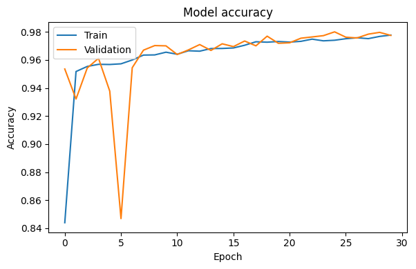
The experimental results demonstrate that the proposed model can achieve a maximum classification accuracy and F1-score of 97.44%, indicating its effectiveness and reliability in virus identification. This approach could provide a fast and dependable way to supplement thorough diagnostic procedures for viral infections, leveraging the power of computer vision and deep learning for automated pathogen detection.
Critical Analysis
The study provides a robust and promising deep learning-based solution for automated virus identification from TEM images. However, the researchers acknowledge that their model was trained and evaluated on a limited dataset, which may not capture the full diversity of viral morphologies encountered in real-world scenarios. Further research is needed to assess the model's performance on larger and more diverse datasets, as well as its generalization capabilities across different imaging modalities and sample preparation techniques.
Additionally, while the reported classification accuracy is impressive, the study does not delve into potential challenges or limitations in deploying such a system in a clinical setting. Factors like processing time, integration with existing workflows, and the need for specialized equipment and expertise should be considered to evaluate the practical feasibility and scalability of the proposed approach.
Conclusion
This study demonstrates the potential of deep learning for fine-grained classification of viral images, which could enable faster and more reliable virus identification compared to manual examination. The high accuracy achieved by the proposed model suggests that such computer-assisted diagnosis tools could significantly improve the efficiency and effectiveness of viral disease detection and management. Further development and validation of this approach may contribute to enhancing our ability to respond to emerging viral threats and mitigate their impact on human, animal, and plant health.
This summary was produced with help from an AI and may contain inaccuracies - check out the links to read the original source documents!
Related Papers
🏷️

0
Heterogeneous virus classification using a functional deep learning model based on transmission electron microscopy images (Preprint)
Niloy Sikder, Md. Al-Masrur Khan, Anupam Kumar Bairagi, Mehedi Masud, Jun Jiat Tiang, Abdullah-Al Nahid
Viruses are submicroscopic agents that can infect all kinds of lifeforms and use their hosts' living cells to replicate themselves. Despite having some of the simplest genetic structures among all living beings, viruses are highly adaptable, resilient, and given the right conditions, are capable of causing unforeseen complications in their hosts' bodies. Due to their multiple transmission pathways, high contagion rate, and lethality, viruses are the biggest biological threat faced by animal and plant species. It is often challenging to promptly detect the presence of a virus in a possible host's body and accurately determine its type using manual examination techniques; however, it can be done using computer-based automatic diagnosis methods. Most notably, the analysis of Transmission Electron Microscopy (TEM) images has been proven to be quite successful in instant virus identification. Using TEM images collected from a recently published dataset, this article proposes a deep learning-based classification model to identify the type of virus within those images correctly. The methodology of this study includes two coherent image processing techniques to reduce the noise present in the raw microscopy images. Experimental results show that it can differentiate among the 14 types of viruses present in the dataset with a maximum of 97.44% classification accuracy and F1-score, which asserts the effectiveness and reliability of the proposed method. Implementing this scheme will impart a fast and dependable way of virus identification subsidiary to the thorough diagnostic procedures.
Read more5/27/2024
🔎

0
Automated Web-Based Malaria Detection System with Machine Learning and Deep Learning Techniques
Abraham G Taye, Sador Yemane, Eshetu Negash, Yared Minwuyelet, Moges Abebe, Melkamu Hunegnaw Asmare
Malaria parasites pose a significant global health burden, causing widespread suffering and mortality. Detecting malaria infection accurately is crucial for effective treatment and control. However, existing automated detection techniques have shown limitations in terms of accuracy and generalizability. Many studies have focused on specific features without exploring more comprehensive approaches. In our case, we formulate a deep learning technique for malaria-infected cell classification using traditional CNNs and transfer learning models notably VGG19, InceptionV3, and Xception. The models were trained using NIH datasets and tested using different performance metrics such as accuracy, precision, recall, and F1-score. The test results showed that deep CNNs achieved the highest accuracy -- 97%, followed by Xception with an accuracy of 95%. A machine learning model SVM achieved an accuracy of 83%, while an Inception-V3 achieved an accuracy of 94%. Furthermore, the system can be accessed through a web interface, where users can upload blood smear images for malaria detection.
Read more7/2/2024


0
Malaria Cell Detection Using Deep Neural Networks
Saurabh Sawant, Anurag Singh
Malaria remains one of the most pressing public health concerns globally, causing significant morbidity and mortality, especially in sub-Saharan Africa. Rapid and accurate diagnosis is crucial for effective treatment and disease management. Traditional diagnostic methods, such as microscopic examination of blood smears, are labor-intensive and require significant expertise, which may not be readily available in resource-limited settings. This project aims to automate the detection of malaria-infected cells using a deep learning approach. We employed a convolutional neural network (CNN) based on the ResNet50 architecture, leveraging transfer learning to enhance performance. The Malaria Cell Images Dataset from Kaggle, containing 27,558 images categorized into infected and uninfected cells, was used for training and evaluation. Our model demonstrated high accuracy, precision, and recall, indicating its potential as a reliable tool for assisting in malaria diagnosis. Additionally, a web application was developed using Streamlit to allow users to upload cell images and receive predictions about malaria infection, making the technology accessible and user-friendly. This paper provides a comprehensive overview of the methodology, experiments, and results, highlighting the effectiveness of deep learning in medical image analysis.
Read more7/1/2024


0
Histopathological Image Classification with Cell Morphology Aware Deep Neural Networks
Andrey Ignatov, Josephine Yates, Valentina Boeva
Histopathological images are widely used for the analysis of diseased (tumor) tissues and patient treatment selection. While the majority of microscopy image processing was previously done manually by pathologists, recent advances in computer vision allow for accurate recognition of lesion regions with deep learning-based solutions. Such models, however, usually require extensive annotated datasets for training, which is often not the case in the considered task, where the number of available patient data samples is very limited. To deal with this problem, we propose a novel DeepCMorph model pre-trained to learn cell morphology and identify a large number of different cancer types. The model consists of two modules: the first one performs cell nuclei segmentation and annotates each cell type, and is trained on a combination of 8 publicly available datasets to ensure its high generalizability and robustness. The second module combines the obtained segmentation map with the original microscopy image and is trained for the downstream task. We pre-trained this module on the Pan-Cancer TCGA dataset consisting of over 270K tissue patches extracted from 8736 diagnostic slides from 7175 patients. The proposed solution achieved a new state-of-the-art performance on the dataset under consideration, detecting 32 cancer types with over 82% accuracy and outperforming all previously proposed solutions by more than 4%. We demonstrate that the resulting pre-trained model can be easily fine-tuned on smaller microscopy datasets, yielding superior results compared to the current top solutions and models initialized with ImageNet weights. The codes and pre-trained models presented in this paper are available at: https://github.com/aiff22/DeepCMorph
Read more7/12/2024