Inter-slice Super-resolution of Magnetic Resonance Images by Pre-training and Self-supervised Fine-tuning
2406.05974
0
0
Abstract
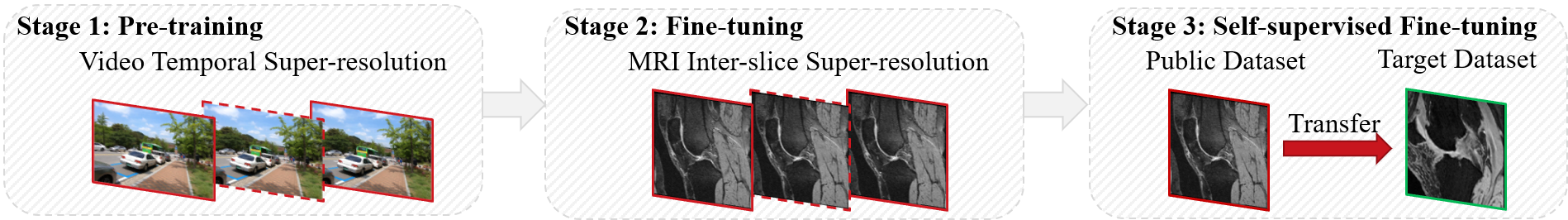
In clinical practice, 2D magnetic resonance (MR) sequences are widely adopted. While individual 2D slices can be stacked to form a 3D volume, the relatively large slice spacing can pose challenges for both image visualization and subsequent analysis tasks, which often require isotropic voxel spacing. To reduce slice spacing, deep-learning-based super-resolution techniques are widely investigated. However, most current solutions require a substantial number of paired high-resolution and low-resolution images for supervised training, which are typically unavailable in real-world scenarios. In this work, we propose a self-supervised super-resolution framework for inter-slice super-resolution of MR images. Our framework is first featured by pre-training on video dataset, as temporal correlation of videos is found beneficial for modeling the spatial relation among MR slices. Then, we use public high-quality MR dataset to fine-tune our pre-trained model, for enhancing awareness of our model to medical data. Finally, given a target dataset at hand, we utilize self-supervised fine-tuning to further ensure our model works well with user-specific super-resolution tasks. The proposed method demonstrates superior performance compared to other self-supervised methods and also holds the potential to benefit various downstream applications.
Create account to get full access
Overview
- This paper presents a method for improving the resolution of magnetic resonance imaging (MRI) scans, specifically focusing on enhancing the resolution between slices (inter-slice super-resolution).
- The approach involves pre-training a deep learning model on a large dataset of MRI scans, then fine-tuning the model using a self-supervised learning technique to further improve its performance on the target task.
- The proposed method is compared to existing super-resolution techniques for MRI, demonstrating improved performance in terms of both quantitative metrics and visual quality.
Plain English Explanation
MRI scans are a widely used medical imaging technique that allows doctors to see inside the human body without invasive procedures. However, the resolution of MRI scans can sometimes be limited, especially in the direction between slices (the "inter-slice" direction). This can make it harder for doctors to see small details or accurately diagnose certain conditions.
The researchers in this paper developed a new way to improve the resolution of MRI scans in the inter-slice direction. They started by training a deep learning model on a large dataset of MRI scans, which helped the model learn general patterns and features in MRI data. Then, they fine-tuned the model using a self-supervised learning technique, which means the model learned to enhance the resolution of the scans without being explicitly told how to do it.
The final model was able to take low-resolution MRI scans and intelligently "fill in the gaps" between slices, resulting in higher-quality images that can help doctors see more detail. The researchers compared their method to other super-resolution techniques and found that it outperformed them, producing MRI scans with better visual quality and more accurate measurements.
Technical Explanation
The paper proposes a two-stage approach for improving the inter-slice super-resolution of MRI scans. The first stage involves pre-training a deep learning model on a large dataset of MRI scans using a standard supervised learning objective. This helps the model learn general features and patterns in MRI data, which can then be leveraged for the super-resolution task.
In the second stage, the pre-trained model is fine-tuned using a self-supervised learning technique. Specifically, the model is trained to predict the missing slices in a partially-observed MRI volume, using the available slices as input. This self-supervised fine-tuning allows the model to further specialize its learned representations for the inter-slice super-resolution problem, without requiring additional manual annotation of the training data.
The authors compare their proposed method to several existing super-resolution techniques for MRI, including Enhancing Super-Resolution Networks through Realistic Thick-Slice Synthesis, Super-Resolution of Biomedical Volumes Using 2D Supervision, and CL-MRI: Self-Supervised Contrastive Learning for MRI Reconstruction. They demonstrate superior performance in terms of both quantitative metrics (such as peak signal-to-noise ratio and structural similarity index) and visual quality of the super-resolved MRI scans.
Critical Analysis
The paper provides a compelling approach for improving the inter-slice super-resolution of MRI scans, leveraging pre-training and self-supervised fine-tuning to enhance the model's performance. One potential limitation is the reliance on the availability of a large dataset of MRI scans for the pre-training stage, which may not always be the case, especially for more specialized medical imaging modalities.
Additionally, the paper does not explore the model's generalization to different types of MRI acquisitions or pathological cases, which could be an important consideration for real-world clinical applications. Further research may be needed to assess the robustness and adaptability of the proposed method to a wider range of MRI data.
The authors also do not provide a detailed discussion of the computational complexity or inference time of their approach, which could be an important factor for practical deployment in clinical settings. Exploring ways to optimize the model's efficiency could further enhance its applicability.
Conclusion
This paper presents a novel method for improving the inter-slice super-resolution of MRI scans, combining pre-training on a large dataset and self-supervised fine-tuning. The proposed approach outperforms existing super-resolution techniques, demonstrating superior performance in terms of both quantitative metrics and visual quality. While the method has some potential limitations, it represents a valuable contribution to the field of medical image super-resolution and could have significant implications for improving the diagnostic capabilities of MRI technology.
This summary was produced with help from an AI and may contain inaccuracies - check out the links to read the original source documents!
Related Papers
🏷️
Enhance the Image: Super Resolution using Artificial Intelligence in MRI
Ziyu Li, Zihan Li, Haoxiang Li, Qiuyun Fan, Karla L. Miller, Wenchuan Wu, Akshay S. Chaudhari, Qiyuan Tian
0
0
This chapter provides an overview of deep learning techniques for improving the spatial resolution of MRI, ranging from convolutional neural networks, generative adversarial networks, to more advanced models including transformers, diffusion models, and implicit neural representations. Our exploration extends beyond the methodologies to scrutinize the impact of super-resolved images on clinical and neuroscientific assessments. We also cover various practical topics such as network architectures, image evaluation metrics, network loss functions, and training data specifics, including downsampling methods for simulating low-resolution images and dataset selection. Finally, we discuss existing challenges and potential future directions regarding the feasibility and reliability of deep learning-based MRI super-resolution, with the aim to facilitate its wider adoption to benefit various clinical and neuroscientific applications.
6/21/2024
🌿
Enhancing Super-Resolution Networks through Realistic Thick-Slice CT Simulation
Zeyu Tang, Xiaodan Xing, Guang Yang
0
0
Deep learning-based Generative Models have the potential to convert low-resolution CT images into high-resolution counterparts without long acquisition times and increased radiation exposure in thin-slice CT imaging. However, procuring appropriate training data for these Super-Resolution (SR) models is challenging. Previous SR research has simulated thick-slice CT images from thin-slice CT images to create training pairs. However, these methods either rely on simplistic interpolation techniques that lack realism or sinogram reconstruction, which require the release of raw data and complex reconstruction algorithms. Thus, we introduce a simple yet realistic method to generate thick CT images from thin-slice CT images, facilitating the creation of training pairs for SR algorithms. The training pairs produced by our method closely resemble real data distributions (PSNR=49.74 vs. 40.66, p$<$0.05). A multivariate Cox regression analysis involving thick slice CT images with lung fibrosis revealed that only the radiomics features extracted using our method demonstrated a significant correlation with mortality (HR=1.19 and HR=1.14, p$<$0.005). This paper represents the first to identify and address the challenge of generating appropriate paired training data for Deep Learning-based CT SR models, which enhances the efficacy and applicability of SR models in real-world scenarios.
6/4/2024

Super-resolution of biomedical volumes with 2D supervision
Cheng Jiang, Alexander Gedeon, Yiwei Lyu, Eric Landgraf, Yufeng Zhang, Xinhai Hou, Akhil Kondepudi, Asadur Chowdury, Honglak Lee, Todd Hollon
0
0
Volumetric biomedical microscopy has the potential to increase the diagnostic information extracted from clinical tissue specimens and improve the diagnostic accuracy of both human pathologists and computational pathology models. Unfortunately, barriers to integrating 3-dimensional (3D) volumetric microscopy into clinical medicine include long imaging times, poor depth / z-axis resolution, and an insufficient amount of high-quality volumetric data. Leveraging the abundance of high-resolution 2D microscopy data, we introduce masked slice diffusion for super-resolution (MSDSR), which exploits the inherent equivalence in the data-generating distribution across all spatial dimensions of biological specimens. This intrinsic characteristic allows for super-resolution models trained on high-resolution images from one plane (e.g., XY) to effectively generalize to others (XZ, YZ), overcoming the traditional dependency on orientation. We focus on the application of MSDSR to stimulated Raman histology (SRH), an optical imaging modality for biological specimen analysis and intraoperative diagnosis, characterized by its rapid acquisition of high-resolution 2D images but slow and costly optical z-sectioning. To evaluate MSDSR's efficacy, we introduce a new performance metric, SliceFID, and demonstrate MSDSR's superior performance over baseline models through extensive evaluations. Our findings reveal that MSDSR not only significantly enhances the quality and resolution of 3D volumetric data, but also addresses major obstacles hindering the broader application of 3D volumetric microscopy in clinical diagnostics and biomedical research.
4/16/2024
🎯
CL-MRI: Self-Supervised Contrastive Learning to Improve the Accuracy of Undersampled MRI Reconstruction
Mevan Ekanayake, Zhifeng Chen, Mehrtash Harandi, Gary Egan, Zhaolin Chen
0
0
In Magnetic Resonance Imaging (MRI), image acquisitions are often undersampled in the measurement domain to accelerate the scanning process, at the expense of image quality. However, image quality is a crucial factor that influences the accuracy of clinical diagnosis; hence, high-quality image reconstruction from undersampled measurements has been a key area of research. Recently, deep learning (DL) methods have emerged as the state-of-the-art for MRI reconstruction, typically involving deep neural networks to transform undersampled MRI images into high-quality MRI images through data-driven processes. Nevertheless, there is clear and significant room for improvement in undersampled DL MRI reconstruction to meet the high standards required for clinical diagnosis, in terms of eliminating aliasing artifacts and reducing image noise. In this paper, we introduce a self-supervised pretraining procedure using contrastive learning to improve the accuracy of undersampled DL MRI reconstruction. We use contrastive learning to transform the MRI image representations into a latent space that maximizes mutual information among different undersampled representations and optimizes the information content at the input of the downstream DL reconstruction models. Our experiments demonstrate improved reconstruction accuracy across a range of acceleration factors and datasets, both quantitatively and qualitatively. Furthermore, our extended experiments validate the proposed framework's robustness under adversarial conditions, such as measurement noise, different k-space sampling patterns, and pathological abnormalities, and also prove the transfer learning capabilities on MRI datasets with completely different anatomy. Additionally, we conducted experiments to visualize and analyze the properties of the proposed MRI contrastive learning latent space.
5/31/2024