Leveraging Persistent Homology for Differential Diagnosis of Mild Cognitive Impairment

0

Sign in to get full access
Overview
- This paper investigates using persistent homology, a branch of topological data analysis, to help diagnose mild cognitive impairment (MCI) from functional magnetic resonance imaging (fMRI) data.
- The researchers developed a framework that combines sliding window embeddings, persistent homology, and deep learning to classify subjects as either healthy or having MCI.
- The proposed approach showed promising results in distinguishing MCI patients from healthy controls, suggesting its potential as a tool for early diagnosis and intervention.
Plain English Explanation
Mild cognitive impairment (MCI) is a condition where a person has slight problems with their thinking and memory, but not severe enough to be considered dementia. Diagnosing MCI can be challenging, as the symptoms can be subtle. This paper explores using a technique called persistent homology to help identify MCI from brain scans.
Persistent homology is a way to analyze the shape and structure of data, like the patterns in brain activity measured by fMRI. The researchers used this to capture changes in brain dynamics over time that could distinguish healthy brains from those with MCI. They developed a model that combined this topological analysis with deep learning to accurately classify whether a person had MCI or not.
The key idea is that the way the brain's activity changes over time may reveal subtle differences between healthy people and those with MCI, which could help with early diagnosis and intervention. This approach provides a new way to analyze brain imaging data that goes beyond just looking at average activity levels, with the potential to uncover more complex biomarkers of cognitive impairment.
Technical Explanation
The researchers proposed a framework that leverages persistent homology, a topological data analysis technique, to capture dynamic changes in fMRI data that can distinguish healthy individuals from those with mild cognitive impairment (MCI).
The main components of their approach are:
-
Sliding Window Embeddings: The fMRI time series data is segmented into overlapping time windows, and each window is embedded into a high-dimensional space using a neural network.
-
Persistent Homology: For each embedded window, the researchers compute the persistent homology, which provides a topological signature of the data's shape and structure. This captures the evolution of topological features across different spatial and temporal scales.
-
Wasserstein Distance: The researchers then compute the Wasserstein distance between the persistent homology representations of each pair of time windows. This distance metric quantifies the dissimilarity between the topological structures.
-
Deep Learning Classification: Finally, the Wasserstein distance matrix is fed into a deep neural network to classify the subject as either healthy or having MCI. The network learns to recognize the patterns in the topological signatures that differentiate the two groups.
The experiments showed that this framework achieved promising results in distinguishing MCI patients from healthy controls, outperforming several baseline methods. The topological features extracted from the fMRI data appear to capture subtle differences in brain dynamics that are indicative of mild cognitive impairment.
Critical Analysis
The paper presents a novel and interesting approach to leveraging topological data analysis for the differential diagnosis of mild cognitive impairment. The use of persistent homology to capture the temporal dynamics of brain activity is a unique and promising avenue of research.
One potential limitation of the study is the relatively small sample size, which may limit the generalizability of the findings. Additionally, the paper does not provide much insight into the specific topological features that were most discriminative between the MCI and healthy groups. Further analysis of the learned representations could shed light on the underlying neurophysiological mechanisms that distinguish these two populations.
It would also be valuable to see how the proposed framework compares to other state-of-the-art approaches for MCI diagnosis, such as those that incorporate multimodal data (e.g., combining fMRI with other neuroimaging or cognitive/behavioral measures). Exploring the complementarity of the topological features with other biomarkers could lead to more robust and comprehensive diagnostic tools.
Overall, this work demonstrates the potential of persistent homology and topological data analysis for advancing our understanding of brain function and dysfunction. As the field continues to evolve, it will be important to carefully evaluate the strengths, limitations, and practical implications of these novel analytical techniques.
Conclusion
This paper presents a promising framework that leverages persistent homology and deep learning to distinguish individuals with mild cognitive impairment from healthy controls based on fMRI data. By capturing the temporal dynamics of brain activity through topological data analysis, the proposed approach shows potential as a tool for early diagnosis and intervention of cognitive decline.
The use of persistent homology to extract meaningful features from complex neuroimaging data represents an exciting direction in the field of computational neuroscience. As this line of research continues to mature, it may lead to better understanding of the underlying neurophysiological changes associated with MCI and other neurological conditions, ultimately enabling more effective clinical decision-making and patient care.
This summary was produced with help from an AI and may contain inaccuracies - check out the links to read the original source documents!
Related Papers


0
Leveraging Persistent Homology for Differential Diagnosis of Mild Cognitive Impairment
Ninad Aithal, Debanjali Bhattacharya, Neelam Sinha, Thomas Gregor Issac
Mild cognitive impairment (MCI) is characterized by subtle changes in cognitive functions, often associated with disruptions in brain connectivity. The present study introduces a novel fine-grained analysis to examine topological alterations in neurodegeneration pertaining to six different brain networks of MCI subjects (Early/Late MCI). To achieve this, fMRI time series from two distinct populations are investigated: (i) the publicly accessible ADNI dataset and (ii) our in-house dataset. The study utilizes sliding window embedding to convert each fMRI time series into a sequence of 3-dimensional vectors, facilitating the assessment of changes in regional brain topology. Distinct persistence diagrams are computed for Betti descriptors of dimension-0, 1, and 2. Wasserstein distance metric is used to quantify differences in topological characteristics. We have examined both (i) ROI-specific inter-subject interactions and (ii) subject-specific inter-ROI interactions. Further, a new deep learning model is proposed for classification, achieving a maximum classification accuracy of 95% for the ADNI dataset and 85% for the in-house dataset. This methodology is further adapted for the differential diagnosis of MCI sub-types, resulting in a peak accuracy of 76.5%, 91.1% and 80% in classifying HC Vs. EMCI, HC Vs. LMCI and EMCI Vs. LMCI, respectively. We showed that the proposed approach surpasses current state-of-the-art techniques designed for classifying MCI and its sub-types using fMRI.
Read more8/29/2024
🌿

0
A Machine Learning Approach for Identifying Anatomical Biomarkers of Early Mild Cognitive Impairment
Alwani Liyana Ahmad, Jose Sanchez-Bornot, Roberto C. Sotero, Damien Coyle, Zamzuri Idris, Ibrahima Faye
Alzheimer Disease poses a significant challenge, necessitating early detection for effective intervention. MRI is a key neuroimaging tool due to its ease of use and cost effectiveness. This study analyzes machine learning methods for MRI based biomarker selection and classification to distinguish between healthy controls and those who develop mild cognitive impairment within five years. Using 3 Tesla MRI data from ADNI and OASIS 3, we applied various machine learning techniques, including MATLAB Classification Learner app, nested cross validation, and Bayesian optimization. Data harmonization with polynomial regression improved performance. Consistent features identified were the entorhinal, hippocampus, lateral ventricle, and lateral orbitofrontal regions. For balanced ADNI data, Naive Bayes with z score harmonization performed best. For balanced OASIS 3, SVM with z score correction excelled. In imbalanced data, RUSBoost showed strong performance on ADNI and OASIS 3. Z score harmonization highlighted the potential of a semi automatic pipeline for early AD detection using MRI.
Read more8/12/2024
🛸

0
Brain Imaging-to-Graph Generation using Adversarial Hierarchical Diffusion Models for MCI Causality Analysis
Qiankun Zuo, Hao Tian, Chi-Man Pun, Hongfei Wang, Yudong Zhang, Jin Hong
Effective connectivity can describe the causal patterns among brain regions. These patterns have the potential to reveal the pathological mechanism and promote early diagnosis and effective drug development for cognitive disease. However, the current methods utilize software toolkits to extract empirical features from brain imaging to estimate effective connectivity. These methods heavily rely on manual parameter settings and may result in large errors during effective connectivity estimation. In this paper, a novel brain imaging-to-graph generation (BIGG) framework is proposed to map functional magnetic resonance imaging (fMRI) into effective connectivity for mild cognitive impairment (MCI) analysis. To be specific, the proposed BIGG framework is based on the diffusion denoising probabilistic models (DDPM), where each denoising step is modeled as a generative adversarial network (GAN) to progressively translate the noise and conditional fMRI to effective connectivity. The hierarchical transformers in the generator are designed to estimate the noise at multiple scales. Each scale concentrates on both spatial and temporal information between brain regions, enabling good quality in noise removal and better inference of causal relations. Meanwhile, the transformer-based discriminator constrains the generator to further capture global and local patterns for improving high-quality and diversity generation. By introducing the diffusive factor, the denoising inference with a large sampling step size is more efficient and can maintain high-quality results for effective connectivity generation. Evaluations of the ADNI dataset demonstrate the feasibility and efficacy of the proposed model. The proposed model not only achieves superior prediction performance compared with other competing methods but also predicts MCI-related causal connections that are consistent with clinical studies.
Read more6/4/2024


0
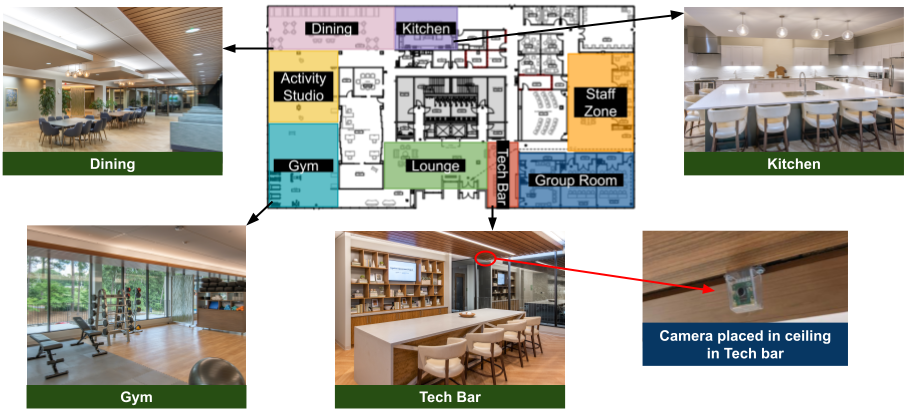
Feasibility of assessing cognitive impairment via distributed camera network and privacy-preserving edge computing
Chaitra Hegde, Yashar Kiarashi, Allan I Levey, Amy D Rodriguez, Hyeokhyen Kwon, Gari D Clifford
INTRODUCTION: Mild cognitive impairment (MCI) is characterized by a decline in cognitive functions beyond typical age and education-related expectations. Since, MCI has been linked to reduced social interactions and increased aimless movements, we aimed to automate the capture of these behaviors to enhance longitudinal monitoring. METHODS: Using a privacy-preserving distributed camera network, we collected movement and social interaction data from groups of individuals with MCI undergoing therapy within a 1700$m^2$ space. We developed movement and social interaction features, which were then used to train a series of machine learning algorithms to distinguish between higher and lower cognitive functioning MCI groups. RESULTS: A Wilcoxon rank-sum test revealed statistically significant differences between high and low-functioning cohorts in features such as linear path length, walking speed, change in direction while walking, entropy of velocity and direction change, and number of group formations in the indoor space. Despite lacking individual identifiers to associate with specific levels of MCI, a machine learning approach using the most significant features provided a 71% accuracy. DISCUSSION: We provide evidence to show that a privacy-preserving low-cost camera network using edge computing framework has the potential to distinguish between different levels of cognitive impairment from the movements and social interactions captured during group activities.
Read more8/21/2024