Meply: A Large-scale Dataset and Baseline Evaluations for Metastatic Perirectal Lymph Node Detection and Segmentation

0

Sign in to get full access
Overview
- This paper introduces Meply, a large-scale dataset and benchmark for evaluating metastatic perirectal lymph node detection and segmentation in medical images.
- Metastatic perirectal lymph nodes are a critical indicator of colorectal cancer progression, and accurate detection and segmentation is essential for diagnosis and treatment.
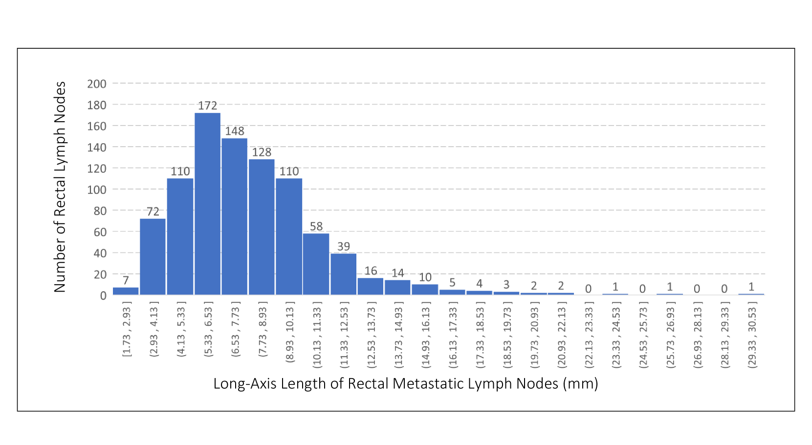
- The Meply dataset contains over 10,000 annotated CT scans with metastatic perirectal lymph node labels, making it the largest of its kind.
- The paper also presents baseline evaluations of several deep learning models for lymph node detection and segmentation tasks.
Plain English Explanation
Meply is a new dataset that can help improve the detection and analysis of a specific type of lymph node that is important for identifying the spread of colorectal cancer. Lymph nodes are small glands that are part of the body's immune system, and when cancer spreads, it can reach these lymph nodes near the rectum.
The Meply dataset contains over 10,000 CT scans (a type of medical imaging) that have been carefully labeled to show where these metastatic perirectal lymph nodes are located. This is the largest dataset of its kind, making it a valuable resource for training and testing new AI models to detect and segment these lymph nodes automatically.
The paper also evaluates several deep learning models - a type of AI that is particularly good at analyzing medical images - to establish baseline performance on the lymph node detection and segmentation tasks using the Meply dataset. This provides a benchmark for comparing the capabilities of different AI approaches on this important problem.
Having a large, high-quality dataset like Meply can accelerate the development of better AI tools for colorectal cancer diagnosis and treatment planning, which could ultimately improve patient outcomes.
Technical Explanation
The paper introduces the Meply dataset, a large-scale collection of over 10,000 annotated CT scans for the task of metastatic perirectal lymph node detection and segmentation. Metastatic perirectal lymph nodes are a crucial indicator of colorectal cancer progression, and accurate localization is essential for disease staging and treatment planning.
The Meply dataset was curated from multiple clinical sites, with each scan manually annotated by experienced radiologists to identify the locations of metastatic perirectal lymph nodes. This comprehensive dataset represents a significant expansion over previous lymph node benchmarks, which have been limited in scale and scope.
To establish baseline performance on the Meply dataset, the authors evaluate several state-of-the-art deep learning models for both lymph node detection and segmentation tasks. The detection models are based on object detection architectures like Faster R-CNN and YOLOX, while the segmentation models leverage popular semantic segmentation frameworks like U-Net and Mask R-CNN.
The results of these baseline experiments demonstrate the challenges of the Meply task, with detection and segmentation performance lagging behind human-level accuracy. The authors identify key areas for improvement, such as better handling of small and occluded lymph nodes, as well as opportunities to leverage the rich contextual information in the CT scans.
By providing this large-scale, high-quality dataset and benchmark evaluations, the Meply paper aims to catalyze further research and development of advanced AI-based tools for colorectal cancer diagnosis and treatment planning.
Critical Analysis
The Meply dataset and benchmark represent a significant contribution to the field of medical image analysis for colorectal cancer. The large scale and comprehensive annotations make it a valuable resource for training and evaluating AI models for lymph node detection and segmentation.
However, the authors acknowledge several limitations of the current dataset and baseline evaluations. For example, the CT scans in Meply were acquired from multiple clinical sites, leading to potential variability in imaging protocols and quality. Additionally, the distribution of lymph node sizes and locations may not fully reflect real-world clinical scenarios.
The authors also note that the baseline deep learning models, while state-of-the-art, still fall short of human-level performance on the Meply tasks. This highlights the need for further advancements in model architectures, training strategies, and incorporating domain-specific knowledge to improve detection and segmentation accuracy.
Future research could also explore ways to leverage the rich contextual information in the CT scans, such as the spatial relationships between lymph nodes and other anatomical structures, to further enhance the AI models' understanding and decision-making.
Additionally, while the Meply dataset is a valuable resource for the research community, its utility in clinical practice will depend on thorough validation and demonstration of improved patient outcomes. Integrating these AI-based tools into the clinical workflow and evaluating their real-world impact will be an important next step.
Conclusion
The Meply dataset and benchmark evaluations presented in this paper represent a significant advancement in the field of medical image analysis for colorectal cancer. By providing a large-scale, high-quality dataset and establishing baseline performance for lymph node detection and segmentation, the authors have laid the groundwork for the development of more accurate and reliable AI-based tools for colorectal cancer diagnosis and treatment planning.
The insights gained from the baseline experiments, as well as the opportunities for future research identified in the paper, suggest that Meply has the potential to catalyze significant progress in this important domain. As the research community continues to build upon this foundation, the ultimate goal is to develop AI systems that can assist clinicians in making more informed decisions, leading to improved patient outcomes and better quality of life for individuals affected by colorectal cancer.
This summary was produced with help from an AI and may contain inaccuracies - check out the links to read the original source documents!
Related Papers
0
Meply: A Large-scale Dataset and Baseline Evaluations for Metastatic Perirectal Lymph Node Detection and Segmentation
Weidong Guo, Hantao Zhang, Shouhong Wan, Bingbing Zou, Wanqin Wang, Chenyang Qiu, Jun Li, Peiquan Jin
Accurate segmentation of metastatic lymph nodes in rectal cancer is crucial for the staging and treatment of rectal cancer. However, existing segmentation approaches face challenges due to the absence of pixel-level annotated datasets tailored for lymph nodes around the rectum. Additionally, metastatic lymph nodes are characterized by their relatively small size, irregular shapes, and lower contrast compared to the background, further complicating the segmentation task. To address these challenges, we present the first large-scale perirectal metastatic lymph node CT image dataset called Meply, which encompasses pixel-level annotations of 269 patients diagnosed with rectal cancer. Furthermore, we introduce a novel lymph-node segmentation model named CoSAM. The CoSAM utilizes sequence-based detection to guide the segmentation of metastatic lymph nodes in rectal cancer, contributing to improved localization performance for the segmentation model. It comprises three key components: sequence-based detection module, segmentation module, and collaborative convergence unit. To evaluate the effectiveness of CoSAM, we systematically compare its performance with several popular segmentation methods using the Meply dataset. Our code and dataset will be publicly available at: https://github.com/kanydao/CoSAM.
Read more4/16/2024

0
LNQ Challenge 2023: Learning Mediastinal Lymph Node Segmentation with a Probabilistic Lymph Node Atlas
Sofija Engelson, Jan Ehrhardt, Timo Kepp, Joshua Niemeijer, Heinz Handels
The evaluation of lymph node metastases plays a crucial role in achieving precise cancer staging, influencing subsequent decisions regarding treatment options. Lymph node detection poses challenges due to the presence of unclear boundaries and the diverse range of sizes and morphological characteristics, making it a resource-intensive process. As part of the LNQ 2023 MICCAI challenge, we propose the use of anatomical priors as a tool to address the challenges that persist in mediastinal lymph node segmentation in combination with the partial annotation of the challenge training data. The model ensemble using all suggested modifications yields a Dice score of 0.6033 and segments 57% of the ground truth lymph nodes, compared to 27% when training on CT only. Segmentation accuracy is improved significantly by incorporating a probabilistic lymph node atlas in loss weighting and post-processing. The largest performance gains are achieved by oversampling fully annotated data to account for the partial annotation of the challenge training data, as well as adding additional data augmentation to address the high heterogeneity of the CT images and lymph node appearance. Our code is available at https://github.com/MICAI-IMI-UzL/LNQ2023.
Read more6/7/2024
🔎
0
A Classification-Based Adaptive Segmentation Pipeline: Feasibility Study Using Polycystic Liver Disease and Metastases from Colorectal Cancer CT Images
Peilong Wang, Timothy L. Kline, Andy D. Missert, Cole J. Cook, Matthew R. Callstrom, Alex Chan, Robert P. Hartman, Zachary S. Kelm, Panagiotis Korfiatis
Automated segmentation tools often encounter accuracy and adaptability issues when applied to images of different pathology. The purpose of this study is to explore the feasibility of building a workflow to efficiently route images to specifically trained segmentation models. By implementing a deep learning classifier to automatically classify the images and route them to appropriate segmentation models, we hope that our workflow can segment the images with different pathology accurately. The data we used in this study are 350 CT images from patients affected by polycystic liver disease and 350 CT images from patients presenting with liver metastases from colorectal cancer. All images had the liver manually segmented by trained imaging analysts. Our proposed adaptive segmentation workflow achieved a statistically significant improvement for the task of total liver segmentation compared to the generic single segmentation model (non-parametric Wilcoxon signed rank test, n=100, p-value << 0.001). This approach is applicable in a wide range of scenarios and should prove useful in clinical implementations of segmentation pipelines.
Read more5/6/2024
0
LN-Gen: Rectal Lymph Nodes Generation via Anatomical Features
Weidong Guo, Hantao Zhang, Shouhong Wan, Bingbing Zou, Wanqin Wang, Peiquan Jin
Accurate segmentation of rectal lymph nodes is crucial for the staging and treatment planning of rectal cancer. However, the complexity of the surrounding anatomical structures and the scarcity of annotated data pose significant challenges. This study introduces a novel lymph node synthesis technique aimed at generating diverse and realistic synthetic rectal lymph node samples to mitigate the reliance on manual annotation. Unlike direct diffusion methods, which often produce masks that are discontinuous and of suboptimal quality, our approach leverages an implicit SDF-based method for mask generation, ensuring the production of continuous, stable, and morphologically diverse masks. Experimental results demonstrate that our synthetic data significantly improves segmentation performance. Our work highlights the potential of diffusion model for accurately synthesizing structurally complex lesions, such as lymph nodes in rectal cancer, alleviating the challenge of limited annotated data in this field and aiding in advancements in rectal cancer diagnosis and treatment.
Read more8/28/2024