Modeling the Neonatal Brain Development Using Implicit Neural Representations

0

Sign in to get full access
Overview
- This paper proposes a novel method for modeling neonatal brain development using implicit neural representations.
- The method represents the brain as a continuous 3D function, allowing for high-fresolution reconstruction and analysis.
- Experiments demonstrate the ability to capture key developmental milestones and structures with high accuracy.
Plain English Explanation
Modeling the development of a baby's brain is an important task in medical research. This paper introduces a new way to approach this challenge. Instead of representing the brain as a collection of discrete points or regions, the researchers propose using implicit neural representations. This means modeling the brain as a continuous 3D function, like a mathematical equation that can describe the entire organ.
The advantage of this approach is that it allows for very detailed, high-resolution reconstruction and analysis of the brain's structure and changes over time. The researchers demonstrate that their method can accurately capture key developmental milestones and features, such as the formation of important brain regions. This could provide valuable insights into the complex process of brain maturation in early life.
Technical Explanation
The paper presents a framework for modeling neonatal brain development using implicit neural representations. The brain is represented as a continuous 3D function, parameterized by a multilayer perceptron neural network. This allows for high-resolution reconstruction and analysis of brain structure and morphology.
The researchers train their model on MRI scans of neonatal brains, capturing developmental changes over time. Experiments show the model's ability to faithfully reconstruct key anatomical features and capture important milestones in brain maturation. For example, the model can accurately trace the formation of the cortex, white matter, and other critical structures.
By leveraging the continuous, high-fidelity representation provided by implicit neural networks, this approach offers advantages over traditional discrete volumetric representations. It enables detailed analysis and potentially opens new avenues for understanding early brain development.
Critical Analysis
The paper presents a promising new technique for modeling neonatal brain development, but it also acknowledges several limitations and areas for further research.
One key limitation is the reliance on MRI data, which can be challenging to acquire, especially for fragile newborns. The authors note that expanding the method to leverage other imaging modalities, such as ultrasound, could broaden its applicability.
Additionally, the current model is trained on a relatively small dataset of neonatal brain scans. Evaluating the method's performance on larger, more diverse cohorts would be an important next step to ensure its robustness and generalizability.
The paper also does not delve deeply into the clinical implications and potential applications of this work. Further research is needed to understand how this model could inform medical decision-making, guide interventions, or enhance our understanding of atypical brain development in conditions like autism or cerebral palsy.
Overall, this research represents an intriguing advance in the field of computational neuroscience, with the potential to shed new light on the complex process of neonatal brain maturation. However, continued development and validation will be crucial to translate these findings into meaningful clinical impact.
Conclusion
This paper introduces a novel method for modeling neonatal brain development using implicit neural representations. By representing the brain as a continuous 3D function, the approach enables high-resolution reconstruction and analysis of key anatomical features and developmental milestones.
The experimental results demonstrate the model's ability to faithfully capture important aspects of brain maturation, laying the groundwork for further research and potential clinical applications. While the current work has some limitations, it represents a promising step forward in our understanding of early brain development and could ultimately contribute to improved diagnostic, prognostic, and therapeutic approaches in pediatric neuroscience.
This summary was produced with help from an AI and may contain inaccuracies - check out the links to read the original source documents!
Related Papers
0
Modeling the Neonatal Brain Development Using Implicit Neural Representations
Florentin Bieder, Paul Friedrich, H'el`ene Corbaz, Alicia Durrer, Julia Wolleb, Philippe C. Cattin
The human brain undergoes rapid development during the third trimester of pregnancy. In this work, we model the neonatal development of the infant brain in this age range. As a basis, we use MR images of preterm- and term-birth neonates from the developing human connectome project (dHCP). We propose a neural network, specifically an implicit neural representation (INR), to predict 2D- and 3D images of varying time points. In order to model a subject-specific development process, it is necessary to disentangle the age from the subjects' identity in the latent space of the INR. We propose two methods, Subject Specific Latent Vectors (SSL) and Stochastic Global Latent Augmentation (SGLA), enabling this disentanglement. We perform an analysis of the results and compare our proposed model to an age-conditioned denoising diffusion model as a baseline. We also show that our method can be applied in a memory-efficient way, which is especially important for 3D data.
Read more8/19/2024

0
The Developing Human Connectome Project: A Fast Deep Learning-based Pipeline for Neonatal Cortical Surface Reconstruction
Qiang Ma, Kaili Liang, Liu Li, Saga Masui, Yourong Guo, Chiara Nosarti, Emma C. Robinson, Bernhard Kainz, Daniel Rueckert
The Developing Human Connectome Project (dHCP) aims to explore developmental patterns of the human brain during the perinatal period. An automated processing pipeline has been developed to extract high-quality cortical surfaces from structural brain magnetic resonance (MR) images for the dHCP neonatal dataset. However, the current implementation of the pipeline requires more than 6.5 hours to process a single MRI scan, making it expensive for large-scale neuroimaging studies. In this paper, we propose a fast deep learning (DL) based pipeline for dHCP neonatal cortical surface reconstruction, incorporating DL-based brain extraction, cortical surface reconstruction and spherical projection, as well as GPU-accelerated cortical surface inflation and cortical feature estimation. We introduce a multiscale deformation network to learn diffeomorphic cortical surface reconstruction end-to-end from T2-weighted brain MRI. A fast unsupervised spherical mapping approach is integrated to minimize metric distortions between cortical surfaces and projected spheres. The entire workflow of our DL-based dHCP pipeline completes within only 24 seconds on a modern GPU, which is nearly 1000 times faster than the original dHCP pipeline. Manual quality control demonstrates that for 82.5% of the test samples, our DL-based pipeline produces superior (54.2%) or equal quality (28.3%) cortical surfaces compared to the original dHCP pipeline.
Read more5/15/2024

0
Detailed delineation of the fetal brain in diffusion MRI via multi-task learning
Davood Karimi, Camilo Calixto, Haykel Snoussi, Maria Camila Cortes-Albornoz, Clemente Velasco-Annis, Caitlin Rollins, Camilo Jaimes, Ali Gholipour, Simon K. Warfield
Diffusion-weighted MRI is increasingly used to study the normal and abnormal development of fetal brain in-utero. Recent studies have shown that dMRI can offer invaluable insights into the neurodevelopmental processes in the fetal stage. However, because of the low data quality and rapid brain development, reliable analysis of fetal dMRI data requires dedicated computational methods that are currently unavailable. The lack of automated methods for fast, accurate, and reproducible data analysis has seriously limited our ability to tap the potential of fetal brain dMRI for medical and scientific applications. In this work, we developed and validated a unified computational framework to (1) segment the brain tissue into white matter, cortical/subcortical gray matter, and cerebrospinal fluid, (2) segment 31 distinct white matter tracts, and (3) parcellate the brain's cortex and delineate the deep gray nuclei and white matter structures into 96 anatomically meaningful regions. We utilized a set of manual, semi-automatic, and automatic approaches to annotate 97 fetal brains. Using these labels, we developed and validated a multi-task deep learning method to perform the three computations. Our evaluations show that the new method can accurately carry out all three tasks, achieving a mean Dice similarity coefficient of 0.865 on tissue segmentation, 0.825 on white matter tract segmentation, and 0.819 on parcellation. The proposed method can greatly advance the field of fetal neuroimaging as it can lead to substantial improvements in fetal brain tractography, tract-specific analysis, and structural connectivity assessment.
Read more9/14/2024
0
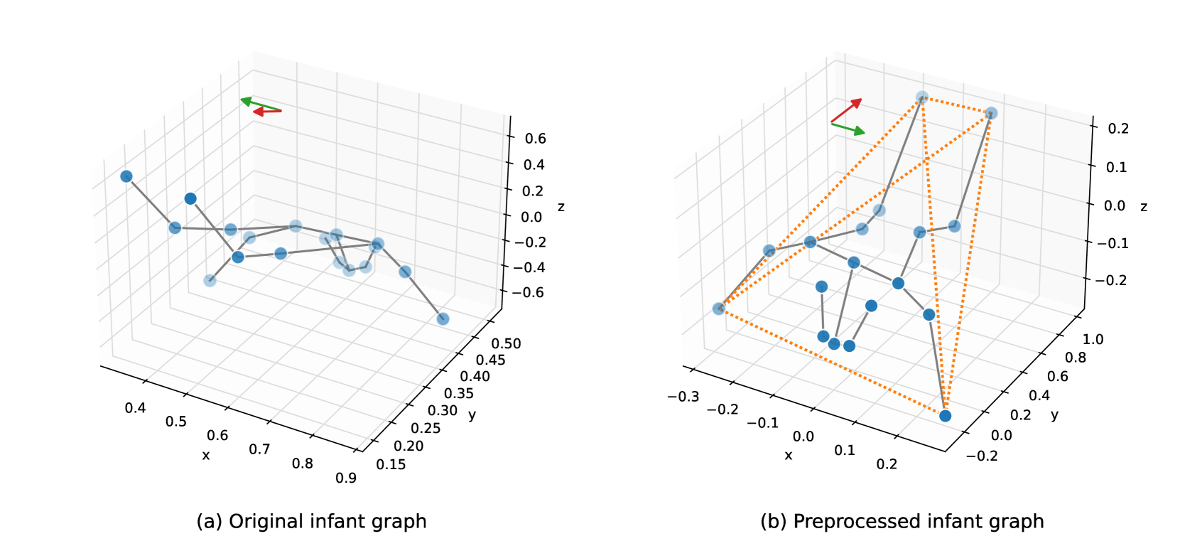
Modeling 3D Infant Kinetics Using Adaptive Graph Convolutional Networks
Daniel Holmberg, Manu Airaksinen, Viviana Marchi, Andrea Guzzetta, Anna Kivi, Leena Haataja, Sampsa Vanhatalo, Teemu Roos
Reliable methods for the neurodevelopmental assessment of infants are essential for early detection of medical issues that may need prompt interventions. Spontaneous motor activity, or 'kinetics', is shown to provide a powerful surrogate measure of upcoming neurodevelopment. However, its assessment is by and large qualitative and subjective, focusing on visually identified, age-specific gestures. Here, we follow an alternative approach, predicting infants' neurodevelopmental maturation based on data-driven evaluation of individual motor patterns. We utilize 3D video recordings of infants processed with pose-estimation to extract spatio-temporal series of anatomical landmarks, and apply adaptive graph convolutional networks to predict the actual age. We show that our data-driven approach achieves improvement over traditional machine learning baselines based on manually engineered features.
Read more6/21/2024