nnU-Net Revisited: A Call for Rigorous Validation in 3D Medical Image Segmentation
2404.09556
0
0

Abstract
The release of nnU-Net marked a paradigm shift in 3D medical image segmentation, demonstrating that a properly configured U-Net architecture could still achieve state-of-the-art results. Despite this, the pursuit of novel architectures, and the respective claims of superior performance over the U-Net baseline, continued. In this study, we demonstrate that many of these recent claims fail to hold up when scrutinized for common validation shortcomings, such as the use of inadequate baselines, insufficient datasets, and neglected computational resources. By meticulously avoiding these pitfalls, we conduct a thorough and comprehensive benchmarking of current segmentation methods including CNN-based, Transformer-based, and Mamba-based approaches. In contrast to current beliefs, we find that the recipe for state-of-the-art performance is 1) employing CNN-based U-Net models, including ResNet and ConvNeXt variants, 2) using the nnU-Net framework, and 3) scaling models to modern hardware resources. These results indicate an ongoing innovation bias towards novel architectures in the field and underscore the need for more stringent validation standards in the quest for scientific progress.
Create account to get full access
Overview
• This paper critically examines the validation practices in 3D medical image segmentation, focusing on the popular nnU-Net framework.
• It highlights several common pitfalls in the validation of these models, including the use of inappropriate baselines, data leakage, and the lack of cross-dataset evaluation.
Plain English Explanation
The paper discusses the importance of rigorous validation in the field of 3D medical image segmentation, which is a critical task for accurately identifying and analyzing different structures within medical scans. The authors specifically look at the validation practices used with the nnU-Net framework, a widely-adopted deep learning model for this purpose.
The paper identifies several common issues that can arise when validating these types of models, such as using inappropriate baseline comparisons, allowing data to "leak" between the training and validation sets, and failing to test the models on diverse datasets beyond the ones they were trained on. These validation pitfalls can lead to overly optimistic performance estimates and make it difficult to truly assess the capabilities and limitations of the models.
By highlighting these validation challenges, the paper aims to encourage researchers in this field to be more rigorous and transparent in their evaluation methods, in order to ensure the models developed are truly robust and generalizable. Proper validation is essential for building confidence in the use of these systems in real-world medical applications.
Technical Explanation
The paper focuses on the validation practices used with the nnU-Net framework, a popular deep learning model for 3D medical image segmentation. The authors identify several common pitfalls in the validation process:
-
Baseline Selection: The authors note that many papers use weak or inappropriate baselines for comparison, such as traditional segmentation methods or simpler neural network architectures. This can make the performance of nnU-Net appear more impressive than it truly is.
-
Data Leakage: The authors find that researchers often fail to properly separate training and validation data, allowing information to "leak" between the sets. This can lead to overly optimistic performance metrics that do not reflect the model's true generalization capabilities.
-
Cross-Dataset Evaluation: The paper emphasizes the importance of testing models on diverse datasets beyond the ones they were trained on. Many papers only evaluate on a single dataset, which makes it difficult to assess how well the models will perform in real-world clinical settings.
To address these issues, the authors recommend that researchers adopt more rigorous validation practices, such as using strong and relevant baselines, implementing strict data separation protocols, and conducting thorough cross-dataset evaluations. They argue that this will lead to a more accurate and reliable understanding of the capabilities and limitations of 3D medical image segmentation models like nnU-Net.
Critical Analysis
The paper raises valid concerns about the validation practices in 3D medical image segmentation research, particularly around the use of nnU-Net. The authors make a compelling case for the need to address these issues, as overoptimistic performance claims can lead to unrealistic expectations and potentially put patients at risk if these models are deployed in clinical settings without proper evaluation.
However, the paper could have delved deeper into the specific reasons why these validation pitfalls are so common in this field. For example, the authors could have discussed the challenges of obtaining diverse, high-quality medical imaging datasets, or the pressure researchers may feel to produce "impressive" results that are more likely to be published.
Additionally, the paper could have provided more concrete recommendations for how researchers can implement rigorous validation practices, such as specific techniques for data partitioning, guidelines for selecting appropriate baselines, and best practices for cross-dataset evaluations.
Overall, the paper successfully highlights the importance of validation in 3D medical image segmentation and serves as a valuable call to action for the research community to adopt more rigorous evaluation methods. Addressing these concerns will be crucial for ensuring the development of reliable and trustworthy AI systems for clinical applications.
Conclusion
This paper provides a critical analysis of the validation practices commonly used in 3D medical image segmentation, with a focus on the nnU-Net framework. The authors identify several key pitfalls, such as the use of weak baselines, data leakage, and the lack of cross-dataset evaluation, that can lead to overly optimistic performance claims.
By highlighting these issues, the paper emphasizes the need for researchers in this field to adopt more rigorous validation protocols. Proper evaluation is essential for building confidence in the capabilities and limitations of these AI-powered medical imaging tools, which will be crucial for their safe and effective deployment in real-world clinical settings.
The authors' call for increased validation rigor is a timely and important contribution to the ongoing efforts to ensure the responsible development and deployment of advanced medical imaging technologies.
This summary was produced with help from an AI and may contain inaccuracies - check out the links to read the original source documents!
Related Papers
🤿
Deep Learning-Based Brain Image Segmentation for Automated Tumour Detection
Suman Sourabh, Murugappan Valliappan, Narayana Darapaneni, Anwesh R P
0
0
Introduction: The present study on the development and evaluation of an automated brain tumor segmentation technique based on deep learning using the 3D U-Net model. Objectives: The objective is to leverage state-of-the-art convolutional neural networks (CNNs) on a large dataset of brain MRI scans for segmentation. Methods: The proposed methodology applies pre-processing techniques for enhanced performance and generalizability. Results: Extensive validation on an independent dataset confirms the model's robustness and potential for integration into clinical workflows. The study emphasizes the importance of data pre-processing and explores various hyperparameters to optimize the model's performance. The 3D U-Net, has given IoUs for training and validation dataset have been 0.8181 and 0.66 respectively. Conclusion: Ultimately, this comprehensive framework showcases the efficacy of deep learning in automating brain tumour detection, offering valuable support in clinical practice.
4/10/2024
Hybrid Multihead Attentive Unet-3D for Brain Tumor Segmentation
Muhammad Ansab Butt, Absaar Ul Jabbar
0
0
Brain tumor segmentation is a critical task in medical image analysis, aiding in the diagnosis and treatment planning of brain tumor patients. The importance of automated and accurate brain tumor segmentation cannot be overstated. It enables medical professionals to precisely delineate tumor regions, assess tumor growth or regression, and plan targeted treatments. Various deep learning-based techniques proposed in the literature have made significant progress in this field, however, they still face limitations in terms of accuracy due to the complex and variable nature of brain tumor morphology. In this research paper, we propose a novel Hybrid Multihead Attentive U-Net architecture, to address the challenges in accurate brain tumor segmentation, and to capture complex spatial relationships and subtle tumor boundaries. The U-Net architecture has proven effective in capturing contextual information and feature representations, while attention mechanisms enhance the model's ability to focus on informative regions and refine the segmentation boundaries. By integrating these two components, our proposed architecture improves accuracy in brain tumor segmentation. We test our proposed model on the BraTS 2020 benchmark dataset and compare its performance with the state-of-the-art well-known SegNet, FCN-8s, and Dense121 U-Net architectures. The results show that our proposed model outperforms the others in terms of the evaluated performance metrics.
5/24/2024
ViM-UNet: Vision Mamba for Biomedical Segmentation
Anwai Archit, Constantin Pape
0
0
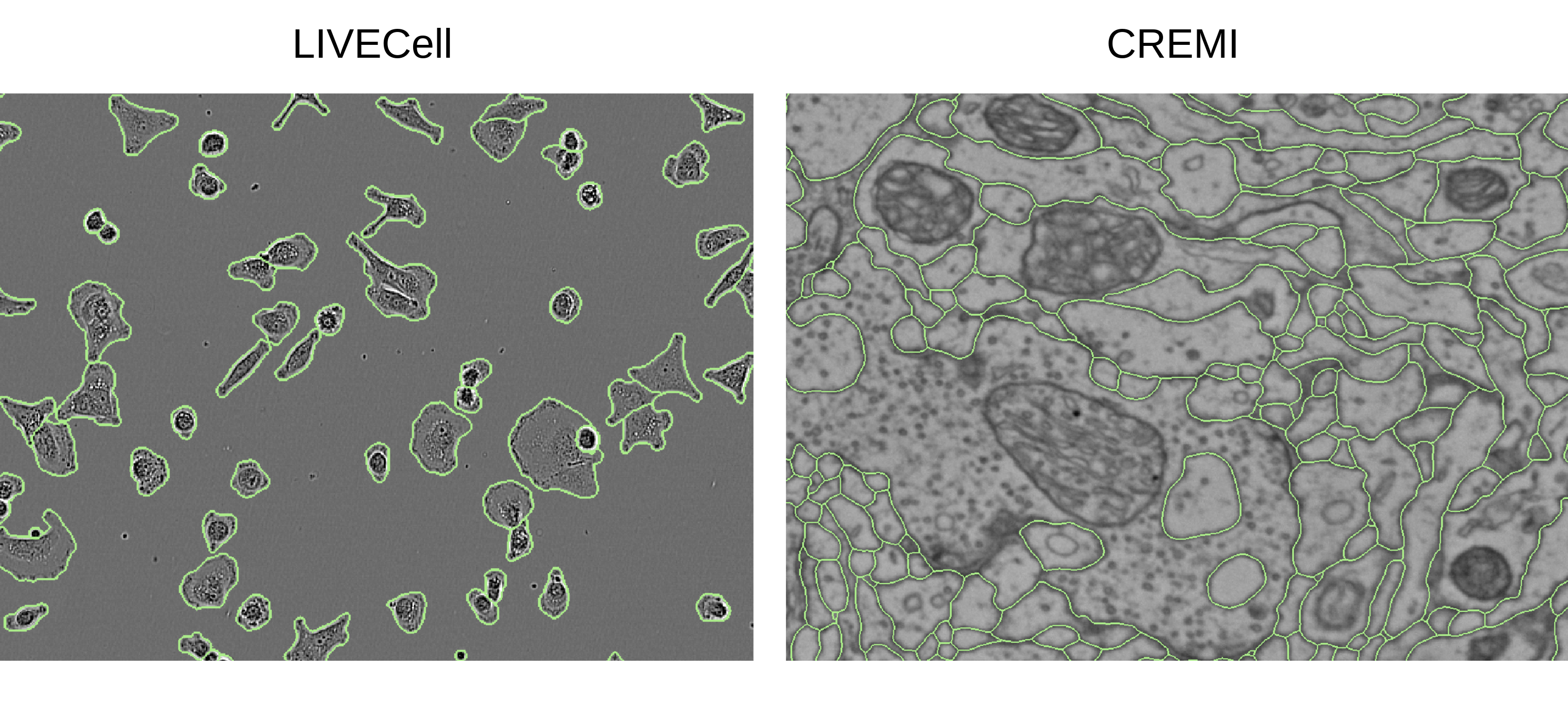
CNNs, most notably the UNet, are the default architecture for biomedical segmentation. Transformer-based approaches, such as UNETR, have been proposed to replace them, benefiting from a global field of view, but suffering from larger runtimes and higher parameter counts. The recent Vision Mamba architecture offers a compelling alternative to transformers, also providing a global field of view, but at higher efficiency. Here, we introduce ViM-UNet, a novel segmentation architecture based on it and compare it to UNet and UNETR for two challenging microscopy instance segmentation tasks. We find that it performs similarly or better than UNet, depending on the task, and outperforms UNETR while being more efficient. Our code is open source and documented at https://github.com/constantinpape/torch-em/blob/main/vimunet.md.
5/16/2024
🎲
Efficient Bayesian Uncertainty Estimation for nnU-Net
Yidong Zhao, Changchun Yang, Artur Schweidtmann, Qian Tao
0
0
The self-configuring nnU-Net has achieved leading performance in a large range of medical image segmentation challenges. It is widely considered as the model of choice and a strong baseline for medical image segmentation. However, despite its extraordinary performance, nnU-Net does not supply a measure of uncertainty to indicate its possible failure. This can be problematic for large-scale image segmentation applications, where data are heterogeneous and nnU-Net may fail without notice. In this work, we introduce a novel method to estimate nnU-Net uncertainty for medical image segmentation. We propose a highly effective scheme for posterior sampling of weight space for Bayesian uncertainty estimation. Different from previous baseline methods such as Monte Carlo Dropout and mean-field Bayesian Neural Networks, our proposed method does not require a variational architecture and keeps the original nnU-Net architecture intact, thereby preserving its excellent performance and ease of use. Additionally, we boost the segmentation performance over the original nnU-Net via marginalizing multi-modal posterior models. We applied our method on the public ACDC and M&M datasets of cardiac MRI and demonstrated improved uncertainty estimation over a range of baseline methods. The proposed method further strengthens nnU-Net for medical image segmentation in terms of both segmentation accuracy and quality control.
5/2/2024