Oral squamous cell detection using deep learning

0
Sign in to get full access
Overview
- This paper presents a deep learning approach for detecting oral squamous cell carcinoma, a type of oral cancer, from histological images.
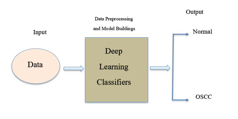
- The researchers developed a convolutional neural network (CNN) model to classify images as either cancerous or non-cancerous.
- The model was trained and evaluated on a dataset of oral tissue biopsy images.
Plain English Explanation
The researchers in this study wanted to develop a way to automatically detect oral cancer from images of tissue samples. Oral squamous cell carcinoma is a type of oral cancer that starts in the flat cells lining the mouth and throat.
To do this, they used a deep learning technique called a convolutional neural network (CNN). CNNs are a type of artificial intelligence algorithm that can learn to recognize patterns in images. The researchers trained the CNN model on a dataset of images taken from oral tissue biopsies, some of which showed cancer and some of which did not.
After training, the model was able to look at a new image and determine whether it showed cancerous or healthy tissue. This could potentially help doctors diagnose oral cancer more quickly and accurately, without having to manually review every tissue sample under a microscope.
Technical Explanation
The researchers developed a convolutional neural network (CNN) model for the task of oral squamous cell carcinoma detection. CNNs are a type of deep learning architecture well-suited for image recognition tasks.
The CNN model consisted of several convolutional layers to extract image features, followed by fully connected layers to perform the final classification. The model was trained on a dataset of histological images from oral tissue biopsies, some of which were confirmed to contain cancerous cells and others that were healthy.
During training, the model learned to recognize the visual patterns associated with cancerous and non-cancerous tissue. It was then evaluated on a test set of images to measure its performance in correctly identifying the presence of oral cancer.
The researchers reported that their CNN model achieved high accuracy, sensitivity, and specificity in differentiating between cancerous and healthy oral tissue samples. This suggests the technique could be a valuable tool to assist pathologists in the diagnosis of oral squamous cell carcinoma.
Critical Analysis
The researchers acknowledged some limitations in their study. The dataset used for training and evaluation was relatively small, which could restrict the model's ability to generalize to a wider range of real-world clinical scenarios. Expanding the dataset with more diverse samples may help improve the model's robustness.
Additionally, the study did not compare the CNN model's performance to that of human pathologists. Further research is needed to assess whether the automated system can match or exceed the diagnostic accuracy of expert clinicians when examining oral tissue samples.
Another area for potential improvement is the model's interpretability. Deep learning models like CNNs can be difficult to interpret, making it challenging to understand the specific visual features the model is using to make its predictions. Incorporating explainable AI techniques could enhance the transparency of the model's decision-making process.
Conclusion
This study demonstrates the potential of deep learning, and specifically convolutional neural networks, to assist in the detection of oral squamous cell carcinoma from histological images. The high performance achieved by the researchers' CNN model suggests it could be a valuable tool to support pathologists in the diagnosis of oral cancer.
However, further research is needed to address limitations around dataset size, comparison to human experts, and model interpretability. Continued advancements in this area could lead to more accurate, efficient, and transparent computer-aided diagnosis systems for oral cancer, potentially improving patient outcomes.
This summary was produced with help from an AI and may contain inaccuracies - check out the links to read the original source documents!
Related Papers
0
Oral squamous cell detection using deep learning
Samrat Kumar Dev Sharma
Oral squamous cell carcinoma (OSCC) represents a significant global health concern, with increasing incidence rates and challenges in early diagnosis and treatment planning. Early detection is crucial for improving patient outcomes and survival rates. Deep learning, a subset of machine learning, has shown remarkable progress in extracting and analyzing crucial information from medical imaging data.EfficientNetB3, an advanced convolutional neural network architecture, has emerged as a leading model for image classification tasks, including medical imaging. Its superior performance, characterized by high accuracy, precision, and recall, makes it particularly promising for OSCC detection and diagnosis. EfficientNetB3 achieved an accuracy of 0.9833, precision of 0.9782, and recall of 0.9782 in our analysis. By leveraging EfficientNetB3 and other deep learning technologies, clinicians can potentially improve the accuracy and efficiency of OSCC diagnosis, leading to more timely interventions and better patient outcomes. This article also discusses the role of deep learning in advancing precision medicine for OSCC and provides insights into prospects and challenges in leveraging this technology for enhanced cancer care.
Read more8/20/2024

0
Cervical Cancer Detection Using Multi-Branch Deep Learning Model
Tatsuhiro Baba, Abu Saleh Musa Miah, Jungpil Shin, Md. Al Mehedi Hasan
Cervical cancer is a crucial global health concern for women, and the persistent infection of High-risk HPV mainly triggers this remains a global health challenge, with young women diagnosis rates soaring from 10% to 40% over three decades. While Pap smear screening is a prevalent diagnostic method, visual image analysis can be lengthy and often leads to mistakes. Early detection of the disease can contribute significantly to improving patient outcomes. In recent decades, many researchers have employed machine learning techniques that achieved promise in cervical cancer detection processes based on medical images. In recent years, many researchers have employed various deep-learning techniques to achieve high-performance accuracy in detecting cervical cancer but are still facing various challenges. This research proposes an innovative and novel approach to automate cervical cancer image classification using Multi-Head Self-Attention (MHSA) and convolutional neural networks (CNNs). The proposed method leverages the strengths of both MHSA mechanisms and CNN to effectively capture both local and global features within cervical images in two streams. MHSA facilitates the model's ability to focus on relevant regions of interest, while CNN extracts hierarchical features that contribute to accurate classification. Finally, we combined the two stream features and fed them into the classification module to refine the feature and the classification. To evaluate the performance of the proposed approach, we used the SIPaKMeD dataset, which classifies cervical cells into five categories. Our model achieved a remarkable accuracy of 98.522%. This performance has high recognition accuracy of medical image classification and holds promise for its applicability in other medical image recognition tasks.
Read more8/21/2024
🤷
0
Evaluating Machine Learning-based Skin Cancer Diagnosis
Tanish Jain
This study evaluates the reliability of two deep learning models for skin cancer detection, focusing on their explainability and fairness. Using the HAM10000 dataset of dermatoscopic images, the research assesses two convolutional neural network architectures: a MobileNet-based model and a custom CNN model. Both models are evaluated for their ability to classify skin lesions into seven categories and to distinguish between dangerous and benign lesions. Explainability is assessed using Saliency Maps and Integrated Gradients, with results interpreted by a dermatologist. The study finds that both models generally highlight relevant features for most lesion types, although they struggle with certain classes like seborrheic keratoses and vascular lesions. Fairness is evaluated using the Equalized Odds metric across sex and skin tone groups. While both models demonstrate fairness across sex groups, they show significant disparities in false positive and false negative rates between light and dark skin tones. A Calibrated Equalized Odds postprocessing strategy is applied to mitigate these disparities, resulting in improved fairness, particularly in reducing false negative rate differences. The study concludes that while the models show promise in explainability, further development is needed to ensure fairness across different skin tones. These findings underscore the importance of rigorous evaluation of AI models in medical applications, particularly in diverse population groups.
Read more9/9/2024
🤿
0
Hybrid Deep Learning Framework for Enhanced Melanoma Detection
Peng Zhang, Divya Chaudhary
Cancer is a leading cause of death worldwide, necessitating advancements in early detection and treatment technologies. In this paper, we present a novel and highly efficient melanoma detection framework that synergistically combines the strengths of U-Net for segmentation and EfficientNet for the classification of skin images. The primary objective of our study is to enhance the accuracy and efficiency of melanoma detection through an innovative hybrid approach. We utilized the HAM10000 dataset to meticulously train the U-Net model, enabling it to precisely segment cancerous regions. Concurrently, we employed the ISIC 2020 dataset to train the EfficientNet model, optimizing it for the binary classification of skin cancer. Our hybrid model demonstrates a significant improvement in performance, achieving a remarkable accuracy of 99.01% on the ISIC 2020 dataset. This exceptional result underscores the superiority of our approach compared to existing model structures. By integrating the precise segmentation capabilities of U-Net with the advanced classification prowess of EfficientNet, our framework offers a comprehensive solution for melanoma detection. The results of our extensive experiments highlight the high accuracy and reliability of our method in both segmentation and classification tasks. This indicates the potential of our hybrid approach to significantly enhance cancer detection, providing a robust tool for medical professionals in the early diagnosis and treatment of melanoma. We believe that our framework can set a new benchmark in the field of automated skin cancer detection, encouraging further research and development in this crucial area of medical imaging.
Read more8/6/2024