Prior-guided Diffusion Model for Cell Segmentation in Quantitative Phase Imaging

0

Sign in to get full access
Overview
- This paper presents a new method for cell segmentation in quantitative phase imaging using a prior-guided diffusion model.
- The proposed approach leverages a diffusion model to generate segmentation maps, incorporating prior knowledge about cell shapes and locations to improve performance.
- The authors demonstrate the effectiveness of their method on various cell imaging datasets, showing improved segmentation accuracy compared to existing techniques.
Plain English Explanation
Cells are the basic building blocks of living organisms, and understanding their structure and behavior is crucial for many scientific and medical applications. Quantitative phase imaging is a technique that allows researchers to capture detailed images of cells without the need for staining or labeling.
However, accurately segmenting individual cells within these images can be a challenging task. This paper introduces a new method that uses a type of artificial intelligence called a "diffusion model" to help with cell segmentation. Diffusion models work by gradually transforming random noise into more structured images, and the researchers have found a way to guide this process using prior information about the shapes and locations of cells.
By incorporating this prior knowledge, the researchers were able to generate more accurate segmentation maps compared to other approaches. This could be valuable for a wide range of applications, from studying cell biology to developing new medical treatments.
Technical Explanation
The paper presents a "Prior-guided Diffusion Model for Cell Segmentation in Quantitative Phase Imaging." The key elements of their approach are:
-
Diffusion Model: The researchers use a diffusion model, which is a type of generative AI model that can transform random noise into more structured images. This is a departure from traditional segmentation methods that rely on hand-crafted features or supervised learning.
-
Prior Guidance: To improve the performance of the diffusion model, the researchers incorporate prior knowledge about the shapes and locations of cells in the images. This prior information is used to guide the diffusion process, leading to more accurate segmentation maps.
-
Experiment Design: The authors evaluate their method on several cell imaging datasets, including fluorescence microscopy and quantitative phase imaging data. They compare the performance of their prior-guided diffusion model to other state-of-the-art segmentation techniques, demonstrating improved accuracy.
-
Insights: The researchers find that their prior-guided diffusion approach outperforms other methods, particularly in cases where the cell shapes or locations are more complex or variable. They also observe that the diffusion model is able to capture subtle details and produce smooth, coherent segmentation maps.
Critical Analysis
The paper presents a novel and promising approach to cell segmentation, but there are a few potential limitations and areas for further research:
-
Dataset Diversity: While the authors evaluate their method on multiple datasets, these may not capture the full range of cell types, imaging modalities, and experimental conditions encountered in real-world applications. Further testing on a more diverse set of data would be valuable.
-
Prior Knowledge: The reliance on prior knowledge about cell shapes and locations could be a potential limitation, as this information may not always be readily available or easy to define. Exploring ways to automatically learn or extract these priors from the data could be an area for future research.
-
Computational Complexity: Diffusion models can be computationally intensive, which could be a concern for real-time or high-throughput applications. Investigating ways to improve the efficiency of the approach would be an important next step.
Overall, this paper presents a compelling new approach to cell segmentation that leverages the power of diffusion models and prior knowledge. While there are some potential areas for improvement, the results demonstrate the potential of this technique to advance quantitative cell imaging and analysis.
Conclusion
This paper introduces a novel "Prior-guided Diffusion Model for Cell Segmentation in Quantitative Phase Imaging." The key innovation is the use of a diffusion model, which is guided by prior information about cell shapes and locations, to generate accurate segmentation maps. The authors demonstrate the effectiveness of their approach on multiple cell imaging datasets, showing improved performance compared to existing segmentation methods.
The findings of this research could have important implications for a wide range of applications, from basic cell biology studies to the development of new medical treatments. While there are some potential limitations and areas for further research, the paper represents a significant contribution to the field of quantitative cell imaging and analysis.
This summary was produced with help from an AI and may contain inaccuracies - check out the links to read the original source documents!
Related Papers
0
Prior-guided Diffusion Model for Cell Segmentation in Quantitative Phase Imaging
Zhuchen Shao, Mark A. Anastasio, Hua Li
Purpose: Quantitative phase imaging (QPI) is a label-free technique that provides high-contrast images of tissues and cells without the use of chemicals or dyes. Accurate semantic segmentation of cells in QPI is essential for various biomedical applications. While DM-based segmentation has demonstrated promising results, the requirement for multiple sampling steps reduces efficiency. This study aims to enhance DM-based segmentation by introducing prior-guided content information into the starting noise, thereby minimizing inefficiencies associated with multiple sampling. Approach: A prior-guided mechanism is introduced into DM-based segmentation, replacing randomly sampled starting noise with noise informed by content information. This mechanism utilizes another trained DM and DDIM inversion to incorporate content information from the to-be-segmented images into the starting noise. An evaluation method is also proposed to assess the quality of the starting noise, considering both content and distribution information. Results: Extensive experiments on various QPI datasets for cell segmentation showed that the proposed method achieved superior performance in DM-based segmentation with only a single sampling. Ablation studies and visual analysis further highlighted the significance of content priors in DM-based segmentation. Conclusion: The proposed method effectively leverages prior content information to improve DM-based segmentation, providing accurate results while reducing the need for multiple samplings. The findings emphasize the importance of integrating content priors into DM-based segmentation methods for optimal performance.
Read more5/13/2024
0
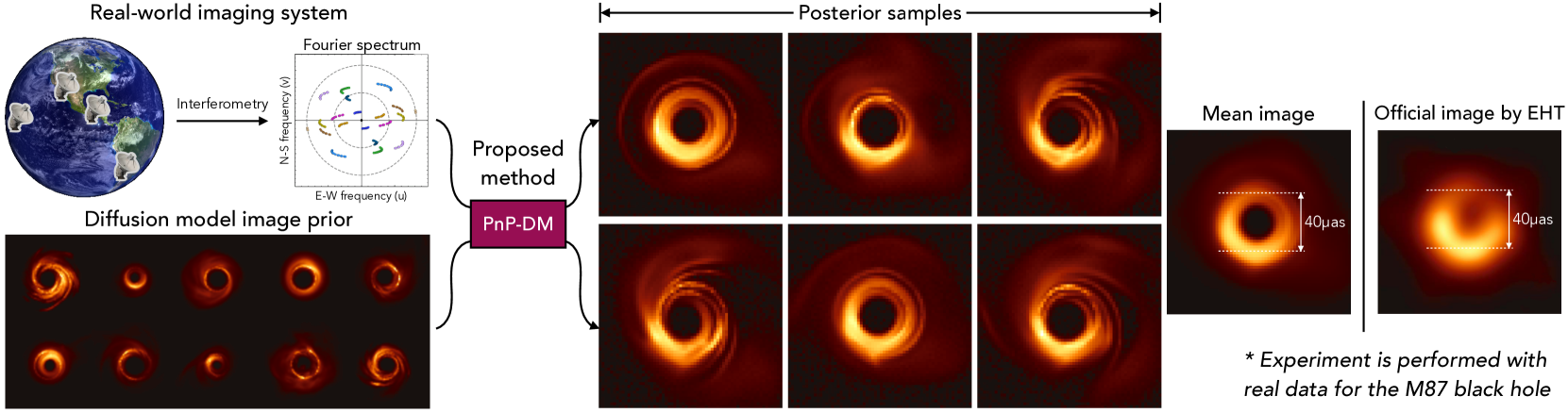
Principled Probabilistic Imaging using Diffusion Models as Plug-and-Play Priors
Zihui Wu, Yu Sun, Yifan Chen, Bingliang Zhang, Yisong Yue, Katherine L. Bouman
Diffusion models (DMs) have recently shown outstanding capability in modeling complex image distributions, making them expressive image priors for solving Bayesian inverse problems. However, most existing DM-based methods rely on approximations in the generative process to be generic to different inverse problems, leading to inaccurate sample distributions that deviate from the target posterior defined within the Bayesian framework. To harness the generative power of DMs while avoiding such approximations, we propose a Markov chain Monte Carlo algorithm that performs posterior sampling for general inverse problems by reducing it to sampling the posterior of a Gaussian denoising problem. Crucially, we leverage a general DM formulation as a unified interface that allows for rigorously solving the denoising problem with a range of state-of-the-art DMs. We demonstrate the effectiveness of the proposed method on six inverse problems (three linear and three nonlinear), including a real-world black hole imaging problem. Experimental results indicate that our proposed method offers more accurate reconstructions and posterior estimation compared to existing DM-based imaging inverse methods.
Read more5/30/2024

0
DP-IQA: Utilizing Diffusion Prior for Blind Image Quality Assessment in the Wild
Honghao Fu, Yufei Wang, Wenhan Yang, Bihan Wen
Blind image quality assessment (IQA) in the wild, which assesses the quality of images with complex authentic distortions and no reference images, presents significant challenges. Given the difficulty in collecting large-scale training data, leveraging limited data to develop a model with strong generalization remains an open problem. Motivated by the robust image perception capabilities of pre-trained text-to-image (T2I) diffusion models, we propose a novel IQA method, diffusion priors-based IQA (DP-IQA), to utilize the T2I model's prior for improved performance and generalization ability. Specifically, we utilize pre-trained Stable Diffusion as the backbone, extracting multi-level features from the denoising U-Net guided by prompt embeddings through a tunable text adapter. Simultaneously, an image adapter compensates for information loss introduced by the lossy pre-trained encoder. Unlike T2I models that require full image distribution modeling, our approach targets image quality assessment, which inherently requires fewer parameters. To improve applicability, we distill the knowledge into a lightweight CNN-based student model, significantly reducing parameters while maintaining or even enhancing generalization performance. Experimental results demonstrate that DP-IQA achieves state-of-the-art performance on various in-the-wild datasets, highlighting the superior generalization capability of T2I priors in blind IQA tasks. To our knowledge, DP-IQA is the first method to apply pre-trained diffusion priors in blind IQA. Codes and checkpoints are available at https://github.com/RomGai/DP-IQA.
Read more8/20/2024

0
Single Exposure Quantitative Phase Imaging with a Conventional Microscope using Diffusion Models
Gabriel della Maggiora, Luis Alberto Croquevielle, Harry Horsley, Thomas Heinis, Artur Yakimovich
Phase imaging is gaining importance due to its applications in fields like biomedical imaging and material characterization. In biomedical applications, it can provide quantitative information missing in label-free microscopy modalities. One of the most prominent methods in phase quantification is the Transport-of-Intensity Equation (TIE). TIE often requires multiple acquisitions at different defocus distances, which is not always feasible in a clinical setting. To address this issue, we propose to use chromatic aberrations to induce the required through-focus images with a single exposure, effectively generating a through-focus stack. Since the defocus distance induced by the aberrations is small, conventional TIE solvers are insufficient to address the resulting artifacts. We propose Zero-Mean Diffusion, a modified version of diffusion models designed for quantitative image prediction, and train it with synthetic data to ensure robust phase retrieval. Our contributions offer an alternative TIE approach that leverages chromatic aberrations, achieving accurate single-exposure phase measurement with white light and thus improving the efficiency of phase imaging. Moreover, we present a new class of diffusion models that are well-suited for quantitative data and have a sound theoretical basis. To validate our approach, we employ a widespread brightfield microscope equipped with a commercially available color camera. We apply our model to clinical microscopy of patients' urine, obtaining accurate phase measurements.
Read more6/10/2024