Realism in Action: Anomaly-Aware Diagnosis of Brain Tumors from Medical Images Using YOLOv8 and DeiT

0
Sign in to get full access
Overview
- The paper presents an approach for diagnosing brain tumors from medical images using the YOLOv8 object detection model and the DeiT vision transformer.
- The model aims to detect and classify brain tumor types while also identifying anomalies that could indicate other medical issues.
- The researchers evaluated the model's performance on a dataset of brain MRI scans and found it achieved high accuracy in tumor detection and classification.
Plain English Explanation
The researchers developed a machine learning model to help doctors identify and diagnose brain tumors from medical images like MRI scans. Their model uses two powerful AI techniques - object detection with YOLOv8 and image classification with a vision transformer called DeiT.
The key idea is that the model can not only detect the presence of a brain tumor, but also identify the specific type of tumor it is. This is important because different types of brain tumors require different treatment approaches. Additionally, the model is designed to detect anomalies in the scan that could indicate other medical issues beyond just the tumor.
The researchers tested their model on a dataset of MRI scans of brain cancer patients and found that it was able to accurately detect and classify the brain tumors. This could be a valuable tool to help doctors make more informed diagnoses and provide better treatment for their patients.
Technical Explanation
The researchers approached the problem of brain tumor diagnosis using a combination of the popular YOLOv8 object detection model and the DeiT vision transformer.
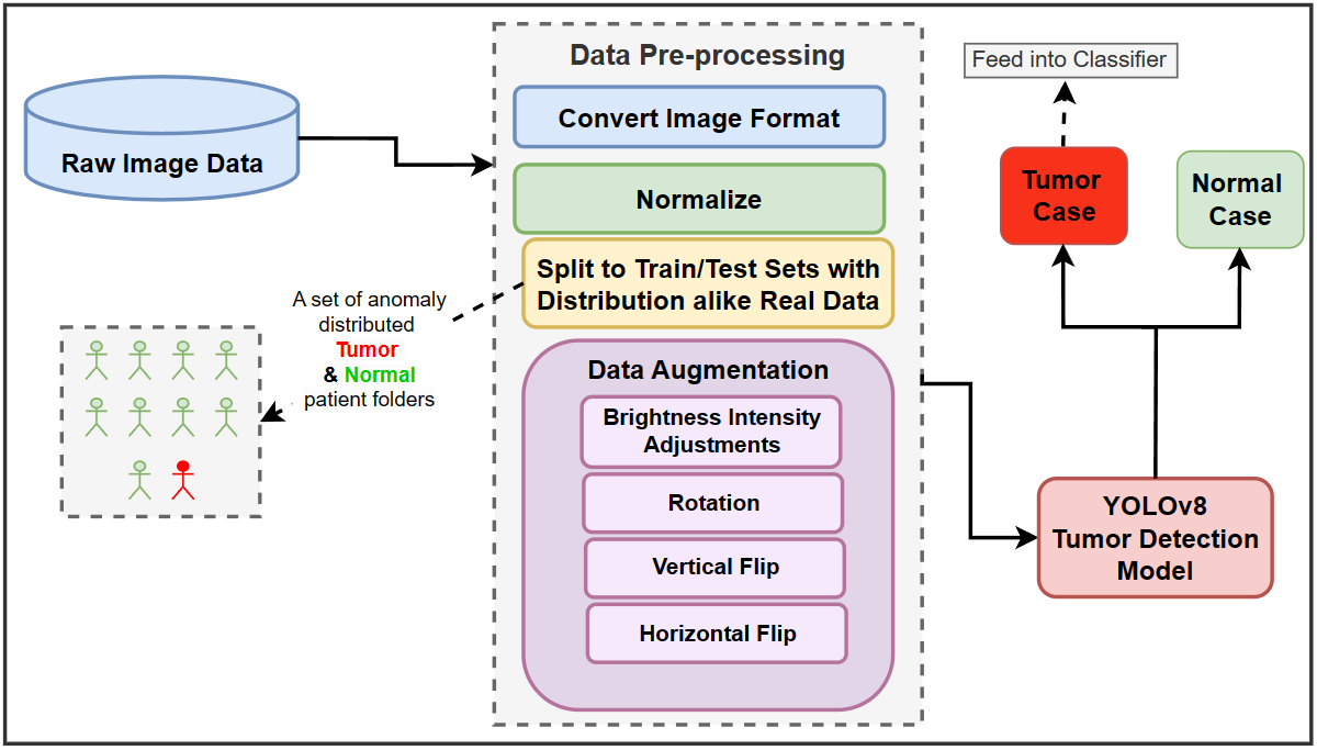
YOLOv8 was used as the backbone for detecting the presence and location of brain tumors within the MRI scans. This allowed the model to not only identify that a tumor was present, but also precisely pinpoint its position in the image.
The DeiT vision transformer was then used to classify the detected tumors into different types, such as glioma, meningioma, and pituitary adenoma. This two-stage approach - detection followed by classification - enabled the model to provide a comprehensive brain tumor diagnosis.
Importantly, the researchers also trained the model to detect anomalies in the MRI scans that could indicate other medical issues beyond just the brain tumor. This "anomaly-aware" capability is a key innovation of their approach, as it allows the model to provide a more holistic assessment of the patient's condition.
The researchers evaluated their model on a dataset of 3,929 brain MRI scans and found that it achieved high accuracy in both tumor detection and classification. This demonstrates the potential of their approach to assist clinicians in making more informed and reliable diagnoses of brain tumors.
Critical Analysis
The researchers have presented a promising approach for automating the diagnosis of brain tumors from medical images. By leveraging the strengths of both object detection and image classification techniques, their model is able to provide a comprehensive assessment that goes beyond just tumor identification.
One notable aspect of their work is the inclusion of anomaly detection, which allows the model to flag potential issues beyond the primary tumor. This is an important consideration, as brain scans may reveal other abnormalities that could impact treatment or management decisions.
However, the paper does not provide much detail on the specific types of anomalies the model was trained to detect, or how it performed in identifying these additional medical concerns. Further research and evaluation in this area could help strengthen the model's clinical utility.
Additionally, while the researchers report high accuracy on their test dataset, it's unclear how the model would perform on more diverse or challenging real-world cases. Validation on a broader range of patient populations and imaging modalities would be necessary to fully assess the model's robustness and generalizability.
Overall, the researchers have presented a compelling approach that combines cutting-edge machine learning techniques to tackle the complex problem of brain tumor diagnosis. With further development and validation, their work could contribute to more accurate and efficient clinical decision-making in this critical domain.
Conclusion
The paper demonstrates the potential of using AI models like YOLOv8 and DeiT to assist in the diagnosis of brain tumors from medical images. By integrating object detection and image classification capabilities, the model can not only identify the presence and location of a tumor, but also determine its type.
Importantly, the model is also trained to detect anomalies in the scans that could indicate other medical issues, providing a more comprehensive assessment. While further research is needed to fully validate the model's performance and generalizability, this work represents an important step towards leveraging advanced AI to improve brain tumor diagnosis and ultimately patient outcomes.
This summary was produced with help from an AI and may contain inaccuracies - check out the links to read the original source documents!
Related Papers
0
Realism in Action: Anomaly-Aware Diagnosis of Brain Tumors from Medical Images Using YOLOv8 and DeiT
Seyed Mohammad Hossein Hashemi, Leila Safari, Amirhossein Dadashzadeh Taromi
In the field of medical sciences, reliable detection and classification of brain tumors from images remains a formidable challenge due to the rarity of tumors within the population of patients. Therefore, the ability to detect tumors in anomaly scenarios is paramount for ensuring timely interventions and improved patient outcomes. This study addresses the issue by leveraging deep learning (DL) techniques to detect and classify brain tumors in challenging situations. The curated data set from the National Brain Mapping Lab (NBML) comprises 81 patients, including 30 Tumor cases and 51 Normal cases. The detection and classification pipelines are separated into two consecutive tasks. The detection phase involved comprehensive data analysis and pre-processing to modify the number of image samples and the number of patients of each class to anomaly distribution (9 Normal per 1 Tumor) to comply with real world scenarios. Next, in addition to common evaluation metrics for the testing, we employed a novel performance evaluation method called Patient to Patient (PTP), focusing on the realistic evaluation of the model. In the detection phase, we fine-tuned a YOLOv8n detection model to detect the tumor region. Subsequent testing and evaluation yielded competitive performance both in Common Evaluation Metrics and PTP metrics. Furthermore, using the Data Efficient Image Transformer (DeiT) module, we distilled a Vision Transformer (ViT) model from a fine-tuned ResNet152 as a teacher in the classification phase. This approach demonstrates promising strides in reliable tumor detection and classification, offering potential advancements in tumor diagnosis for real-world medical imaging scenarios.
Read more9/26/2024
🖼️
0
MedYOLO: A Medical Image Object Detection Framework
Joseph Sobek, Jose R. Medina Inojosa, Betsy J. Medina Inojosa, S. M. Rassoulinejad-Mousavi, Gian Marco Conte, Francisco Lopez-Jimenez, Bradley J. Erickson
Artificial intelligence-enhanced identification of organs, lesions, and other structures in medical imaging is typically done using convolutional neural networks (CNNs) designed to make voxel-accurate segmentations of the region of interest. However, the labels required to train these CNNs are time-consuming to generate and require attention from subject matter experts to ensure quality. For tasks where voxel-level precision is not required, object detection models offer a viable alternative that can reduce annotation effort. Despite this potential application, there are few options for general purpose object detection frameworks available for 3-D medical imaging. We report on MedYOLO, a 3-D object detection framework using the one-shot detection method of the YOLO family of models and designed for use with medical imaging. We tested this model on four different datasets: BRaTS, LIDC, an abdominal organ Computed Tomography (CT) dataset, and an ECG-gated heart CT dataset. We found our models achieve high performance on commonly present medium and large-sized structures such as the heart, liver, and pancreas even without hyperparameter tuning. However, the models struggle with very small or rarely present structures.
Read more6/10/2024

0
Intraoperative Glioma Segmentation with YOLO + SAM for Improved Accuracy in Tumor Resection
Samir Kassam, Angelo Markham, Katie Vo, Yashas Revanakara, Michael Lam, Kevin Zhu
Gliomas, a common type of malignant brain tumor, present significant surgical challenges due to their similarity to healthy tissue. Preoperative Magnetic Resonance Imaging (MRI) images are often ineffective during surgery due to factors such as brain shift, which alters the position of brain structures and tumors. This makes real-time intraoperative MRI (ioMRI) crucial, as it provides updated imaging that accounts for these shifts, ensuring more accurate tumor localization and safer resections. This paper presents a deep learning pipeline combining You Only Look Once Version 8 (YOLOv8) and Segment Anything Model Vision Transformer-base (SAM ViT-b) to enhance glioma detection and segmentation during ioMRI. Our model was trained using the Brain Tumor Segmentation 2021 (BraTS 2021) dataset, which includes standard magnetic resonance imaging (MRI) images, and noise-augmented MRI images that simulate ioMRI images. Noised MRI images are harder for a deep learning pipeline to segment, but they are more representative of surgical conditions. Achieving a Dice Similarity Coefficient (DICE) score of 0.79, our model performs comparably to state-of-the-art segmentation models tested on noiseless data. This performance demonstrates the model's potential to assist surgeons in maximizing tumor resection and improving surgical outcomes.
Read more8/28/2024
🤿
0
An Optimized Ensemble Deep Learning Model For Brain Tumor Classification
Md. Alamin Talukder, Md. Manowarul Islam, Md Ashraf Uddin
Brain tumors present a grave risk to human life, demanding precise and timely diagnosis for effective treatment. Inaccurate identification of brain tumors can significantly diminish life expectancy, underscoring the critical need for precise diagnostic methods. Manual identification of brain tumors within vast Magnetic Resonance Imaging (MRI) image datasets is arduous and time-consuming. Thus, the development of a reliable deep learning (DL) model is essential to enhance diagnostic accuracy and ultimately save lives. This study introduces an innovative optimization-based deep ensemble approach employing transfer learning (TL) to efficiently classify brain tumors. Our methodology includes meticulous preprocessing, reconstruction of TL architectures, fine-tuning, and ensemble DL models utilizing weighted optimization techniques such as Genetic Algorithm-based Weight Optimization (GAWO) and Grid Search-based Weight Optimization (GSWO). Experimentation is conducted on the Figshare Contrast-Enhanced MRI (CE-MRI) brain tumor dataset, comprising 3064 images. Our approach achieves notable accuracy scores, with Xception, ResNet50V2, ResNet152V2, InceptionResNetV2, GAWO, and GSWO attaining 99.42%, 98.37%, 98.22%, 98.26%, 99.71%, and 99.76% accuracy, respectively. Notably, GSWO demonstrates superior accuracy, averaging 99.76% accuracy across five folds on the Figshare CE-MRI brain tumor dataset. The comparative analysis highlights the significant performance enhancement of our proposed model over existing counterparts. In conclusion, our optimized deep ensemble model exhibits exceptional accuracy in swiftly classifying brain tumors. Furthermore, it has the potential to assist neurologists and clinicians in making accurate and immediate diagnostic decisions.
Read more5/7/2024