The RSNA Abdominal Traumatic Injury CT (RATIC) Dataset

0
🌀
Sign in to get full access
Overview
- The RSNA Abdominal Traumatic Injury CT (RATIC) dataset is the largest publicly available collection of adult abdominal CT studies annotated for traumatic injuries.
- The dataset includes 4,274 studies from 23 institutions across 14 countries and is freely available for non-commercial use via Kaggle.
- The dataset was created for the RSNA 2023 Abdominal Trauma Detection competition, aiming to encourage the development of advanced machine learning models for detecting abdominal injuries on CT scans.
Plain English Explanation
The RSNA Abdominal Traumatic Injury CT (RATIC) dataset is a massive collection of CT scans of adult abdominal injuries, made available for researchers and developers to work on. This dataset can be used to train advanced AI models to automatically detect and identify different types of abdominal injuries, like damage to the liver, spleen, kidneys, bowel, and other organs. The dataset was created for a competition run by the Radiological Society of North America (RSNA) in 2023, with the goal of improving medical care and outcomes for patients with abdominal trauma. By making this large and annotated dataset publicly available, the researchers hope to spur new innovations in machine learning that can help doctors better diagnose and treat these types of injuries.
Technical Explanation
The RSNA Abdominal Traumatic Injury CT (RATIC) dataset is the largest publicly available collection of adult abdominal CT studies annotated for traumatic injuries. It includes 4,274 studies from 23 institutions across 14 countries, freely available for non-commercial use via Kaggle.
The dataset was created for the RSNA 2023 Abdominal Trauma Detection competition, with the goal of encouraging the development of advanced machine learning models for detecting abdominal injuries on CT scans. The annotations cover a range of traumatic injuries across multiple organs, including the liver, spleen, kidneys, bowel, and mesentery. These annotations were created by expert radiologists from the American Society of Emergency Radiology (ASER) and Society of Abdominal Radiology (SAR).
The dataset provides annotations at multiple levels, including the presence of injuries in three solid organs with injury grading, image-level annotations for active extravasations and bowel injury, and voxelwise segmentations of each of the potentially injured organs. This rich set of annotations is intended to facilitate research and development in machine learning for abdominal trauma detection, which could lead to improved patient care and outcomes.
Critical Analysis
The RSNA Abdominal Traumatic Injury CT (RATIC) dataset represents a valuable resource for the medical imaging and machine learning research communities. By making this large, annotated dataset publicly available, the researchers have opened up new avenues for developing advanced AI models that can assist clinicians in the diagnosis and treatment of abdominal injuries.
However, it is important to note that the dataset is limited to adult patients, and the generalizability of models trained on this data to pediatric or other patient populations may need further investigation. Additionally, the dataset only covers a subset of potential abdominal injuries, and there may be opportunities to expand the annotation scope in the future.
Researchers and developers using this dataset should also be mindful of potential biases in the data, as the studies were collected from 23 institutions across 14 countries. Differences in imaging protocols, patient demographics, and clinical practices may need to be accounted for when training and evaluating models.
Despite these potential limitations, the RATIC dataset represents a significant step forward in the field of medical imaging and AI-assisted diagnosis. By encouraging the development of more accurate and reliable tools for detecting abdominal injuries, this dataset has the potential to improve patient outcomes and enhance the delivery of emergency medical care.
Conclusion
The RSNA Abdominal Traumatic Injury CT (RATIC) dataset is a groundbreaking resource for researchers and developers working in the field of medical imaging and machine learning. By providing a large, annotated dataset of adult abdominal CT studies, the researchers have created an opportunity for the development of advanced AI models that can assist clinicians in the diagnosis and treatment of traumatic abdominal injuries.
The dataset's comprehensive annotations, covering a range of organ-specific injuries and other relevant findings, are aimed at facilitating research and innovation in this critical area of emergency medicine. While the dataset has some limitations, it represents a significant step forward in the quest to improve patient care and outcomes through the power of cutting-edge machine learning techniques.
By making this dataset freely available, the researchers have opened the door for a broader community of researchers and developers to contribute to the advancement of this field, potentially leading to breakthroughs that could save lives and enhance the delivery of emergency medical services around the world.
This summary was produced with help from an AI and may contain inaccuracies - check out the links to read the original source documents!
Related Papers
🌀
0
The RSNA Abdominal Traumatic Injury CT (RATIC) Dataset
Jeffrey D. Rudie, Hui-Ming Lin, Robyn L. Ball, Sabeena Jalal, Luciano M. Prevedello, Savvas Nicolaou, Brett S. Marinelli, Adam E. Flanders, Kirti Magudia, George Shih, Melissa A. Davis, John Mongan, Peter D. Chang, Ferco H. Berger, Sebastiaan Hermans, Meng Law, Tyler Richards, Jan-Peter Grunz, Andreas Steven Kunz, Shobhit Mathur, Sandro Galea-Soler, Andrew D. Chung, Saif Afat, Chin-Chi Kuo, Layal Aweidah, Ana Villanueva Campos, Arjuna Somasundaram, Felipe Antonio Sanchez Tijmes, Attaporn Jantarangkoon, Leonardo Kayat Bittencourt, Michael Brassil, Ayoub El Hajjami, Hakan Dogan, Muris Becircic, Agrahara G. Bharatkumar, Eduardo Moreno J'udice de Mattos Farina, Dataset Curator Group, Dataset Contributor Group, Dataset Annotator Group, Errol Colak
The RSNA Abdominal Traumatic Injury CT (RATIC) dataset is the largest publicly available collection of adult abdominal CT studies annotated for traumatic injuries. This dataset includes 4,274 studies from 23 institutions across 14 countries. The dataset is freely available for non-commercial use via Kaggle at https://www.kaggle.com/competitions/rsna-2023-abdominal-trauma-detection. Created for the RSNA 2023 Abdominal Trauma Detection competition, the dataset encourages the development of advanced machine learning models for detecting abdominal injuries on CT scans. The dataset encompasses detection and classification of traumatic injuries across multiple organs, including the liver, spleen, kidneys, bowel, and mesentery. Annotations were created by expert radiologists from the American Society of Emergency Radiology (ASER) and Society of Abdominal Radiology (SAR). The dataset is annotated at multiple levels, including the presence of injuries in three solid organs with injury grading, image-level annotations for active extravasations and bowel injury, and voxelwise segmentations of each of the potentially injured organs. With the release of this dataset, we hope to facilitate research and development in machine learning and abdominal trauma that can lead to improved patient care and outcomes.
Read more5/31/2024

0
AbdomenAtlas: A Large-Scale, Detailed-Annotated, & Multi-Center Dataset for Efficient Transfer Learning and Open Algorithmic Benchmarking
Wenxuan Li, Chongyu Qu, Xiaoxi Chen, Pedro R. A. S. Bassi, Yijia Shi, Yuxiang Lai, Qian Yu, Huimin Xue, Yixiong Chen, Xiaorui Lin, Yutong Tang, Yining Cao, Haoqi Han, Zheyuan Zhang, Jiawei Liu, Tiezheng Zhang, Yujiu Ma, Jincheng Wang, Guang Zhang, Alan Yuille, Zongwei Zhou
We introduce the largest abdominal CT dataset (termed AbdomenAtlas) of 20,460 three-dimensional CT volumes sourced from 112 hospitals across diverse populations, geographies, and facilities. AbdomenAtlas provides 673K high-quality masks of anatomical structures in the abdominal region annotated by a team of 10 radiologists with the help of AI algorithms. We start by having expert radiologists manually annotate 22 anatomical structures in 5,246 CT volumes. Following this, a semi-automatic annotation procedure is performed for the remaining CT volumes, where radiologists revise the annotations predicted by AI, and in turn, AI improves its predictions by learning from revised annotations. Such a large-scale, detailed-annotated, and multi-center dataset is needed for two reasons. Firstly, AbdomenAtlas provides important resources for AI development at scale, branded as large pre-trained models, which can alleviate the annotation workload of expert radiologists to transfer to broader clinical applications. Secondly, AbdomenAtlas establishes a large-scale benchmark for evaluating AI algorithms -- the more data we use to test the algorithms, the better we can guarantee reliable performance in complex clinical scenarios. An ISBI & MICCAI challenge named BodyMaps: Towards 3D Atlas of Human Body was launched using a subset of our AbdomenAtlas, aiming to stimulate AI innovation and to benchmark segmentation accuracy, inference efficiency, and domain generalizability. We hope our AbdomenAtlas can set the stage for larger-scale clinical trials and offer exceptional opportunities to practitioners in the medical imaging community. Codes, models, and datasets are available at https://www.zongweiz.com/dataset
Read more7/24/2024

0
Advanced AI Framework for Enhanced Detection and Assessment of Abdominal Trauma: Integrating 3D Segmentation with 2D CNN and RNN Models
Liheng Jiang, Xuechun yang, Chang Yu, Zhizhong Wu, Yuting Wang
Trauma is a significant cause of mortality and disability, particularly among individuals under forty. Traditional diagnostic methods for traumatic injuries, such as X-rays, CT scans, and MRI, are often time-consuming and dependent on medical expertise, which can delay critical interventions. This study explores the application of artificial intelligence (AI) and machine learning (ML) to improve the speed and accuracy of abdominal trauma diagnosis. We developed an advanced AI-based model combining 3D segmentation, 2D Convolutional Neural Networks (CNN), and Recurrent Neural Networks (RNN) to enhance diagnostic performance. Our model processes abdominal CT scans to provide real-time, precise assessments, thereby improving clinical decision-making and patient outcomes. Comprehensive experiments demonstrated that our approach significantly outperforms traditional diagnostic methods, as evidenced by rigorous evaluation metrics. This research sets a new benchmark for automated trauma detection, leveraging the strengths of AI and ML to revolutionize trauma care.
Read more7/24/2024
0
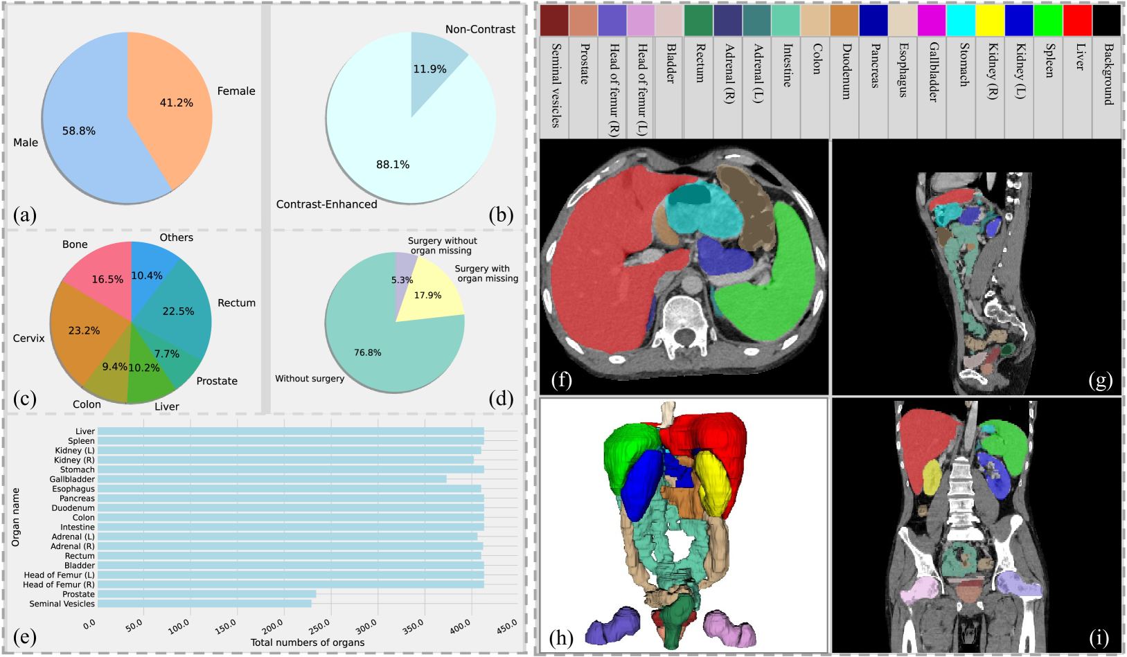
Rethinking Abdominal Organ Segmentation (RAOS) in the clinical scenario: A robustness evaluation benchmark with challenging cases
Xiangde Luo, Zihan Li, Shaoting Zhang, Wenjun Liao, Guotai Wang
Deep learning has enabled great strides in abdominal multi-organ segmentation, even surpassing junior oncologists on common cases or organs. However, robustness on corner cases and complex organs remains a challenging open problem for clinical adoption. To investigate model robustness, we collected and annotated the RAOS dataset comprising 413 CT scans ($sim$80k 2D images, $sim$8k 3D organ annotations) from 413 patients each with 17 (female) or 19 (male) labelled organs, manually delineated by oncologists. We grouped scans based on clinical information into 1) diagnosis/radiotherapy (317 volumes), 2) partial excision without the whole organ missing (22 volumes), and 3) excision with the whole organ missing (74 volumes). RAOS provides a potential benchmark for evaluating model robustness including organ hallucination. It also includes some organs that can be very hard to access on public datasets like the rectum, colon, intestine, prostate and seminal vesicles. We benchmarked several state-of-the-art methods in these three clinical groups to evaluate performance and robustness. We also assessed cross-generalization between RAOS and three public datasets. This dataset and comprehensive analysis establish a potential baseline for future robustness research: url{https://github.com/Luoxd1996/RAOS}.
Read more6/21/2024