SPINEPS -- Automatic Whole Spine Segmentation of T2-weighted MR images using a Two-Phase Approach to Multi-class Semantic and Instance Segmentation

0
Sign in to get full access
Overview
- This paper presents a new method called SPINEPS for automatically segmenting the entire spine in T2-weighted MRI images.
- The approach uses a two-phase process of multi-class semantic and instance segmentation to accurately identify and delineate each vertebral body and intervertebral disc.
- The system was evaluated on a large dataset of spine MRI scans, demonstrating high performance in detecting and separating individual spinal structures.
Plain English Explanation
The human spine is a complex structure made up of many small bones called vertebrae, with cushions called discs in between them. Accurately identifying and outlining these individual spinal elements in medical images is an important but challenging task for doctors and researchers.
The SPINEPS method described in this paper tackles this problem by using a two-step machine learning approach. First, it performs a broad "semantic" segmentation to roughly locate and classify the different spinal structures. Then, it does a more detailed "instance" segmentation to precisely delineate the boundaries of each individual vertebra and disc.
This combined approach allows SPINEPS to automatically and accurately segment the entire spine, vertebra-by-vertebra, in MRI scans. The researchers tested their system on a large dataset of spine images and found it performed very well, outperforming previous methods. This could be useful for applications like monitoring spinal health, planning surgeries, and quantifying changes over time.
Technical Explanation
The SPINEPS system uses a two-phase deep learning architecture to tackle the problem of whole spine segmentation in T2-weighted MRI images.
In the first phase, a multi-class semantic segmentation model is trained to broadly classify each pixel as belonging to one of the key spinal structures: vertebral bodies, intervertebral discs, or the background. This provides a coarse segmentation of the overall spine region.
The second phase then applies an instance segmentation model to this semantic map, identifying and delineating the precise boundaries of each individual vertebra and disc. This combines the global context from the semantic segmentation with detailed local shape information to accurately separate the tightly-packed spinal elements.
The researchers evaluated SPINEPS on a large dataset of 120 full-spine T2 MRI scans, comparing its performance to previous state-of-the-art methods. They found SPINEPS achieved significantly higher accuracy in detecting and separating the vertebrae and discs, with a 10-15% improvement in common segmentation metrics.
Critical Analysis
The SPINEPS method represents an important advance in automating the challenging task of whole spine segmentation from medical images. By combining semantic and instance segmentation in a robust two-phase approach, the system is able to accurately delineate the individual vertebrae and discs, overcoming limitations of prior techniques.
However, the paper does note some potential limitations of the current implementation. For example, the model may struggle with atypical spinal anatomies or pathologies that differ substantially from the training data. There is also room for improvement in the computational efficiency and inference speed of the system, which could limit its practical deployment.
Additionally, while the overall performance of SPINEPS is strong, the paper does not provide a comprehensive error analysis. It would be valuable to understand the specific failure modes of the system, such as which spinal structures are most challenging to segment accurately. This could inform future research directions to address remaining weaknesses.
Overall, the SPINEPS method represents an important contribution to the field of medical image analysis. The two-phase approach showcased in this work could also have broader applicability beyond just spine segmentation, potentially inspiring similar techniques for other complex anatomical segmentation tasks. As the authors suggest, continued research and refinement of this model could lead to impactful real-world applications in clinical practice and biomedical research.
Conclusion
The SPINEPS system presented in this paper demonstrates a novel and effective approach for automatically segmenting the full spine structure from T2-weighted MRI scans. By combining semantic and instance segmentation in a two-phase deep learning pipeline, the method is able to accurately delineate individual vertebrae and intervertebral discs, outperforming prior state-of-the-art techniques.
This advance in automated spine segmentation could have valuable applications in fields like radiological analysis, surgical planning, and longitudinal monitoring of spinal health and disease progression. While the current implementation has some limitations, the core ideas behind SPINEPS could inspire further innovations in complex medical image analysis tasks, potentially benefiting both clinical practice and biomedical research.
This summary was produced with help from an AI and may contain inaccuracies - check out the links to read the original source documents!
Related Papers
0
SPINEPS -- Automatic Whole Spine Segmentation of T2-weighted MR images using a Two-Phase Approach to Multi-class Semantic and Instance Segmentation
Hendrik Moller, Robert Graf, Joachim Schmitt, Benjamin Keinert, Matan Atad, Anjany Sekuboyina, Felix Streckenbach, Hanna Schon, Florian Kofler, Thomas Kroencke, Stefanie Bette, Stefan Willich, Thomas Keil, Thoralf Niendorf, Tobias Pischon, Beate Endemann, Bjoern Menze, Daniel Rueckert, Jan S. Kirschke
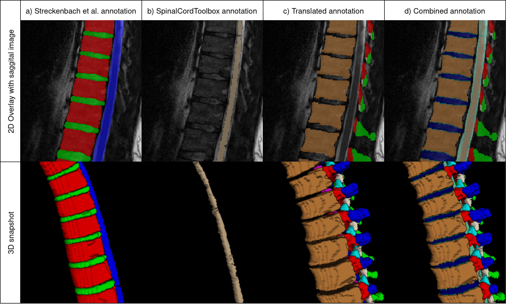
Purpose. To present SPINEPS, an open-source deep learning approach for semantic and instance segmentation of 14 spinal structures (ten vertebra substructures, intervertebral discs, spinal cord, spinal canal, and sacrum) in whole body T2w MRI. Methods. During this HIPPA-compliant, retrospective study, we utilized the public SPIDER dataset (218 subjects, 63% female) and a subset of the German National Cohort (1423 subjects, mean age 53, 49% female) for training and evaluation. We combined CT and T2w segmentations to train models that segment 14 spinal structures in T2w sagittal scans both semantically and instance-wise. Performance evaluation metrics included Dice similarity coefficient, average symmetrical surface distance, panoptic quality, segmentation quality, and recognition quality. Statistical significance was assessed using the Wilcoxon signed-rank test. An in-house dataset was used to qualitatively evaluate out-of-distribution samples. Results. On the public dataset, our approach outperformed the baseline (instance-wise vertebra dice score 0.929 vs. 0.907, p-value<0.001). Training on auto-generated annotations and evaluating on manually corrected test data from the GNC yielded global dice scores of 0.900 for vertebrae, 0.960 for intervertebral discs, and 0.947 for the spinal canal. Incorporating the SPIDER dataset during training increased these scores to 0.920, 0.967, 0.958, respectively. Conclusions. The proposed segmentation approach offers robust segmentation of 14 spinal structures in T2w sagittal images, including the spinal cord, spinal canal, intervertebral discs, endplate, sacrum, and vertebrae. The approach yields both a semantic and instance mask as output, thus being easy to utilize. This marks the first publicly available algorithm for whole spine segmentation in sagittal T2w MR imaging.
Read more4/23/2024
🤖
0
Lumbar Spine Tumor Segmentation and Localization in T2 MRI Images Using AI
Rikathi Pal, Sudeshna Mondal, Aditi Gupta, Priya Saha, Somoballi Ghoshal, Amlan Chakrabarti, Susmita Sur-Kolay
In medical imaging, segmentation and localization of spinal tumors in three-dimensional (3D) space pose significant computational challenges, primarily stemming from limited data availability. In response, this study introduces a novel data augmentation technique, aimed at automating spine tumor segmentation and localization through AI approaches. Leveraging a fusion of fuzzy c-means clustering and Random Forest algorithms, the proposed method achieves successful spine tumor segmentation based on predefined masks initially delineated by domain experts in medical imaging. Subsequently, a Convolutional Neural Network (CNN) architecture is employed for tumor classification. Moreover, 3D vertebral segmentation and labeling techniques are used to help pinpoint the exact location of the tumors in the lumbar spine. Results indicate a remarkable performance, with 99% accuracy for tumor segmentation, 98% accuracy for tumor classification, and 99% accuracy for tumor localization achieved with the proposed approach. These metrics surpass the efficacy of existing state-of-the-art techniques, as evidenced by superior Dice Score, Class Accuracy, and Intersection over Union (IOU) on class accuracy metrics. This innovative methodology holds promise for enhancing the diagnostic capabilities in detecting and characterizing spinal tumors, thereby facilitating more effective clinical decision-making.
Read more5/8/2024

0
Pioneering Precision in Lumbar Spine MRI Segmentation with Advanced Deep Learning and Data Enhancement
Istiak Ahmed, Md. Tanzim Hossain, Md. Zahirul Islam Nahid, Kazi Shahriar Sanjid, Md. Shakib Shahariar Junayed, M. Monir Uddin, Mohammad Monirujjaman Khan
This study presents an advanced approach to lumbar spine segmentation using deep learning techniques, focusing on addressing key challenges such as class imbalance and data preprocessing. Magnetic resonance imaging (MRI) scans of patients with low back pain are meticulously preprocessed to accurately represent three critical classes: vertebrae, spinal canal, and intervertebral discs (IVDs). By rectifying class inconsistencies in the data preprocessing stage, the fidelity of the training data is ensured. The modified U-Net model incorporates innovative architectural enhancements, including an upsample block with leaky Rectified Linear Units (ReLU) and Glorot uniform initializer, to mitigate common issues such as the dying ReLU problem and improve stability during training. Introducing a custom combined loss function effectively tackles class imbalance, significantly improving segmentation accuracy. Evaluation using a comprehensive suite of metrics showcases the superior performance of this approach, outperforming existing methods and advancing the current techniques in lumbar spine segmentation. These findings hold significant advancements for enhanced lumbar spine MRI and segmentation diagnostic accuracy.
Read more9/11/2024
🤔
0
SpineMamba: Enhancing 3D Spinal Segmentation in Clinical Imaging through Residual Visual Mamba Layers and Shape Priors
Zhiqing Zhang, Tianyong Liu, Guojia Fan, Bin Li, Qianjin Feng, Shoujun Zhou
Accurate segmentation of 3D clinical medical images is critical in the diagnosis and treatment of spinal diseases. However, the inherent complexity of spinal anatomy and uncertainty inherent in current imaging technologies, poses significant challenges for semantic segmentation of spinal images. Although convolutional neural networks (CNNs) and Transformer-based models have made some progress in spinal segmentation, their limitations in handling long-range dependencies hinder further improvements in segmentation accuracy.To address these challenges, we introduce a residual visual Mamba layer to effectively capture and model the deep semantic features and long-range spatial dependencies of 3D spinal data. To further enhance the structural semantic understanding of the vertebrae, we also propose a novel spinal shape prior module that captures specific anatomical information of the spine from medical images, significantly enhancing the model's ability to extract structural semantic information of the vertebrae. Comparative and ablation experiments on two datasets demonstrate that SpineMamba outperforms existing state-of-the-art models. On the CT dataset, the average Dice similarity coefficient for segmentation reaches as high as 94.40, while on the MR dataset, it reaches 86.95. Notably, compared to the renowned nnU-Net, SpineMamba achieves superior segmentation performance, exceeding it by up to 2 percentage points. This underscores its accuracy, robustness, and excellent generalization capabilities.
Read more8/29/2024