Supervised Contrastive Vision Transformer for Breast Histopathological Image Classification

0

Sign in to get full access
Overview
- This paper proposes a supervised contrastive vision transformer for breast histopathological image classification.
- The goal is to improve the accuracy of classifying breast cancer images, specifically invasive ductal carcinoma (IDC).
- The approach leverages supervised contrastive learning and transfer learning from a pre-trained vision transformer model.
Plain English Explanation
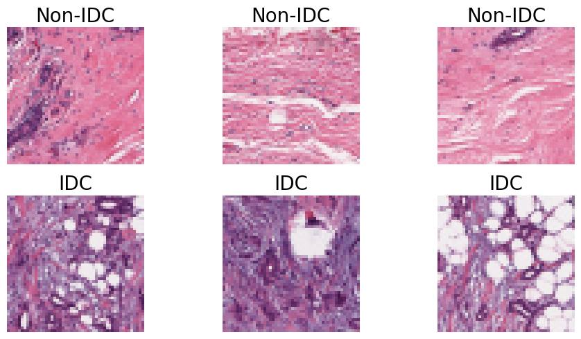
Breast cancer is a serious health issue that affects many people. One type of breast cancer is invasive ductal carcinoma (IDC), which can be detected by examining tissue samples under a microscope. This process, called histopathology, is important for diagnosing and treating breast cancer.
The researchers in this study wanted to make the process of classifying breast histopathology images more accurate. They developed a new machine learning model called a "supervised contrastive vision transformer." This model uses a technique called "supervised contrastive learning" to improve its ability to identify the differences between cancerous and non-cancerous breast tissue.
The researchers also used a pre-trained "vision transformer" model as the starting point for their new model. This allowed them to take advantage of the knowledge the vision transformer had already learned about general visual patterns, which helped the new model perform better on the breast histopathology images.
Overall, this research aims to provide a more accurate and reliable way to classify breast cancer images, which could ultimately lead to earlier detection and better treatment outcomes for patients.
Technical Explanation
The researchers proposed a supervised contrastive vision transformer for breast histopathological image classification. The key components of their approach include:
-
Supervised Contrastive Learning: The model was trained using a supervised contrastive loss function, which encourages the model to learn representations that maximize the similarity between images of the same class (e.g., IDC) and minimize the similarity between images of different classes.
-
Transfer Learning: The researchers initialized their model with a pre-trained vision transformer (ViT) [https://aimodels.fyi/papers/arxiv/breast-cancer-image-classification-method-based-deep], which had been trained on a large general-purpose image dataset. This allowed the model to leverage knowledge from the pre-trained ViT to improve performance on the breast histopathology task.
-
Architecture: The supervised contrastive vision transformer consists of a ViT backbone, followed by a supervised contrastive learning head and a classification head. The ViT backbone extracts visual features from the input images, the supervised contrastive learning head learns class-discriminative representations, and the classification head produces the final predictions.
The researchers evaluated their model on a breast histopathology dataset and compared its performance to other state-of-the-art approaches, such as convolutional neural networks and mammography-based breast cancer diagnosis methods. Their results showed that the supervised contrastive vision transformer outperformed these baselines, demonstrating the effectiveness of their approach.
Critical Analysis
The researchers acknowledge several limitations of their work:
-
Dataset Size: The breast histopathology dataset used in the study is relatively small, which may limit the generalization of the model to larger and more diverse datasets.
-
Interpretability: The supervised contrastive vision transformer, like many deep learning models, can be considered a "black box" in terms of interpretability. It may be difficult to understand the specific visual features and decision-making process of the model.
-
Potential Bias: The dataset used in the study may be subject to biases, such as demographic biases in the patient population. This could lead to the model learning biases, which could impact its performance on more diverse datasets.
-
Segmentation vs. Classification: The study focuses on the classification of breast histopathology images, but segmentation of the relevant regions within the images could potentially further improve the model's performance and interpretability.
Despite these limitations, the supervised contrastive vision transformer proposed in this study represents a promising approach for improving the accuracy of breast cancer classification from histopathological images. Further research is needed to address the limitations and explore the broader applicability of this technique.
Conclusion
This paper presents a novel supervised contrastive vision transformer for breast histopathological image classification. The key contributions include the use of supervised contrastive learning to improve class discrimination and the leverage of transfer learning from a pre-trained vision transformer model.
The results demonstrate that this approach outperforms other state-of-the-art methods, suggesting its potential to enhance the accuracy and reliability of breast cancer diagnosis from histopathology images. While the study has some limitations, it represents an important step forward in the development of advanced machine learning techniques for medical image analysis and disease diagnosis.
Overall, this research has significant implications for improving breast cancer detection and treatment, which could ultimately lead to better health outcomes for patients.
This summary was produced with help from an AI and may contain inaccuracies - check out the links to read the original source documents!
Related Papers
0
Supervised Contrastive Vision Transformer for Breast Histopathological Image Classification
Mohammad Shiri, Monalika Padma Reddy, Jiangwen Sun
Invasive ductal carcinoma (IDC) is the most prevalent form of breast cancer. Breast tissue histopathological examination is critical in diagnosing and classifying breast cancer. Although existing methods have shown promising results, there is still room for improvement in the classification accuracy and generalization of IDC using histopathology images. We present a novel approach, Supervised Contrastive Vision Transformer (SupCon-ViT), for improving the classification of invasive ductal carcinoma in terms of accuracy and generalization by leveraging the inherent strengths and advantages of both transfer learning, i.e., pre-trained vision transformer, and supervised contrastive learning. Our results on a benchmark breast cancer dataset demonstrate that SupCon-Vit achieves state-of-the-art performance in IDC classification, with an F1-score of 0.8188, precision of 0.7692, and specificity of 0.8971, outperforming existing methods. In addition, the proposed model demonstrates resilience in scenarios with minimal labeled data, making it highly efficient in real-world clinical settings where labelled data is limited. Our findings suggest that supervised contrastive learning in conjunction with pre-trained vision transformers appears to be a viable strategy for an accurate classification of IDC, thus paving the way for a more efficient and reliable diagnosis of breast cancer through histopathological image analysis.
Read more4/19/2024
🏷️
0
Classification of Breast Cancer Histopathology Images using a Modified Supervised Contrastive Learning Method
Matina Mahdizadeh Sani, Ali Royat, Mahdieh Soleymani Baghshah
Deep neural networks have reached remarkable achievements in medical image processing tasks, specifically classifying and detecting various diseases. However, when confronted with limited data, these networks face a critical vulnerability, often succumbing to overfitting by excessively memorizing the limited information available. This work addresses the challenge mentioned above by improving the supervised contrastive learning method to reduce the impact of false positives. Unlike most existing methods that rely predominantly on fully supervised learning, our approach leverages the advantages of self-supervised learning in conjunction with employing the available labeled data. We evaluate our method on the BreakHis dataset, which consists of breast cancer histopathology images, and demonstrate an increase in classification accuracy by 1.45% at the image level and 1.42% at the patient level compared to the state-of-the-art method. This improvement corresponds to 93.63% absolute accuracy, highlighting our approach's effectiveness in leveraging data properties to learn more appropriate representation space.
Read more5/7/2024
0
Comparative Analysis of Transfer Learning Models for Breast Cancer Classification
Sania Eskandari, Ali Eslamian, Qiang Cheng
The classification of histopathological images is crucial for the early and precise detection of breast cancer. This study investigates the efficiency of deep learning models in distinguishing between Invasive Ductal Carcinoma (IDC) and non-IDC in histopathology slides. We conducted a thorough comparison examination of eight sophisticated models: ResNet-50, DenseNet-121, ResNeXt-50, Vision Transformer (ViT), GoogLeNet (Inception v3), EfficientNet, MobileNet, and SqueezeNet. This analysis was carried out using a large dataset of 277,524 image patches. Our research makes a substantial contribution to the field by offering a comprehensive assessment of the performance of each model. We particularly highlight the exceptional efficacy of attention-based mechanisms in the ViT model, which achieved a remarkable validation accuracy of 93%, surpassing conventional convolutional networks. This study highlights the promise of advanced machine learning approaches in clinical settings, offering improved precision as well as efficiency in breast cancer diagnosis.
Read more9/2/2024

0
BCDNet: A Convolutional Neural Network For Breast Cancer Detection
Yujia Lin, Aiwei Lian, Mingyu Liao, Yipeng Liu
Previous research has established that breast cancer is a prevalent cancer type, with Invasive Ductal Carcinoma (IDC) being the most common subtype. The incidence of this dangerous cancer continues to rise, making accurate and rapid diagnosis, particularly in the early stages, critically important. While modern Computer-Aided Diagnosis (CAD) systems can address most cases, medical professionals still face challenges in using them in the field without powerful computing resources. In this paper, we propose a novel CNN model called BCDNet, which effectively detects IDC in histopathological images with an accuracy of up to 89.5% and reduces training time effectively.
Read more8/28/2024