Towards a Benchmark for Colorectal Cancer Segmentation in Endorectal Ultrasound Videos: Dataset and Model Development

0

Sign in to get full access
Overview
- The paper presents a dataset and model for colorectal cancer segmentation in endorectal ultrasound videos.
- The goal is to create a benchmark for this task, which has important clinical applications.
- The dataset includes annotated endorectal ultrasound videos of patients with colorectal cancer.
- The authors develop a transformer-based segmentation model and evaluate its performance on the dataset.
Plain English Explanation
The paper focuses on the problem of automatically detecting and outlining colorectal tumors in endorectal ultrasound videos. Endorectal ultrasound is a medical imaging technique that uses sound waves to create images of the rectum and surrounding areas. This is an important tool for diagnosing and monitoring colorectal cancer.
The researchers created a dataset of annotated endorectal ultrasound videos, where experts have carefully outlined the locations of any tumors present. They then developed a transformer-based machine learning model to automatically segment the tumors in these videos.
The goal is to establish a standard benchmark for evaluating and comparing different algorithms for this task. Having a common dataset and evaluation protocol is crucial for driving progress in this area, which could lead to better tools for colorectal cancer diagnosis and monitoring.
Technical Explanation
The paper first describes the process of collecting and annotating the endorectal ultrasound video dataset. The dataset includes 30 videos from 20 patients with colorectal cancer. Experts manually segmented the tumors in each frame of the videos, providing the ground truth labels.
The authors then present a transformer-based segmentation model that is designed to operate on the 3D spatio-temporal data of the ultrasound videos. The model takes the video frames as input and outputs a segmentation mask identifying the tumor regions.
The model is evaluated on the dataset using standard segmentation metrics like Dice score and Intersection over Union (IoU). The results show that the transformer-based model outperforms a baseline 3D U-Net architecture, demonstrating the potential of this approach for colorectal tumor segmentation.
Critical Analysis
The authors acknowledge several limitations of the current work. The dataset is relatively small, with only 30 videos, which may limit the model's generalization ability. Additionally, the videos were acquired from a single clinical site, so the model's performance on data from other institutions is unclear.
The paper also does not explore the model's ability to generalize to different tumor types or stages of colorectal cancer. Further research is needed to assess the robustness of the approach in real-world clinical settings.
That said, the authors have taken an important step in creating a standardized benchmark for this clinically relevant task. The dataset and evaluation protocol provide a valuable resource for the research community to build upon and advance the state of the art in colorectal tumor segmentation.
Conclusion
This paper presents a dataset and transformer-based model for colorectal cancer segmentation in endorectal ultrasound videos. The goal is to establish a benchmark for this task, which could lead to improved diagnostic tools and better patient outcomes. While the current work has some limitations, it represents a significant contribution to the field and lays the groundwork for future research in this area.
This summary was produced with help from an AI and may contain inaccuracies - check out the links to read the original source documents!
Related Papers
0
Towards a Benchmark for Colorectal Cancer Segmentation in Endorectal Ultrasound Videos: Dataset and Model Development
Yuncheng Jiang, Yiwen Hu, Zixun Zhang, Jun Wei, Chun-Mei Feng, Xuemei Tang, Xiang Wan, Yong Liu, Shuguang Cui, Zhen Li
Endorectal ultrasound (ERUS) is an important imaging modality that provides high reliability for diagnosing the depth and boundary of invasion in colorectal cancer. However, the lack of a large-scale ERUS dataset with high-quality annotations hinders the development of automatic ultrasound diagnostics. In this paper, we collected and annotated the first benchmark dataset that covers diverse ERUS scenarios, i.e. colorectal cancer segmentation, detection, and infiltration depth staging. Our ERUS-10K dataset comprises 77 videos and 10,000 high-resolution annotated frames. Based on this dataset, we further introduce a benchmark model for colorectal cancer segmentation, named the Adaptive Sparse-context TRansformer (ASTR). ASTR is designed based on three considerations: scanning mode discrepancy, temporal information, and low computational complexity. For generalizing to different scanning modes, the adaptive scanning-mode augmentation is proposed to convert between raw sector images and linear scan ones. For mining temporal information, the sparse-context transformer is incorporated to integrate inter-frame local and global features. For reducing computational complexity, the sparse-context block is introduced to extract contextual features from auxiliary frames. Finally, on the benchmark dataset, the proposed ASTR model achieves a 77.6% Dice score in rectal cancer segmentation, largely outperforming previous state-of-the-art methods.
Read more8/20/2024
👁️
0
Expanding the Medical Decathlon dataset: segmentation of colon and colorectal cancer from computed tomography images
I. M. Chernenkiy, Y. A. Drach, S. R. Mustakimova, V. V. Kazantseva, N. A. Ushakov, S. K. Efetov, M. V. Feldsherov
Colorectal cancer is the third-most common cancer in the Western Hemisphere. The segmentation of colorectal and colorectal cancer by computed tomography is an urgent problem in medicine. Indeed, a system capable of solving this problem will enable the detection of colorectal cancer at early stages of the disease, facilitate the search for pathology by the radiologist, and significantly accelerate the process of diagnosing the disease. However, scientific publications on medical image processing mostly use closed, non-public data. This paper presents an extension of the Medical Decathlon dataset with colorectal markups in order to improve the quality of segmentation algorithms. An experienced radiologist validated the data, categorized it into subsets by quality, and published it in the public domain. Based on the obtained results, we trained neural network models of the UNet architecture with 5-part cross-validation and achieved a Dice metric quality of $0.6988 pm 0.3$. The published markups will improve the quality of colorectal cancer detection and simplify the radiologist's job for study description.
Read more8/1/2024

0
The ULS23 Challenge: a Baseline Model and Benchmark Dataset for 3D Universal Lesion Segmentation in Computed Tomography
M. J. J. de Grauw, E. Th. Scholten, E. J. Smit, M. J. C. M. Rutten, M. Prokop, B. van Ginneken, A. Hering
Size measurements of tumor manifestations on follow-up CT examinations are crucial for evaluating treatment outcomes in cancer patients. Efficient lesion segmentation can speed up these radiological workflows. While numerous benchmarks and challenges address lesion segmentation in specific organs like the liver, kidneys, and lungs, the larger variety of lesion types encountered in clinical practice demands a more universal approach. To address this gap, we introduced the ULS23 benchmark for 3D universal lesion segmentation in chest-abdomen-pelvis CT examinations. The ULS23 training dataset contains 38,693 lesions across this region, including challenging pancreatic, colon and bone lesions. For evaluation purposes, we curated a dataset comprising 775 lesions from 284 patients. Each of these lesions was identified as a target lesion in a clinical context, ensuring diversity and clinical relevance within this dataset. The ULS23 benchmark is publicly accessible via uls23.grand-challenge.org, enabling researchers worldwide to assess the performance of their segmentation methods. Furthermore, we have developed and publicly released our baseline semi-supervised 3D lesion segmentation model. This model achieved an average Dice coefficient of 0.703 $pm$ 0.240 on the challenge test set. We invite ongoing submissions to advance the development of future ULS models.
Read more6/24/2024
0
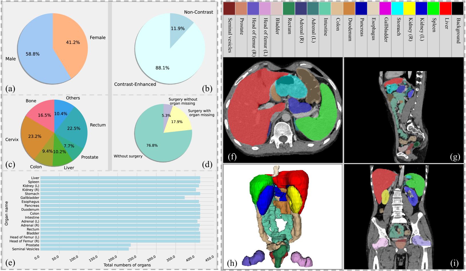
Rethinking Abdominal Organ Segmentation (RAOS) in the clinical scenario: A robustness evaluation benchmark with challenging cases
Xiangde Luo, Zihan Li, Shaoting Zhang, Wenjun Liao, Guotai Wang
Deep learning has enabled great strides in abdominal multi-organ segmentation, even surpassing junior oncologists on common cases or organs. However, robustness on corner cases and complex organs remains a challenging open problem for clinical adoption. To investigate model robustness, we collected and annotated the RAOS dataset comprising 413 CT scans ($sim$80k 2D images, $sim$8k 3D organ annotations) from 413 patients each with 17 (female) or 19 (male) labelled organs, manually delineated by oncologists. We grouped scans based on clinical information into 1) diagnosis/radiotherapy (317 volumes), 2) partial excision without the whole organ missing (22 volumes), and 3) excision with the whole organ missing (74 volumes). RAOS provides a potential benchmark for evaluating model robustness including organ hallucination. It also includes some organs that can be very hard to access on public datasets like the rectum, colon, intestine, prostate and seminal vesicles. We benchmarked several state-of-the-art methods in these three clinical groups to evaluate performance and robustness. We also assessed cross-generalization between RAOS and three public datasets. This dataset and comprehensive analysis establish a potential baseline for future robustness research: url{https://github.com/Luoxd1996/RAOS}.
Read more6/21/2024