Wide-Field, High-Resolution Reconstruction in Computational Multi-Aperture Miniscope Using a Fourier Neural Network

0

Sign in to get full access
Overview
- Describes a computational multi-aperture miniscope system that can capture wide-field, high-resolution images using a Fourier neural network
- Presents a novel approach to image reconstruction that leverages the Fourier domain to enhance spatial resolution
- Demonstrates the system's ability to capture high-quality images over a large field of view, with potential applications in neuroscience and other scientific domains
Plain English Explanation
This research paper introduces a new type of microscope system that can capture wide-field, high-resolution images using a computational approach. Traditional microscopes often struggle to achieve both a large field of view and high spatial resolution, but this system addresses this challenge by using multiple small apertures (or "mini-scopes") to capture different parts of the image.
The key innovation is the use of a Fourier neural network to reconstruct the final high-resolution image from the data collected by the mini-scopes. The Fourier domain, which represents the frequency content of the image, is used to enhance the spatial resolution and stitch the different image segments together seamlessly.
This approach has several potential benefits. It can capture wide-field images with high detail, which could be useful for applications in neuroscience, where researchers need to observe large areas of the brain at high resolution. The computational nature of the system also makes it more flexible and adaptable compared to traditional optical microscopes.
Overall, this research demonstrates a novel way to overcome the trade-off between field of view and spatial resolution in microscopy, with potential impacts on scientific imaging and understanding.
Technical Explanation
The paper presents a computational multi-aperture miniscope system that can capture wide-field, high-resolution images using a Fourier neural network-based reconstruction approach. The system consists of an array of small, individual microscope objectives ("mini-scopes") that each capture a portion of the overall field of view.
To reconstruct the final high-resolution image, the authors developed a Fourier neural network that operates in the frequency domain. This allows the system to effectively stitch together the different image segments captured by the mini-scopes, while also enhancing the spatial resolution beyond what would be possible with a single objective.
Key aspects of the system include:
- Multi-aperture miniscope design to capture a wide field of view
- Fourier-domain image reconstruction using a neural network
- Leveraging the Fourier domain to improve spatial resolution beyond the limits of individual mini-scopes
- Seamless stitching of image segments to create a unified, high-resolution output
The authors demonstrate the effectiveness of their approach through experiments and comparisons to other microscopy techniques, showcasing the system's ability to capture high-quality images over a large field of view.
Critical Analysis
The paper presents a compelling and well-designed approach to overcoming the trade-off between field of view and spatial resolution in microscopy. The use of a Fourier neural network for image reconstruction is a novel and potentially powerful technique that could have broader applications beyond just this specific system.
However, the authors do acknowledge some limitations of their approach. For example, the computational complexity of the neural network-based reconstruction may limit the real-time capabilities of the system, and there could be challenges in scaling the number of mini-scopes beyond the current configuration.
Additionally, while the authors demonstrate the system's performance on various test samples, it would be helpful to see more real-world applications and use cases, particularly in the context of neuroscience or other scientific domains where high-resolution, wide-field imaging is critical.
Further research could also explore ways to optimize the neural network architecture and training process to improve efficiency and robustness, or to investigate alternative approaches to Fourier-domain image reconstruction that could be even more effective.
Overall, this research represents an important step forward in computational microscopy and showcases the potential for neural networks to enhance the capabilities of imaging systems. As the field continues to evolve, it will be interesting to see how this work and similar approaches are further developed and applied in the future.
Conclusion
The paper presents a novel computational multi-aperture miniscope system that can capture wide-field, high-resolution images using a Fourier neural network-based reconstruction approach. This represents an important advancement in microscopy, as it addresses the longstanding challenge of achieving both a large field of view and high spatial resolution.
The key innovation is the use of the Fourier domain to enhance the spatial resolution of the final image, beyond what would be possible with individual mini-scopes. The neural network-based reconstruction also enables seamless stitching of the different image segments, resulting in a unified, high-quality output.
This research has significant potential implications for scientific imaging, particularly in fields like neuroscience, where large-scale, high-resolution observations are critical for understanding complex biological systems. As the authors continue to refine and expand this technology, it could lead to transformative new capabilities in microscopy and imaging more broadly.
This summary was produced with help from an AI and may contain inaccuracies - check out the links to read the original source documents!
Related Papers
0
Wide-Field, High-Resolution Reconstruction in Computational Multi-Aperture Miniscope Using a Fourier Neural Network
Qianwan Yang, Ruipeng Guo, Guorong Hu, Yujia Xue, Yunzhe Li, Lei Tian
Traditional fluorescence microscopy is constrained by inherent trade-offs among resolution, field-of-view, and system complexity. To navigate these challenges, we introduce a simple and low-cost computational multi-aperture miniature microscope, utilizing a microlens array for single-shot wide-field, high-resolution imaging. Addressing the challenges posed by extensive view multiplexing and non-local, shift-variant aberrations in this device, we present SV-FourierNet, a novel multi-channel Fourier neural network. SV-FourierNet facilitates high-resolution image reconstruction across the entire imaging field through its learned global receptive field. We establish a close relationship between the physical spatially-varying point-spread functions and the network's learned effective receptive field. This ensures that SV-FourierNet has effectively encapsulated the spatially-varying aberrations in our system, and learned a physically meaningful function for image reconstruction. Training of SV-FourierNet is conducted entirely on a physics-based simulator. We showcase wide-field, high-resolution video reconstructions on colonies of freely moving C. elegans and imaging of a mouse brain section. Our computational multi-aperture miniature microscope, augmented with SV-FourierNet, represents a major advancement in computational microscopy and may find broad applications in biomedical research and other fields requiring compact microscopy solutions.
Read more5/31/2024

0
Single-shot volumetric fluorescence imaging with neural fields
Oumeng Zhang, Haowen Zhou, Brandon Y. Feng, Elin M. Larsson, Reinaldo E. Alcalde, Siyuan Yin, Catherine Deng, Changhuei Yang
Single-shot volumetric fluorescence (SVF) imaging offers a significant advantage over traditional imaging methods that require scanning across multiple axial planes as it can capture biological processes with high temporal resolution across a large field of view. The key challenges in SVF imaging include requiring sparsity constraints to meet the multiplexing requirements of compressed sensing, eliminating depth ambiguity in the reconstruction, and maintaining high resolution across a large field of view. In this paper, we introduce the QuadraPol point spread function (PSF) combined with neural fields, a novel approach for SVF imaging. This method utilizes a custom polarizer at the back focal plane and a polarization camera to detect fluorescence, effectively encoding the 3D scene within a compact PSF without depth ambiguity. Additionally, we propose a reconstruction algorithm based on the neural fields technique that provides improved reconstruction quality and addresses the inaccuracies of phase retrieval methods used to correct imaging system aberrations. This algorithm combines the accuracy of experimental PSFs with the long depth of field of computationally generated retrieved PSFs. QuadraPol PSF, combined with neural fields, significantly reduces the acquisition time of a conventional fluorescence microscope by approximately 20 times and captures a 100 mm$^3$ cubic volume in one shot. We validate the effectiveness of both our hardware and algorithm through all-in-focus imaging of bacterial colonies on sand surfaces and visualization of plant root morphology. Our approach offers a powerful tool for advancing biological research and ecological studies.
Read more6/6/2024
🧠
0
Coordinate-based neural representations for computational adaptive optics in widefield microscopy
Iksung Kang, Qinrong Zhang, Stella X. Yu, Na Ji
Widefield microscopy is widely used for non-invasive imaging of biological structures at subcellular resolution. When applied to complex specimen, its image quality is degraded by sample-induced optical aberration. Adaptive optics can correct wavefront distortion and restore diffraction-limited resolution but require wavefront sensing and corrective devices, increasing system complexity and cost. Here, we describe a self-supervised machine learning algorithm, CoCoA, that performs joint wavefront estimation and three-dimensional structural information extraction from a single input 3D image stack without the need for external training dataset. We implemented CoCoA for widefield imaging of mouse brain tissues and validated its performance with direct-wavefront-sensing-based adaptive optics. Importantly, we systematically explored and quantitatively characterized the limiting factors of CoCoA's performance. Using CoCoA, we demonstrated the first in vivo widefield mouse brain imaging using machine-learning-based adaptive optics. Incorporating coordinate-based neural representations and a forward physics model, the self-supervised scheme of CoCoA should be applicable to microscopy modalities in general.
Read more6/26/2024
0
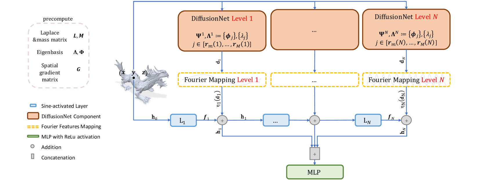
MDNF: Multi-Diffusion-Nets for Neural Fields on Meshes
Avigail Cohen Rimon, Tal Shnitzer, Mirela Ben Chen
We propose a novel framework for representing neural fields on triangle meshes that is multi-resolution across both spatial and frequency domains. Inspired by the Neural Fourier Filter Bank (NFFB), our architecture decomposes the spatial and frequency domains by associating finer spatial resolution levels with higher frequency bands, while coarser resolutions are mapped to lower frequencies. To achieve geometry-aware spatial decomposition we leverage multiple DiffusionNet components, each associated with a different spatial resolution level. Subsequently, we apply a Fourier feature mapping to encourage finer resolution levels to be associated with higher frequencies. The final signal is composed in a wavelet-inspired manner using a sine-activated MLP, aggregating higher-frequency signals on top of lower-frequency ones. Our architecture attains high accuracy in learning complex neural fields and is robust to discontinuities, exponential scale variations of the target field, and mesh modification. We demonstrate the effectiveness of our approach through its application to diverse neural fields, such as synthetic RGB functions, UV texture coordinates, and vertex normals, illustrating different challenges. To validate our method, we compare its performance against two alternatives, showcasing the advantages of our multi-resolution architecture.
Read more9/6/2024