X-Diffusion: Generating Detailed 3D MRI Volumes From a Single Image Using Cross-Sectional Diffusion Models
2404.19604
0
0
🖼️
Abstract
In this work, we present X-Diffusion, a cross-sectional diffusion model tailored for Magnetic Resonance Imaging (MRI) data. X-Diffusion is capable of generating the entire MRI volume from just a single MRI slice or optionally from few multiple slices, setting new benchmarks in the precision of synthesized MRIs from extremely sparse observations. The uniqueness lies in the novel view-conditional training and inference of X-Diffusion on MRI volumes, allowing for generalized MRI learning. Our evaluations span both brain tumour MRIs from the BRATS dataset and full-body MRIs from the UK Biobank dataset. Utilizing the paired pre-registered Dual-energy X-ray Absorptiometry (DXA) and MRI modalities in the UK Biobank dataset, X-Diffusion is able to generate detailed 3D MRI volume from a single full-body DXA. Remarkably, the resultant MRIs not only stand out in precision on unseen examples (surpassing state-of-the-art results by large margins) but also flawlessly retain essential features of the original MRI, including tumour profiles, spine curvature, brain volume, and beyond. Furthermore, the trained X-Diffusion model on the MRI datasets attains a generalization capacity out-of-domain (e.g. generating knee MRIs even though it is trained on brains). The code is available on the project website https://emmanuelleb985.github.io/XDiffusion/ .
Create account to get full access
Overview
- Presents X-Diffusion, a cross-sectional diffusion model for Magnetic Resonance Imaging (MRI) data
- Can generate entire MRI volumes from a single slice or a few slices, outperforming state-of-the-art methods
- Leverages view-conditional training and inference to enable generalized MRI learning
- Evaluated on brain tumor MRIs and full-body MRIs, and can generate detailed 3D MRI volumes from single Dual-energy X-ray Absorptiometry (DXA) scans
- Demonstrates strong generalization capabilities, such as generating knee MRIs despite being trained on brain MRIs
Plain English Explanation
X-Diffusion is a new AI model that can take a single slice or a few slices of an MRI scan and generate a complete 3D MRI volume. This is a significant improvement over existing methods, which typically require much more input data to generate high-quality MRI reconstructions.
The key innovation in X-Diffusion is its "view-conditional" training and inference approach. This means the model learns to understand MRI data from different perspectives, allowing it to generate accurate 3D MRI volumes even from very limited input. For example, the model can take a single 2D slice from a full-body MRI and create a detailed 3D reconstruction of the entire body, including important features like the spine and brain.
The researchers evaluated X-Diffusion on two medical imaging datasets: brain tumor MRIs and full-body MRIs from the UK Biobank study. In both cases, the model demonstrated impressive performance, generating MRI reconstructions that were more accurate than state-of-the-art methods. Remarkably, the model was even able to generate detailed knee MRIs, even though it was trained only on brain and full-body MRI data.
This technology has exciting potential applications in medical imaging, such as reducing the time and cost required to obtain high-quality MRI scans, or enabling more detailed analysis of MRI data. By generating accurate 3D MRI volumes from limited input, X-Diffusion could help make MRI more accessible and widely used in healthcare settings.
Technical Explanation
X-Diffusion is a cross-sectional diffusion model designed to generate complete Magnetic Resonance Imaging (MRI) volumes from extremely sparse observations, such as a single MRI slice or a few slices. This represents a significant advancement over existing methods, which typically require much more input data to produce high-quality MRI reconstructions.
The core innovation in X-Diffusion is its novel view-conditional training and inference approach. Unlike traditional models that learn a single, fixed mapping from input to output, X-Diffusion learns to understand MRI data from multiple perspectives. This allows the model to generate accurate 3D MRI volumes even when only provided with a limited number of 2D slices as input.
The researchers evaluated X-Diffusion on two medical imaging datasets: the BRATS brain tumor MRI dataset and the full-body MRI data from the UK Biobank study. In both cases, X-Diffusion demonstrated remarkable performance, outperforming state-of-the-art methods by a large margin in terms of the precision and accuracy of the generated MRI volumes.
Notably, the researchers leveraged the paired Dual-energy X-ray Absorptiometry (DXA) and MRI modalities available in the UK Biobank dataset. This allowed them to train X-Diffusion to generate detailed 3D MRI volumes from a single full-body DXA scan, which is a much more accessible and lower-cost imaging technique compared to MRI.
Beyond these impressive results, the researchers also found that the trained X-Diffusion model exhibited strong generalization capabilities. For instance, the model was able to generate accurate knee MRIs, even though it had only been trained on brain and full-body MRI data. This suggests X-Diffusion has the potential to be applied to a wide range of MRI reconstruction tasks, beyond the specific datasets used in this study.
Critical Analysis
The X-Diffusion paper presents a compelling approach to MRI volume generation from sparse observations, with impressive results across multiple datasets. However, there are a few potential limitations and areas for further research that are worth considering.
One potential concern is the reliance on paired MRI and DXA data from the UK Biobank dataset for the full-body MRI generation task. While this demonstrates the model's ability to leverage multimodal data, it may limit the generalizability of this approach to settings where such paired data is not available.
Additionally, the paper does not provide a thorough analysis of the model's performance on rare or unusual MRI abnormalities, such as atypical brain tumors or other medical conditions. It would be valuable to understand how well X-Diffusion can handle these edge cases, as accurate MRI reconstruction is particularly important in scenarios involving complex pathologies.
Further research could also explore the interpretability of the X-Diffusion model, shedding light on the specific mechanisms and learned representations that enable its strong performance. This could help advance our understanding of how diffusion models can effectively capture the complexities of medical imaging data.
Despite these potential areas for improvement, the X-Diffusion paper represents a significant advancement in the field of MRI reconstruction and synthesis. The model's ability to generate high-quality 3D MRI volumes from extremely limited input data is a remarkable achievement with promising implications for medical imaging applications.
Conclusion
The X-Diffusion paper presents a novel cross-sectional diffusion model that can generate complete MRI volumes from just a single slice or a few slices, outperforming existing state-of-the-art methods. The key innovation is the model's view-conditional training and inference approach, which allows it to learn a generalized understanding of MRI data from multiple perspectives.
The researchers have demonstrated the effectiveness of X-Diffusion on both brain tumor MRIs and full-body MRIs, including the ability to generate detailed 3D MRI volumes from single Dual-energy X-ray Absorptiometry (DXA) scans. Additionally, the model exhibits impressive generalization capabilities, such as generating accurate knee MRIs despite being trained only on brain and full-body MRI data.
These results have exciting implications for medical imaging applications, as X-Diffusion has the potential to reduce the time, cost, and resource requirements for obtaining high-quality MRI scans. By enabling accurate MRI reconstruction from sparse observations, this technology could help make MRI more accessible and widely used in healthcare settings.
Overall, the X-Diffusion paper represents a significant advancement in the field of MRI synthesis and reconstruction, with the potential to have a transformative impact on medical imaging workflows and patient care.
This summary was produced with help from an AI and may contain inaccuracies - check out the links to read the original source documents!
Related Papers
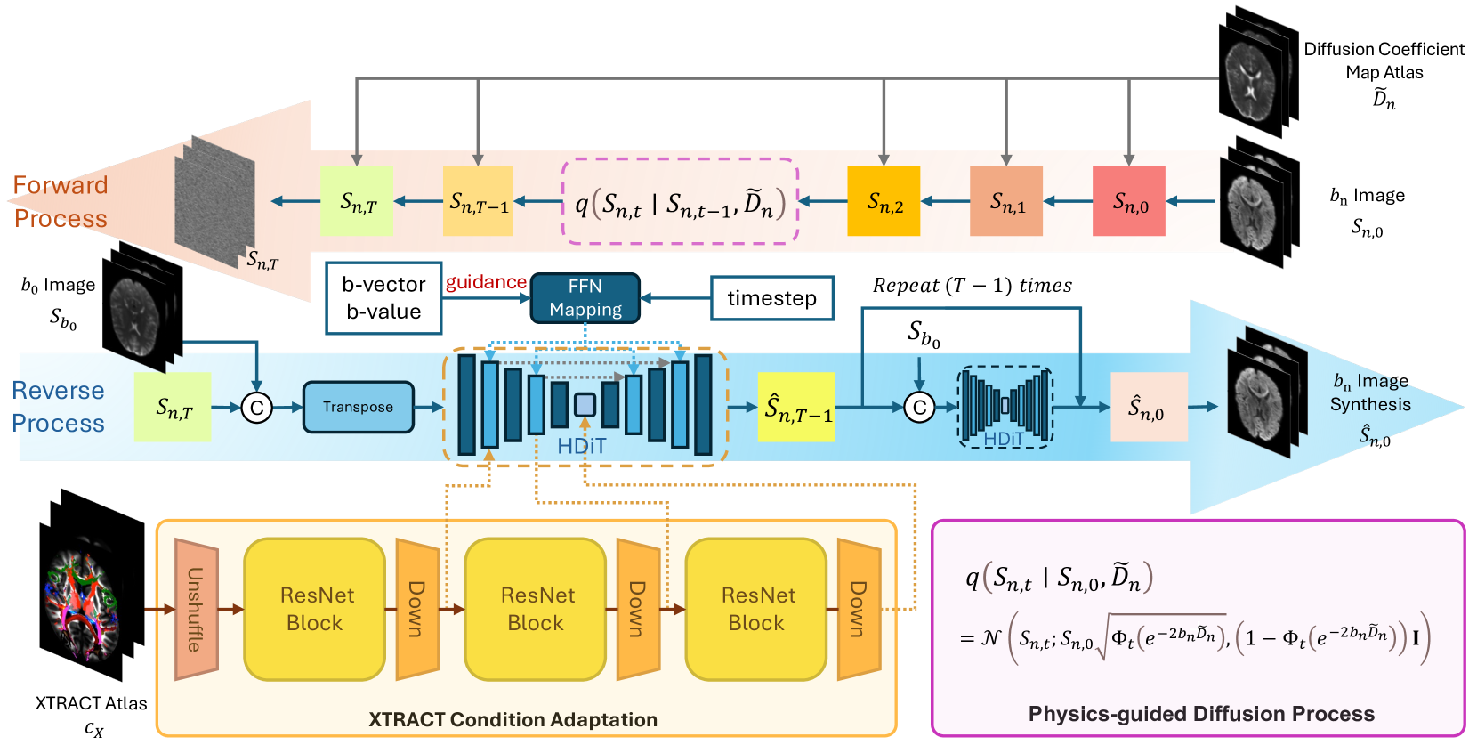
Phy-Diff: Physics-guided Hourglass Diffusion Model for Diffusion MRI Synthesis
Juanhua Zhang, Ruodan Yan, Alessandro Perelli, Xi Chen, Chao Li
0
0
Diffusion MRI (dMRI) is an important neuroimaging technique with high acquisition costs. Deep learning approaches have been used to enhance dMRI and predict diffusion biomarkers through undersampled dMRI. To generate more comprehensive raw dMRI, generative adversarial network based methods are proposed to include b-values and b-vectors as conditions, but they are limited by unstable training and less desirable diversity. The emerging diffusion model (DM) promises to improve generative performance. However, it remains challenging to include essential information in conditioning DM for more relevant generation, i.e., the physical principles of dMRI and white matter tract structures. In this study, we propose a physics-guided diffusion model to generate high-quality dMRI. Our model introduces the physical principles of dMRI in the noise evolution in the diffusion process and introduce a query-based conditional mapping within the difussion model. In addition, to enhance the anatomical fine detials of the generation, we introduce the XTRACT atlas as prior of white matter tracts by adopting an adapter technique. Our experiment results show that our method outperforms other state-of-the-art methods and has the potential to advance dMRI enhancement.
6/6/2024
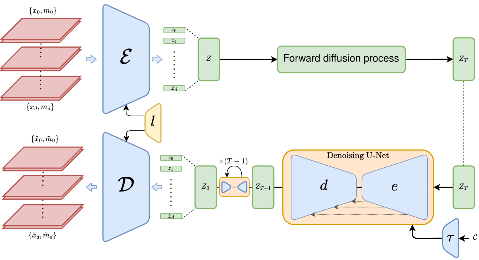
3D MRI Synthesis with Slice-Based Latent Diffusion Models: Improving Tumor Segmentation Tasks in Data-Scarce Regimes
Aghiles Kebaili, J'er^ome Lapuyade-Lahorgue, Pierre Vera, Su Ruan
0
0
Despite the increasing use of deep learning in medical image segmentation, the limited availability of annotated training data remains a major challenge due to the time-consuming data acquisition and privacy regulations. In the context of segmentation tasks, providing both medical images and their corresponding target masks is essential. However, conventional data augmentation approaches mainly focus on image synthesis. In this study, we propose a novel slice-based latent diffusion architecture designed to address the complexities of volumetric data generation in a slice-by-slice fashion. This approach extends the joint distribution modeling of medical images and their associated masks, allowing a simultaneous generation of both under data-scarce regimes. Our approach mitigates the computational complexity and memory expensiveness typically associated with diffusion models. Furthermore, our architecture can be conditioned by tumor characteristics, including size, shape, and relative position, thereby providing a diverse range of tumor variations. Experiments on a segmentation task using the BRATS2022 confirm the effectiveness of the synthesized volumes and masks for data augmentation.
6/11/2024
📈
SPIRiT-Diffusion: Self-Consistency Driven Diffusion Model for Accelerated MRI
Zhuo-Xu Cui, Chentao Cao, Yue Wang, Sen Jia, Jing Cheng, Xin Liu, Hairong Zheng, Dong Liang, Yanjie Zhu
0
0
Diffusion models have emerged as a leading methodology for image generation and have proven successful in the realm of magnetic resonance imaging (MRI) reconstruction. However, existing reconstruction methods based on diffusion models are primarily formulated in the image domain, making the reconstruction quality susceptible to inaccuracies in coil sensitivity maps (CSMs). k-space interpolation methods can effectively address this issue but conventional diffusion models are not readily applicable in k-space interpolation. To overcome this challenge, we introduce a novel approach called SPIRiT-Diffusion, which is a diffusion model for k-space interpolation inspired by the iterative self-consistent SPIRiT method. Specifically, we utilize the iterative solver of the self-consistent term (i.e., k-space physical prior) in SPIRiT to formulate a novel stochastic differential equation (SDE) governing the diffusion process. Subsequently, k-space data can be interpolated by executing the diffusion process. This innovative approach highlights the optimization model's role in designing the SDE in diffusion models, enabling the diffusion process to align closely with the physics inherent in the optimization model, a concept referred to as model-driven diffusion. We evaluated the proposed SPIRiT-Diffusion method using a 3D joint intracranial and carotid vessel wall imaging dataset. The results convincingly demonstrate its superiority over image-domain reconstruction methods, achieving high reconstruction quality even at a substantial acceleration rate of 10.
4/23/2024
✅
Conditional Diffusion Models for Semantic 3D Brain MRI Synthesis
Zolnamar Dorjsembe, Hsing-Kuo Pao, Sodtavilan Odonchimed, Furen Xiao
0
0
Artificial intelligence (AI) in healthcare, especially in medical imaging, faces challenges due to data scarcity and privacy concerns. Addressing these, we introduce Med-DDPM, a diffusion model designed for 3D semantic brain MRI synthesis. This model effectively tackles data scarcity and privacy issues by integrating semantic conditioning. This involves the channel-wise concatenation of a conditioning image to the model input, enabling control in image generation. Med-DDPM demonstrates superior stability and performance compared to existing 3D brain imaging synthesis methods. It generates diverse, anatomically coherent images with high visual fidelity. In terms of dice score accuracy in the tumor segmentation task, Med-DDPM achieves 0.6207, close to the 0.6531 accuracy of real images, and outperforms baseline models. Combined with real images, it further increases segmentation accuracy to 0.6675, showing the potential of our proposed method for data augmentation. This model represents the first use of a diffusion model in 3D semantic brain MRI synthesis, producing high-quality images. Its semantic conditioning feature also shows potential for image anonymization in biomedical imaging, addressing data and privacy issues. We provide the code and model weights for Med-DDPM on our GitHub repository (https://github.com/mobaidoctor/med-ddpm/) to support reproducibility.
4/22/2024