3D MRI Synthesis with Slice-Based Latent Diffusion Models: Improving Tumor Segmentation Tasks in Data-Scarce Regimes
2406.05421
0
0
Abstract
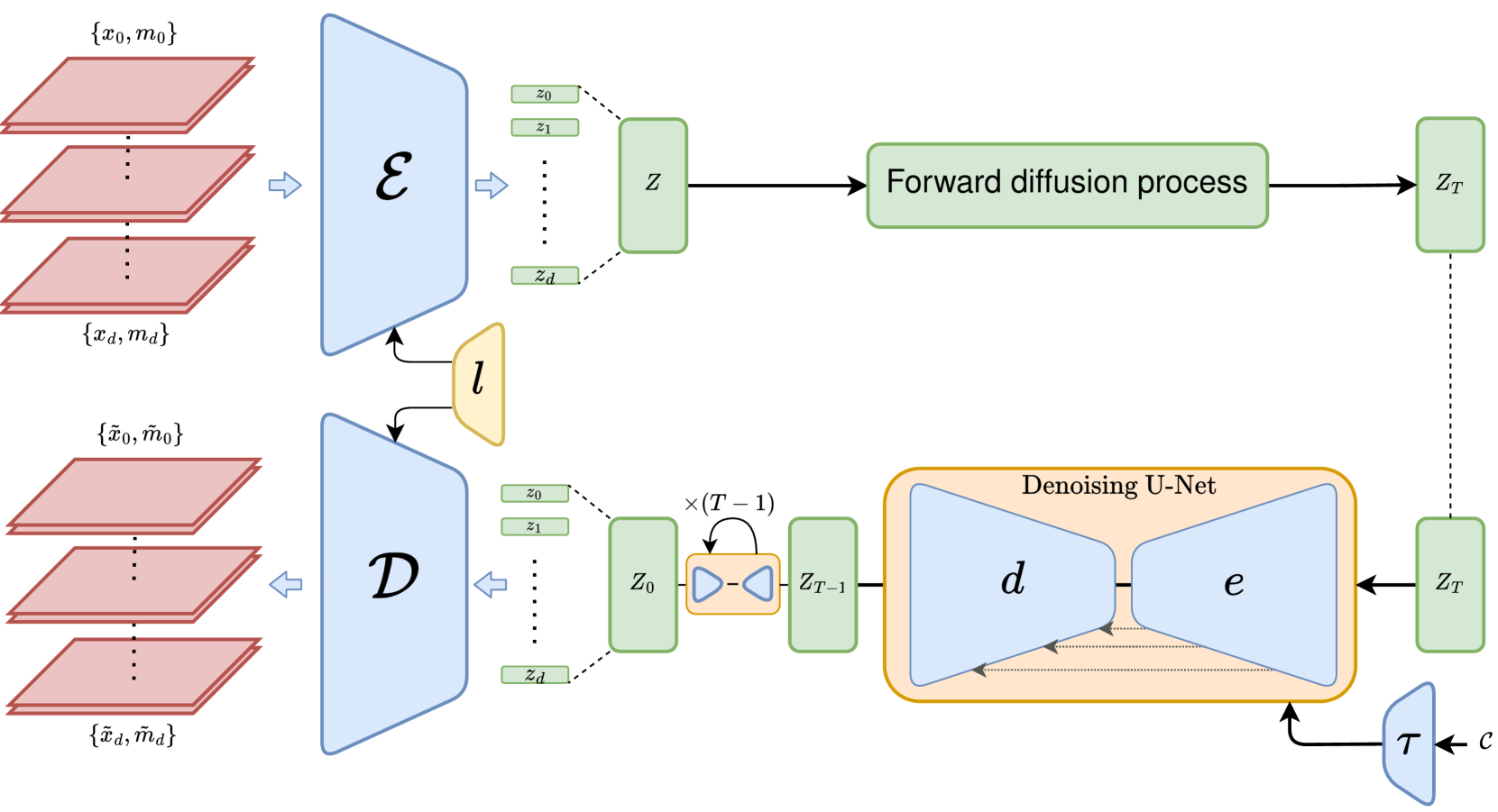
Despite the increasing use of deep learning in medical image segmentation, the limited availability of annotated training data remains a major challenge due to the time-consuming data acquisition and privacy regulations. In the context of segmentation tasks, providing both medical images and their corresponding target masks is essential. However, conventional data augmentation approaches mainly focus on image synthesis. In this study, we propose a novel slice-based latent diffusion architecture designed to address the complexities of volumetric data generation in a slice-by-slice fashion. This approach extends the joint distribution modeling of medical images and their associated masks, allowing a simultaneous generation of both under data-scarce regimes. Our approach mitigates the computational complexity and memory expensiveness typically associated with diffusion models. Furthermore, our architecture can be conditioned by tumor characteristics, including size, shape, and relative position, thereby providing a diverse range of tumor variations. Experiments on a segmentation task using the BRATS2022 confirm the effectiveness of the synthesized volumes and masks for data augmentation.
Create account to get full access
Overview
- The paper introduces a novel 3D MRI synthesis approach using slice-based latent diffusion models to improve tumor segmentation tasks in data-scarce regimes.
- The method leverages diffusion models to generate high-quality 3D MRI scans from 2D slices, overcoming the challenge of limited 3D training data.
- The generated scans are then used to augment the training dataset for tumor segmentation models, enhancing their performance in low-data scenarios.
Plain English Explanation
Magnetic Resonance Imaging (MRI) is a widely used medical imaging technique that provides detailed 3D scans of the body. However, creating high-quality 3D MRI data can be challenging, especially in cases where the available training data is limited. This is a common problem, as collecting and annotating 3D MRI scans is a time-consuming and expensive process.
To address this issue, the researchers in this paper developed a novel approach that leverages diffusion models to generate synthetic 3D MRI scans. Diffusion models are a type of generative AI model that can create new, realistic-looking images by learning from a dataset of existing images.
The key innovation in this paper is the use of "slice-based" diffusion models, which means the models are trained on 2D slices of MRI scans rather than the full 3D volumes. This allows the models to be trained on a larger and more diverse set of 2D slices, which can then be used to generate high-quality 3D scans.
The generated 3D MRI scans are then used to augment the training dataset for tumor segmentation models, which are AI models that can automatically identify and outline tumors in MRI images. By adding the synthetic 3D scans to the training data, the researchers were able to improve the performance of these segmentation models, especially in situations where the original training data was limited.
This approach, called "3D MRI Synthesis with Slice-Based Latent Diffusion Models," has the potential to significantly enhance medical image analysis capabilities in data-scarce regimes, where access to high-quality 3D MRI data may be limited.
Technical Explanation
The researchers' approach, called "3D MRI Synthesis with Slice-Based Latent Diffusion Models," leverages diffusion models to generate synthetic 3D MRI scans from 2D slices. The key steps are as follows:
-
Slice-Based Diffusion Model: The researchers train a diffusion model on 2D slices of MRI scans, rather than the full 3D volumes. This allows the model to be trained on a larger and more diverse dataset of 2D slices.
-
3D Scan Generation: The trained diffusion model is then used to generate high-quality 3D MRI scans by iteratively refining a noisy 3D volume, one slice at a time, based on the learned 2D slice representations.
-
Tumor Segmentation Augmentation: The synthetic 3D MRI scans are used to augment the training dataset for tumor segmentation models, improving their performance in data-scarce regimes.
The researchers evaluated their approach on several public MRI datasets, demonstrating significant improvements in tumor segmentation accuracy compared to baseline models trained on limited real data. They also showed that the synthetic scans generated by their slice-based diffusion model were highly realistic and could be used effectively for data augmentation.
Critical Analysis
The researchers acknowledge several limitations and areas for further research in their paper:
-
Data Quality: The quality of the generated 3D scans is ultimately dependent on the quality and diversity of the 2D slice data used to train the diffusion model. Ensuring the generated scans are clinically relevant and representative of real-world data remains an ongoing challenge.
-
Computational Complexity: The iterative slice-by-slice generation process used to create the 3D scans can be computationally intensive, potentially limiting the scalability of the approach for large-scale deployment.
-
Generalization Across Modalities: The current work focuses on MRI data, but it would be valuable to explore the potential for applying this approach to other medical imaging modalities, such as CT or PET scans, to further expand its utility.
-
Uncertainty Quantification: The paper does not explicitly address the issue of uncertainty quantification in the generated 3D scans, which could be an important consideration for clinical decision-making and model interpretability.
Additionally, while the results demonstrate the effectiveness of the proposed approach, it would be valuable to see further validation on larger, more diverse datasets and in real-world clinical settings to fully assess its practical impact.
Conclusion
The paper presents a novel 3D MRI synthesis approach using slice-based latent diffusion models that can significantly improve the performance of tumor segmentation models in data-scarce regimes. By leveraging the power of diffusion models to generate high-quality synthetic 3D scans from 2D slices, the researchers have developed a promising solution to the challenge of limited 3D training data in medical imaging.
This work has the potential to enhance computer-aided diagnosis and disease monitoring capabilities, ultimately leading to improved patient outcomes. As the field of medical image synthesis continues to advance, approaches like the one described in this paper will likely play an increasingly important role in advancing the state of the art in medical AI.
This summary was produced with help from an AI and may contain inaccuracies - check out the links to read the original source documents!
Related Papers

Generative Enhancement for 3D Medical Images
Lingting Zhu, Noel Codella, Dongdong Chen, Zhenchao Jin, Lu Yuan, Lequan Yu
0
0
The limited availability of 3D medical image datasets, due to privacy concerns and high collection or annotation costs, poses significant challenges in the field of medical imaging. While a promising alternative is the use of synthesized medical data, there are few solutions for realistic 3D medical image synthesis due to difficulties in backbone design and fewer 3D training samples compared to 2D counterparts. In this paper, we propose GEM-3D, a novel generative approach to the synthesis of 3D medical images and the enhancement of existing datasets using conditional diffusion models. Our method begins with a 2D slice, noted as the informed slice to serve the patient prior, and propagates the generation process using a 3D segmentation mask. By decomposing the 3D medical images into masks and patient prior information, GEM-3D offers a flexible yet effective solution for generating versatile 3D images from existing datasets. GEM-3D can enable dataset enhancement by combining informed slice selection and generation at random positions, along with editable mask volumes to introduce large variations in diffusion sampling. Moreover, as the informed slice contains patient-wise information, GEM-3D can also facilitate counterfactual image synthesis and dataset-level de-enhancement with desired control. Experiments on brain MRI and abdomen CT images demonstrate that GEM-3D is capable of synthesizing high-quality 3D medical images with volumetric consistency, offering a straightforward solution for dataset enhancement during inference. The code is available at https://github.com/HKU-MedAI/GEM-3D.
5/27/2024

Diff3Dformer: Leveraging Slice Sequence Diffusion for Enhanced 3D CT Classification with Transformer Networks
Zihao Jin, Yingying Fang, Jiahao Huang, Caiwen Xu, Simon Walsh, Guang Yang
0
0
The manifestation of symptoms associated with lung diseases can vary in different depths for individual patients, highlighting the significance of 3D information in CT scans for medical image classification. While Vision Transformer has shown superior performance over convolutional neural networks in image classification tasks, their effectiveness is often demonstrated on sufficiently large 2D datasets and they easily encounter overfitting issues on small medical image datasets. To address this limitation, we propose a Diffusion-based 3D Vision Transformer (Diff3Dformer), which utilizes the latent space of the Diffusion model to form the slice sequence for 3D analysis and incorporates clustering attention into ViT to aggregate repetitive information within 3D CT scans, thereby harnessing the power of the advanced transformer in 3D classification tasks on small datasets. Our method exhibits improved performance on two different scales of small datasets of 3D lung CT scans, surpassing the state of the art 3D methods and other transformer-based approaches that emerged during the COVID-19 pandemic, demonstrating its robust and superior performance across different scales of data. Experimental results underscore the superiority of our proposed method, indicating its potential for enhancing medical image classification tasks in real-world scenarios.
6/28/2024
📊
Using Diffusion Models to Generate Synthetic Labelled Data for Medical Image Segmentation
Daniel Saragih, Atsuhiro Hibi, Pascal Tyrrell
0
0
Medical image analysis has become a prominent area where machine learning has been applied. However, high quality, publicly available data is limited either due to patient privacy laws or the time and cost required for experts to annotate images. In this retrospective study, we designed and evaluated a pipeline to generate synthetic labeled polyp images for augmenting medical image segmentation models with the aim of reducing this data scarcity. In particular, we trained diffusion models on the HyperKvasir dataset, comprising 1000 images of polyps in the human GI tract from 2008 to 2016. Qualitative expert review, Fr'echet Inception Distance (FID), and Multi-Scale Structural Similarity (MS-SSIM) were tested for evaluation. Additionally, various segmentation models were trained with the generated data and evaluated using Dice score and Intersection over Union. We found that our pipeline produced images more akin to real polyp images based on FID scores, and segmentation performance also showed improvements over GAN methods when trained entirely, or partially, with synthetic data, despite requiring less compute for training. Moreover, the improvement persists when tested on different datasets, showcasing the transferability of the generated images.
5/13/2024

Similarity-aware Syncretic Latent Diffusion Model for Medical Image Translation with Representation Learning
Tingyi Lin, Pengju Lyu, Jie Zhang, Yuqing Wang, Cheng Wang, Jianjun Zhu
0
0
Non-contrast CT (NCCT) imaging may reduce image contrast and anatomical visibility, potentially increasing diagnostic uncertainty. In contrast, contrast-enhanced CT (CECT) facilitates the observation of regions of interest (ROI). Leading generative models, especially the conditional diffusion model, demonstrate remarkable capabilities in medical image modality transformation. Typical conditional diffusion models commonly generate images with guidance of segmentation labels for medical modal transformation. Limited access to authentic guidance and its low cardinality can pose challenges to the practical clinical application of conditional diffusion models. To achieve an equilibrium of generative quality and clinical practices, we propose a novel Syncretic generative model based on the latent diffusion model for medical image translation (S$^2$LDM), which can realize high-fidelity reconstruction without demand of additional condition during inference. S$^2$LDM enhances the similarity in distinct modal images via syncretic encoding and diffusing, promoting amalgamated information in the latent space and generating medical images with more details in contrast-enhanced regions. However, syncretic latent spaces in the frequency domain tend to favor lower frequencies, commonly locate in identical anatomic structures. Thus, S$^2$LDM applies adaptive similarity loss and dynamic similarity to guide the generation and supplements the shortfall in high-frequency details throughout the training process. Quantitative experiments confirm the effectiveness of our approach in medical image translation. Our code will release lately.
6/21/2024