X-ray2CTPA: Generating 3D CTPA scans from 2D X-ray conditioning

0

Sign in to get full access
Overview
• This paper presents a deep learning-based method called "X-ray2CTPA" that can generate 3D CTPA scans from 2D chest X-ray images.
• The researchers developed a novel network architecture that takes a 2D X-ray image as input and outputs a 3D CTPA scan, allowing for generation of 3D data from more readily available 2D images.
• The method could potentially be useful for applications such as computer-aided diagnosis of thoracic diseases from chest X-rays or bootstrapping chest CT image understanding.
Plain English Explanation
The paper describes a new artificial intelligence (AI) model that can create 3D medical scans from regular 2D X-ray images. Typically, 3D scans like CT (computed tomography) scans provide more detailed information than 2D X-rays, but they also require more specialized and expensive medical equipment to obtain.
The researchers developed a deep learning algorithm called "X-ray2CTPA" that can take a standard 2D chest X-ray image as input and output a simulated 3D CT angiography (CTPA) scan. CTPA scans are used to visualize blood vessels in the lungs and can be useful for diagnosing certain medical conditions.
By generating 3D scans from more commonly available 2D X-rays, this approach could help improve disease detection and monitoring, especially in settings with limited access to advanced imaging technologies. It could also potentially be used to reconstruct 3D images from low-resolution chest X-rays or to better understand chest CT scans by training on the generated 3D data.
Technical Explanation
The X-ray2CTPA model uses a novel neural network architecture that takes a 2D chest X-ray image as input and generates a corresponding 3D CTPA scan as output. The network consists of an encoder that compresses the 2D X-ray input into a low-dimensional latent representation, and a decoder that then expands this latent representation back into a 3D CTPA volume.
To train the model, the researchers used a dataset of paired 2D X-ray and 3D CTPA scans from the same patients. This allowed the network to learn the mapping between the 2D and 3D modalities during the training process. The model was further enhanced by incorporating additional multi-view X-ray image synthesis techniques to improve the quality of the generated 3D outputs.
Experiments demonstrated that the X-ray2CTPA model was able to generate realistic-looking 3D CTPA volumes from input 2D X-ray images, with promising results for applications such as XCTDiff-style reconstruction of consistent anatomical structures and computer-aided diagnosis of thoracic diseases.
Critical Analysis
The paper presents a promising approach for generating 3D medical scans from 2D X-ray images, which could have significant practical benefits. However, there are a few important caveats to consider:
-
The model was trained and evaluated on a relatively small dataset of paired 2D-3D scans, so its performance on larger, more diverse datasets remains to be seen.
-
The generated 3D CTPA scans, while visually plausible, may not fully capture all the anatomical details and nuances present in true CTPA data. Further evaluation is needed to assess the clinical utility of the generated scans.
-
The paper does not explore the potential biases or limitations of the model, such as its performance on non-standard X-ray views or on patients with rare or unusual medical conditions.
Despite these limitations, the X-ray2CTPA approach represents an important step forward in leveraging deep learning to reconstruct 3D medical images from more readily available 2D data. With further research and validation, this technology could potentially improve access to advanced medical imaging in resource-constrained settings or facilitate new applications in computer-aided diagnosis and disease monitoring.
Conclusion
The X-ray2CTPA paper presents a novel deep learning-based method for generating 3D CTPA scans from 2D chest X-ray images. By bridging the gap between these two common medical imaging modalities, this approach could lead to improved disease detection and monitoring, particularly in areas with limited access to advanced imaging technologies. While the current implementation has some limitations, the researchers have demonstrated the potential of this technique to enhance the practical utility of 2D X-ray data through the generation of more informative 3D representations.
This summary was produced with help from an AI and may contain inaccuracies - check out the links to read the original source documents!
Related Papers
0
X-ray2CTPA: Generating 3D CTPA scans from 2D X-ray conditioning
Noa Cahan, Eyal Klang, Galit Aviram, Yiftach Barash, Eli Konen, Raja Giryes, Hayit Greenspan
Chest X-rays or chest radiography (CXR), commonly used for medical diagnostics, typically enables limited imaging compared to computed tomography (CT) scans, which offer more detailed and accurate three-dimensional data, particularly contrast-enhanced scans like CT Pulmonary Angiography (CTPA). However, CT scans entail higher costs, greater radiation exposure, and are less accessible than CXRs. In this work we explore cross-modal translation from a 2D low contrast-resolution X-ray input to a 3D high contrast and spatial-resolution CTPA scan. Driven by recent advances in generative AI, we introduce a novel diffusion-based approach to this task. We evaluate the models performance using both quantitative metrics and qualitative feedback from radiologists, ensuring diagnostic relevance of the generated images. Furthermore, we employ the synthesized 3D images in a classification framework and show improved AUC in a PE categorization task, using the initial CXR input. The proposed method is generalizable and capable of performing additional cross-modality translations in medical imaging. It may pave the way for more accessible and cost-effective advanced diagnostic tools. The code for this project is available: https://github.com/NoaCahan/X-ray2CTPA .
Read more7/15/2024

0
DiffuX2CT: Diffusion Learning to Reconstruct CT Images from Biplanar X-Rays
Xuhui Liu, Zhi Qiao, Runkun Liu, Hong Li, Juan Zhang, Xiantong Zhen, Zhen Qian, Baochang Zhang
Computed tomography (CT) is widely utilized in clinical settings because it delivers detailed 3D images of the human body. However, performing CT scans is not always feasible due to radiation exposure and limitations in certain surgical environments. As an alternative, reconstructing CT images from ultra-sparse X-rays offers a valuable solution and has gained significant interest in scientific research and medical applications. However, it presents great challenges as it is inherently an ill-posed problem, often compromised by artifacts resulting from overlapping structures in X-ray images. In this paper, we propose DiffuX2CT, which models CT reconstruction from orthogonal biplanar X-rays as a conditional diffusion process. DiffuX2CT is established with a 3D global coherence denoising model with a new, implicit conditioning mechanism. We realize the conditioning mechanism by a newly designed tri-plane decoupling generator and an implicit neural decoder. By doing so, DiffuX2CT achieves structure-controllable reconstruction, which enables 3D structural information to be recovered from 2D X-rays, therefore producing faithful textures in CT images. As an extra contribution, we collect a real-world lumbar CT dataset, called LumbarV, as a new benchmark to verify the clinical significance and performance of CT reconstruction from X-rays. Extensive experiments on this dataset and three more publicly available datasets demonstrate the effectiveness of our proposal.
Read more7/19/2024
0
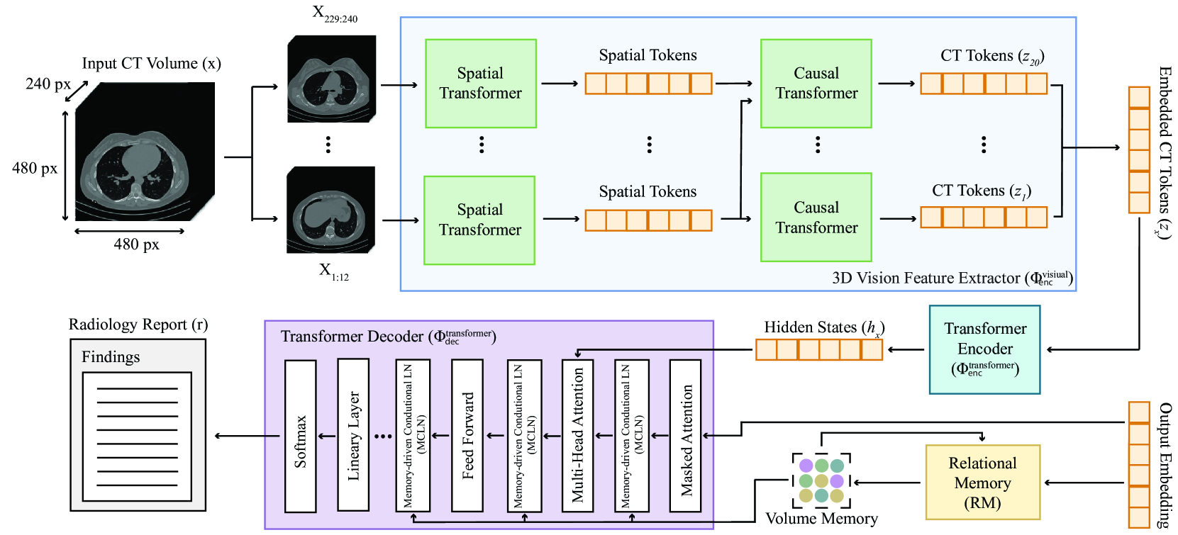
CT2Rep: Automated Radiology Report Generation for 3D Medical Imaging
Ibrahim Ethem Hamamci, Sezgin Er, Bjoern Menze
Medical imaging plays a crucial role in diagnosis, with radiology reports serving as vital documentation. Automating report generation has emerged as a critical need to alleviate the workload of radiologists. While machine learning has facilitated report generation for 2D medical imaging, extending this to 3D has been unexplored due to computational complexity and data scarcity. We introduce the first method to generate radiology reports for 3D medical imaging, specifically targeting chest CT volumes. Given the absence of comparable methods, we establish a baseline using an advanced 3D vision encoder in medical imaging to demonstrate our method's effectiveness, which leverages a novel auto-regressive causal transformer. Furthermore, recognizing the benefits of leveraging information from previous visits, we augment CT2Rep with a cross-attention-based multi-modal fusion module and hierarchical memory, enabling the incorporation of longitudinal multimodal data. Access our code at https://github.com/ibrahimethemhamamci/CT2Rep
Read more7/8/2024

0
New!DX2CT: Diffusion Model for 3D CT Reconstruction from Bi or Mono-planar 2D X-ray(s)
Yun Su Jeong, Hye Bin Yoo, Il Yong Chun
Computational tomography (CT) provides high-resolution medical imaging, but it can expose patients to high radiation. X-ray scanners have low radiation exposure, but their resolutions are low. This paper proposes a new conditional diffusion model, DX2CT, that reconstructs three-dimensional (3D) CT volumes from bi or mono-planar X-ray image(s). Proposed DX2CT consists of two key components: 1) modulating feature maps extracted from two-dimensional (2D) X-ray(s) with 3D positions of CT volume using a new transformer and 2) effectively using the modulated 3D position-aware feature maps as conditions of DX2CT. In particular, the proposed transformer can provide conditions with rich information of a target CT slice to the conditional diffusion model, enabling high-quality CT reconstruction. Our experiments with the bi or mono-planar X-ray(s) benchmark datasets show that proposed DX2CT outperforms several state-of-the-art methods. Our codes and model will be available at: https://www.github.com/intyeger/DX2CT.
Read more9/16/2024