3D Reconstruction of the Human Colon from Capsule Endoscope Video

0
Sign in to get full access
Overview
- The paper presents a method for 3D reconstruction of the human colon from capsule endoscope video.
- It involves using deep learning models to estimate the camera pose and reconstruct the 3D geometry of the colon.
- The goal is to enable better visualization and analysis of the colon during endoscopic procedures.
Plain English Explanation
The human colon, or large intestine, is an important part of the digestive system. Doctors often use a small, swallowable camera called a capsule endoscope to examine the inside of the colon for any issues or abnormalities.
This paper describes a new technique to create a 3D model of the colon from the video captured by the capsule endoscope. By reconstructing the 3D shape of the colon, doctors can get a more detailed and accurate view of the organ. This could help them better identify and diagnose any problems.
The key steps are:
- Estimating the position and orientation of the camera as it moves through the colon (called camera pose estimation).
- Using this camera information to reconstruct the 3D geometry of the colon walls (called 3D reconstruction).
The researchers developed deep learning models to automate these tasks, making the 3D reconstruction process faster and more accurate than previous methods.
Technical Explanation
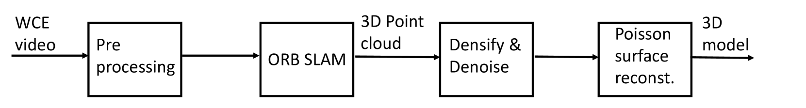
The paper first formulates the problem of 3D reconstruction from capsule endoscope video as estimating the camera pose (position and orientation) and using that to build a 3D model of the colon.
For camera pose estimation, the authors train a deep neural network to predict the 6 degrees of freedom of the camera (3D position and 3D orientation) from the video frames. This allows them to track the camera's movement through the colon.
The 3D reconstruction step then uses the estimated camera poses to triangulate the 3D positions of points on the colon wall, building up a 3D point cloud representation of the colon geometry.
The paper evaluates the accuracy of the 3D reconstruction on a dataset of colon capsule endoscopy videos, showing that it can produce high-quality 3D models that match the true colon anatomy.
Critical Analysis
The paper provides a promising approach for 3D visualization of the colon from capsule endoscopy. However, some potential limitations include:
- The 3D reconstruction accuracy may be affected by factors like image quality, colon deformation, and occlusions in the video.
- The method has only been evaluated on a single dataset, so its generalizability to other patient populations or endoscopy systems is unclear.
- Computational efficiency is not discussed, which could be an important practical consideration for real-time clinical use.
Further research could investigate ways to improve robustness, generalization, and efficiency of the 3D reconstruction pipeline. Validation on larger and more diverse datasets would also strengthen the conclusions.
Conclusion
This paper presents a deep learning-based method for 3D reconstruction of the human colon from capsule endoscope video. By estimating the camera pose and using it to build a 3D model of the colon geometry, the technique can provide doctors with a more detailed and accurate visualization of the organ.
While further research is needed to address potential limitations, this work represents an important step towards improved diagnosis and analysis capabilities for colonoscopy procedures using advanced 3D imaging techniques.
This summary was produced with help from an AI and may contain inaccuracies - check out the links to read the original source documents!
Related Papers
0
3D Reconstruction of the Human Colon from Capsule Endoscope Video
P{aa}l Anders Floor, Ivar Farup, Marius Pedersen
As the number of people affected by diseases in the gastrointestinal system is ever-increasing, a higher demand on preventive screening is inevitable. This will significantly increase the workload on gastroenterologists. To help reduce the workload, tools from computer vision may be helpful. In this paper, we investigate the possibility of constructing 3D models of whole sections of the human colon using image sequences from wireless capsule endoscope video, providing enhanced viewing for gastroenterologists. As capsule endoscope images contain distortion and artifacts non-ideal for many 3D reconstruction algorithms, the problem is challenging. However, recent developments of virtual graphics-based models of the human gastrointestinal system, where distortion and artifacts can be enabled or disabled, makes it possible to ``dissect'' the problem. The graphical model also provides a ground truth, enabling computation of geometric distortion introduced by the 3D reconstruction method. In this paper, most distortions and artifacts are left out to determine if it is feasible to reconstruct whole sections of the human gastrointestinal system by existing methods. We demonstrate that 3D reconstruction is possible using simultaneous localization and mapping. Further, to reconstruct the gastrointestinal wall surface from resulting point clouds, varying greatly in density, Poisson surface reconstruction is a good option. The results are promising, encouraging further research on this problem.
Read more7/23/2024
📉
0
SimCol3D -- 3D Reconstruction during Colonoscopy Challenge
Anita Rau, Sophia Bano, Yueming Jin, Pablo Azagra, Javier Morlana, Rawen Kader, Edward Sanderson, Bogdan J. Matuszewski, Jae Young Lee, Dong-Jae Lee, Erez Posner, Netanel Frank, Varshini Elangovan, Sista Raviteja, Zhengwen Li, Jiquan Liu, Seenivasan Lalithkumar, Mobarakol Islam, Hongliang Ren, Laurence B. Lovat, Jos'e M. M. Montiel, Danail Stoyanov
Colorectal cancer is one of the most common cancers in the world. While colonoscopy is an effective screening technique, navigating an endoscope through the colon to detect polyps is challenging. A 3D map of the observed surfaces could enhance the identification of unscreened colon tissue and serve as a training platform. However, reconstructing the colon from video footage remains difficult. Learning-based approaches hold promise as robust alternatives, but necessitate extensive datasets. Establishing a benchmark dataset, the 2022 EndoVis sub-challenge SimCol3D aimed to facilitate data-driven depth and pose prediction during colonoscopy. The challenge was hosted as part of MICCAI 2022 in Singapore. Six teams from around the world and representatives from academia and industry participated in the three sub-challenges: synthetic depth prediction, synthetic pose prediction, and real pose prediction. This paper describes the challenge, the submitted methods, and their results. We show that depth prediction from synthetic colonoscopy images is robustly solvable, while pose estimation remains an open research question.
Read more7/4/2024

0
Classification of Endoscopy and Video Capsule Images using CNN-Transformer Model
Aliza Subedi, Smriti Regmi, Nisha Regmi, Bhumi Bhusal, Ulas Bagci, Debesh Jha
Gastrointestinal cancer is a leading cause of cancer-related incidence and death, making it crucial to develop novel computer-aided diagnosis systems for early detection and enhanced treatment. Traditional approaches rely on the expertise of gastroenterologists to identify diseases; however, this process is subjective, and interpretation can vary even among expert clinicians. Considering recent advancements in classifying gastrointestinal anomalies and landmarks in endoscopic and video capsule endoscopy images, this study proposes a hybrid model that combines the advantages of Transformers and Convolutional Neural Networks (CNNs) to enhance classification performance. Our model utilizes DenseNet201 as a CNN branch to extract local features and integrates a Swin Transformer branch for global feature understanding, combining both to perform the classification task. For the GastroVision dataset, our proposed model demonstrates excellent performance with Precision, Recall, F1 score, Accuracy, and Matthews Correlation Coefficient (MCC) of 0.8320, 0.8386, 0.8324, 0.8386, and 0.8191, respectively, showcasing its robustness against class imbalance and surpassing other CNNs as well as the Swin Transformer model. Similarly, for the Kvasir-Capsule, a large video capsule endoscopy dataset, our model outperforms all others, achieving overall Precision, Recall, F1 score, Accuracy, and MCC of 0.7007, 0.7239, 0.6900, 0.7239, and 0.3871. Moreover, we generated saliency maps to explain our model's focus areas, demonstrating its reliable decision-making process. The results underscore the potential of our hybrid CNN-Transformer model in aiding the early and accurate detection of gastrointestinal (GI) anomalies.
Read more8/21/2024

0
High-fidelity Endoscopic Image Synthesis by Utilizing Depth-guided Neural Surfaces
Baoru Huang, Yida Wang, Anh Nguyen, Daniel Elson, Francisco Vasconcelos, Danail Stoyanov
In surgical oncology, screening colonoscopy plays a pivotal role in providing diagnostic assistance, such as biopsy, and facilitating surgical navigation, particularly in polyp detection. Computer-assisted endoscopic surgery has recently gained attention and amalgamated various 3D computer vision techniques, including camera localization, depth estimation, surface reconstruction, etc. Neural Radiance Fields (NeRFs) and Neural Implicit Surfaces (NeuS) have emerged as promising methodologies for deriving accurate 3D surface models from sets of registered images, addressing the limitations of existing colon reconstruction approaches stemming from constrained camera movement. However, the inadequate tissue texture representation and confused scale problem in monocular colonoscopic image reconstruction still impede the progress of the final rendering results. In this paper, we introduce a novel method for colon section reconstruction by leveraging NeuS applied to endoscopic images, supplemented by a single frame of depth map. Notably, we pioneered the exploration of utilizing only one frame depth map in photorealistic reconstruction and neural rendering applications while this single depth map can be easily obtainable from other monocular depth estimation networks with an object scale. Through rigorous experimentation and validation on phantom imagery, our approach demonstrates exceptional accuracy in completely rendering colon sections, even capturing unseen portions of the surface. This breakthrough opens avenues for achieving stable and consistently scaled reconstructions, promising enhanced quality in cancer screening procedures and treatment interventions.
Read more4/23/2024