Adaptive Sampling of k-Space in Magnetic Resonance for Rapid Pathology Prediction

0
Sign in to get full access
Overview
• This paper presents an adaptive sampling approach for magnetic resonance imaging (MRI) that can rapidly predict pathology from limited data.
Plain English Explanation
• MRI is a medical imaging technique that uses powerful magnets and radio waves to create detailed images of the inside of the body. However, traditional MRI scans can be slow and require patients to hold still for a long time.
• The researchers in this paper developed a new method that can quickly predict a patient's medical condition (called "pathology") from just a small amount of MRI data. Their approach uses machine learning to intelligently sample the MRI data, focusing on the most important regions that contain the most useful information for diagnosis.
• By adaptively sampling the MRI data, the method can dramatically reduce the time needed to acquire a full scan, while still providing accurate predictions of the patient's pathology. This could lead to faster and more efficient MRI exams, which would benefit both patients and healthcare providers.
Technical Explanation
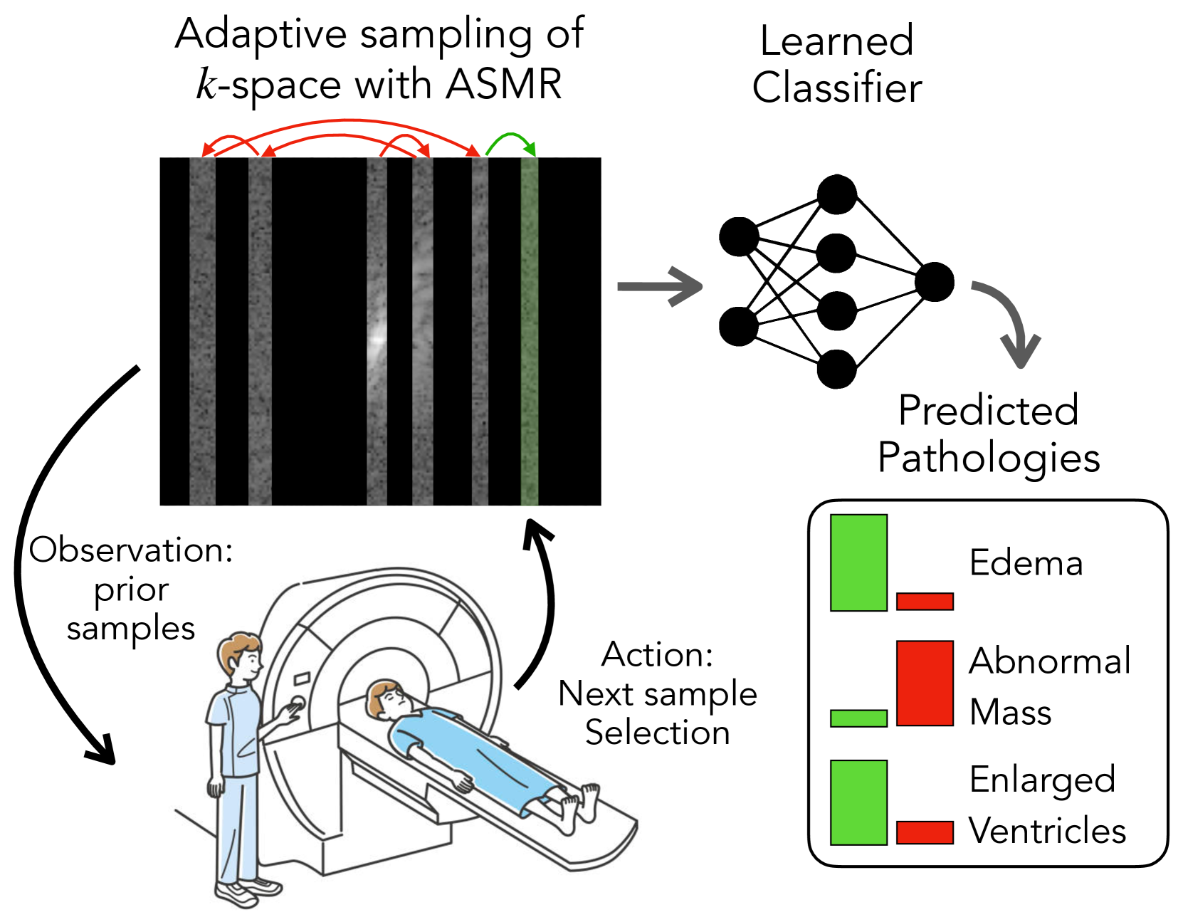
• The paper proposes an adaptive sampling framework for MRI, which uses a deep reinforcement learning agent to intelligently select which regions of the MRI "k-space" (the raw data space) to sample.
• The agent is trained to learn a sampling policy that minimizes the number of k-space samples required to accurately predict the patient's pathology, as determined by a separate neural network classifier.
• The experiments demonstrate that the adaptive sampling method can achieve high prediction accuracy using 2-4x fewer k-space samples compared to traditional uniform sampling approaches. This reduces the overall scan time and patient burden.
• The paper also introduces a novel technique called "Pathology-Aware Reconstruction" that leverages the pathology prediction to guide the reconstruction of the full MRI image from the limited samples.
Critical Analysis
• The paper makes a compelling case for the benefits of adaptive sampling in MRI, but it would be helpful to see more real-world validation of the approach, such as on a larger and more diverse patient population.
• The reliance on a separate pathology prediction network introduces additional complexity that could impact clinical deployment. Integrating the sampling and pathology prediction into a single end-to-end framework may be a valuable area for future research.
• While the results demonstrate significant reductions in scan time, the paper does not quantify the potential impacts on diagnostic accuracy or clinical decision-making. Further research is needed to fully understand the clinical implications of this approach.
Conclusion
• This paper presents a novel adaptive sampling method for MRI that can dramatically reduce scan times while maintaining accurate pathology prediction. If successfully deployed, this technology has the potential to improve the efficiency and accessibility of MRI exams, benefiting both patients and healthcare providers.
This summary was produced with help from an AI and may contain inaccuracies - check out the links to read the original source documents!
Related Papers
0
Adaptive Sampling of k-Space in Magnetic Resonance for Rapid Pathology Prediction
Chen-Yu Yen, Raghav Singhal, Umang Sharma, Rajesh Ranganath, Sumit Chopra, Lerrel Pinto
Magnetic Resonance (MR) imaging, despite its proven diagnostic utility, remains an inaccessible imaging modality for disease surveillance at the population level. A major factor rendering MR inaccessible is lengthy scan times. An MR scanner collects measurements associated with the underlying anatomy in the Fourier space, also known as the k-space. Creating a high-fidelity image requires collecting large quantities of such measurements, increasing the scan time. Traditionally to accelerate an MR scan, image reconstruction from under-sampled k-space data is the method of choice. However, recent works show the feasibility of bypassing image reconstruction and directly learning to detect disease directly from a sparser learned subset of the k-space measurements. In this work, we propose Adaptive Sampling for MR (ASMR), a sampling method that learns an adaptive policy to sequentially select k-space samples to optimize for target disease detection. On 6 out of 8 pathology classification tasks spanning the Knee, Brain, and Prostate MR scans, ASMR reaches within 2% of the performance of a fully sampled classifier while using only 8% of the k-space, as well as outperforming prior state-of-the-art work in k-space sampling such as EMRT, LOUPE, and DPS.
Read more6/7/2024

0
The MRI Scanner as a Diagnostic: Image-less Active Sampling
Yuning Du, Rohan Dharmakumar, Sotirios A. Tsaftaris
Despite the high diagnostic accuracy of Magnetic Resonance Imaging (MRI), using MRI as a Point-of-Care (POC) disease identification tool poses significant accessibility challenges due to the use of high magnetic field strength and lengthy acquisition times. We ask a simple question: Can we dynamically optimise acquired samples, at the patient level, according to an (automated) downstream decision task, while discounting image reconstruction? We propose an ML-based framework that learns an active sampling strategy, via reinforcement learning, at a patient-level to directly infer disease from undersampled k-space. We validate our approach by inferring Meniscus Tear in undersampled knee MRI data, where we achieve diagnostic performance comparable with ML-based diagnosis, using fully sampled k-space data. We analyse task-specific sampling policies, showcasing the adaptability of our active sampling approach. The introduced frugal sampling strategies have the potential to reduce high field strength requirements that in turn strengthen the viability of MRI-based POC disease identification and associated preliminary screening tools.
Read more6/26/2024

0
Tumor likelihood estimation on MRI prostate data by utilizing k-Space information
M. Rempe, F. Horst, C. Seibold, B. Hadaschik, M. Schlimbach, J. Egger, K. Kroninger, F. Breuer, M. Blaimer, J. Kleesiek
We present a novel preprocessing and prediction pipeline for the classification of magnetic resonance imaging (MRI) that takes advantage of the information rich complex valued k-Space. Using a publicly available MRI raw dataset with 312 subject and a total of 9508 slices, we show the advantage of utilizing the k-Space for better prostate cancer likelihood estimation in comparison to just using the magnitudinal information in the image domain, with an AUROC of $86.1%pm1.8%$. Additionally, by using high undersampling rates and a simple principal component analysis (PCA) for coil compression, we reduce the time needed for reconstruction by avoiding the time intensive GRAPPA reconstruction algorithm. By using digital undersampling for our experiments, we show that scanning and reconstruction time could be reduced. Even with an undersampling factor of 16, our approach achieves meaningful results, with an AUROC of $71.4%pm2.9%$, using the PCA coil combination and taking into account the k-Space information. With this study, we were able to show the feasibility of preserving phase and k-Space information, with consistent results. Besides preserving valuable information for further diagnostics, this approach can work without the time intensive ADC and reconstruction calculations, greatly reducing the post processing, as well as potential scanning time, increasing patient comfort and allowing a close to real-time prediction.
Read more7/9/2024

0
Classification, Regression and Segmentation directly from k-Space in Cardiac MRI
Ruochen Li, Jiazhen Pan, Youxiang Zhu, Juncheng Ni, Daniel Rueckert
Cardiac Magnetic Resonance Imaging (CMR) is the gold standard for diagnosing cardiovascular diseases. Clinical diagnoses predominantly rely on magnitude-only Digital Imaging and Communications in Medicine (DICOM) images, omitting crucial phase information that might provide additional diagnostic benefits. In contrast, k-space is complex-valued and encompasses both magnitude and phase information, while humans cannot directly perceive. In this work, we propose KMAE, a Transformer-based model specifically designed to process k-space data directly, eliminating conventional intermediary conversion steps to the image domain. KMAE can handle critical cardiac disease classification, relevant phenotype regression, and cardiac morphology segmentation tasks. We utilize this model to investigate the potential of k-space-based diagnosis in cardiac MRI. Notably, this model achieves competitive classification and regression performance compared to image-domain methods e.g. Masked Autoencoders (MAEs) and delivers satisfactory segmentation performance with a myocardium dice score of 0.884. Last but not least, our model exhibits robust performance with consistent results even when the k-space is 8* undersampled. We encourage the MR community to explore the untapped potential of k-space and pursue end-to-end, automated diagnosis with reduced human intervention.
Read more7/30/2024