Classification, Regression and Segmentation directly from k-Space in Cardiac MRI

0

Sign in to get full access
Overview
- This paper presents a novel deep learning approach for direct cardiac MRI analysis, including classification, regression, and segmentation, directly from the raw k-space data.
- The key idea is to bypass the traditional image reconstruction step and instead learn to extract clinically relevant information directly from the k-space measurements.
- The authors demonstrate the effectiveness of their approach on several cardiac MRI tasks, including left ventricular volume prediction, myocardial scar detection, and left ventricular segmentation.
Plain English Explanation
The paper describes a new way to analyze cardiac MRI (magnetic resonance imaging) data using deep learning. Traditionally, MRI data is first reconstructed into an image, and then analyzed using machine learning techniques. However, the authors of this paper propose a different approach: analyzing the raw MRI data, called k-space data, directly without first reconstructing an image.
The key advantage of this approach is that it can potentially extract more clinically relevant information from the raw data, without the image reconstruction step potentially introducing errors or losing important details. The authors demonstrate that their method can be used for a variety of cardiac MRI tasks, such as predicting the volume of the left ventricle of the heart, detecting myocardial scarring, and segmenting the left ventricle.
Technical Explanation
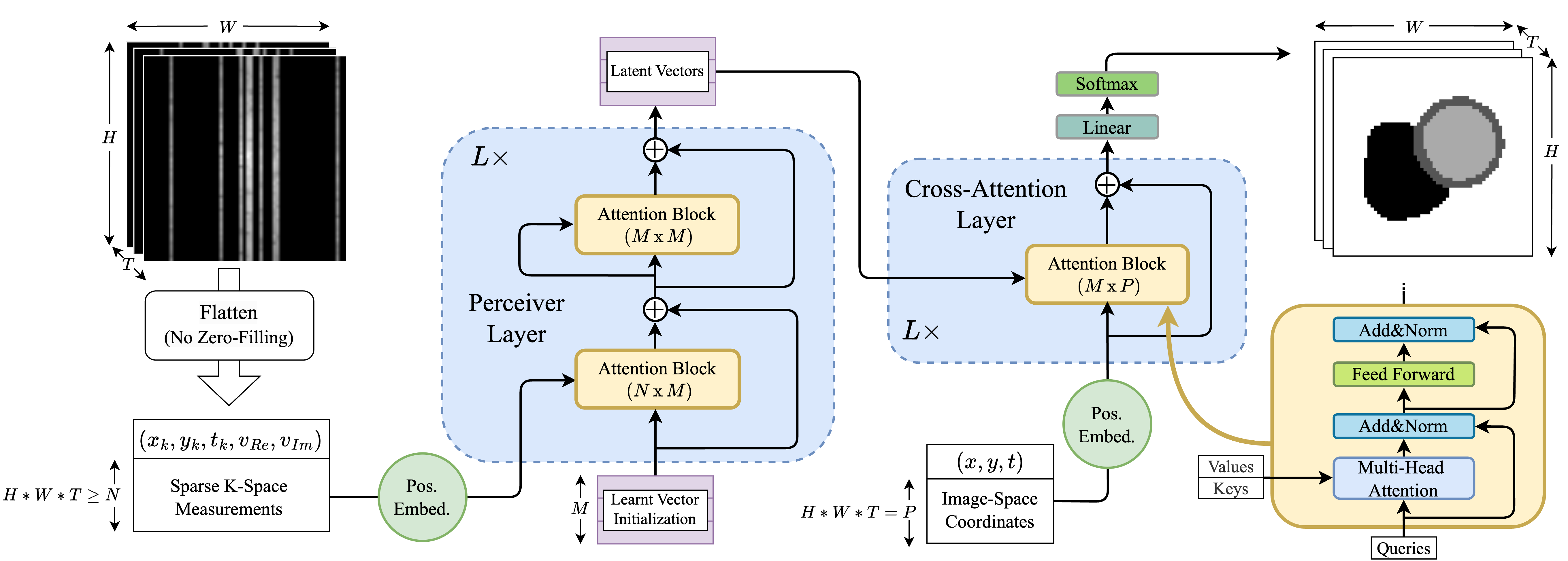
The paper proposes a deep learning framework that can perform classification, regression, and segmentation tasks directly on the k-space data, without the need for an intermediate image reconstruction step. The authors use a convolutional neural network architecture that takes the raw k-space data as input and produces the desired output, such as a classification, regression, or segmentation result.
The key technical innovation is the design of the network architecture, which includes specialized layers and loss functions tailored to the k-space data domain. For example, the network includes layers that can operate directly on the complex-valued k-space data, rather than requiring a separate real and imaginary channel.
The authors evaluate their approach on several cardiac MRI tasks, including left ventricular volume prediction, myocardial scar detection, and left ventricular segmentation. They show that their k-space-based approach outperforms traditional image-based methods, demonstrating the potential benefits of bypassing the image reconstruction step.
Critical Analysis
The paper presents a compelling approach for leveraging the raw k-space data in cardiac MRI analysis. By avoiding the image reconstruction step, the authors are able to potentially extract more clinically relevant information directly from the underlying measurements.
However, the paper does not address some potential limitations or concerns. For example, it is unclear how the approach would scale to more complex cardiac MRI tasks or datasets, or how robust it would be to variations in acquisition protocols or hardware.
Additionally, the paper does not explore the interpretability of the k-space-based models, which could be an important consideration for clinical applications. It would be valuable to understand how the network is making its predictions and whether the insights it derives from the k-space data are readily translatable to clinical practice.
Conclusion
This paper presents a novel deep learning framework for performing classification, regression, and segmentation tasks directly on cardiac MRI k-space data, without the need for an intermediate image reconstruction step. The authors demonstrate the effectiveness of their approach on several clinically relevant tasks, showing that it can outperform traditional image-based methods.
While the paper highlights the potential benefits of this k-space-based approach, further research is needed to address scalability, robustness, and interpretability concerns. Overall, the work represents an important step towards leveraging the full potential of raw MRI data for improved clinical decision-making.
This summary was produced with help from an AI and may contain inaccuracies - check out the links to read the original source documents!
Related Papers
0
Classification, Regression and Segmentation directly from k-Space in Cardiac MRI
Ruochen Li, Jiazhen Pan, Youxiang Zhu, Juncheng Ni, Daniel Rueckert
Cardiac Magnetic Resonance Imaging (CMR) is the gold standard for diagnosing cardiovascular diseases. Clinical diagnoses predominantly rely on magnitude-only Digital Imaging and Communications in Medicine (DICOM) images, omitting crucial phase information that might provide additional diagnostic benefits. In contrast, k-space is complex-valued and encompasses both magnitude and phase information, while humans cannot directly perceive. In this work, we propose KMAE, a Transformer-based model specifically designed to process k-space data directly, eliminating conventional intermediary conversion steps to the image domain. KMAE can handle critical cardiac disease classification, relevant phenotype regression, and cardiac morphology segmentation tasks. We utilize this model to investigate the potential of k-space-based diagnosis in cardiac MRI. Notably, this model achieves competitive classification and regression performance compared to image-domain methods e.g. Masked Autoencoders (MAEs) and delivers satisfactory segmentation performance with a myocardium dice score of 0.884. Last but not least, our model exhibits robust performance with consistent results even when the k-space is 8* undersampled. We encourage the MR community to explore the untapped potential of k-space and pursue end-to-end, automated diagnosis with reduced human intervention.
Read more7/30/2024
0
Direct Cardiac Segmentation from Undersampled K-space Using Transformers
Yundi Zhang, Nil Stolt-Ans'o, Jiazhen Pan, Wenqi Huang, Kerstin Hammernik, Daniel Rueckert
The prevailing deep learning-based methods of predicting cardiac segmentation involve reconstructed magnetic resonance (MR) images. The heavy dependency of segmentation approaches on image quality significantly limits the acceleration rate in fast MR reconstruction. Moreover, the practice of treating reconstruction and segmentation as separate sequential processes leads to artifact generation and information loss in the intermediate stage. These issues pose a great risk to achieving high-quality outcomes. To leverage the redundant k-space information overlooked in this dual-step pipeline, we introduce a novel approach to directly deriving segmentations from sparse k-space samples using a transformer (DiSK). DiSK operates by globally extracting latent features from 2D+time k-space data with attention blocks and subsequently predicting the segmentation label of query points. We evaluate our model under various acceleration factors (ranging from 4 to 64) and compare against two image-based segmentation baselines. Our model consistently outperforms the baselines in Dice and Hausdorff distances across foreground classes for all presented sampling rates.
Read more6/4/2024

0
Tumor likelihood estimation on MRI prostate data by utilizing k-Space information
M. Rempe, F. Horst, C. Seibold, B. Hadaschik, M. Schlimbach, J. Egger, K. Kroninger, F. Breuer, M. Blaimer, J. Kleesiek
We present a novel preprocessing and prediction pipeline for the classification of magnetic resonance imaging (MRI) that takes advantage of the information rich complex valued k-Space. Using a publicly available MRI raw dataset with 312 subject and a total of 9508 slices, we show the advantage of utilizing the k-Space for better prostate cancer likelihood estimation in comparison to just using the magnitudinal information in the image domain, with an AUROC of $86.1%pm1.8%$. Additionally, by using high undersampling rates and a simple principal component analysis (PCA) for coil compression, we reduce the time needed for reconstruction by avoiding the time intensive GRAPPA reconstruction algorithm. By using digital undersampling for our experiments, we show that scanning and reconstruction time could be reduced. Even with an undersampling factor of 16, our approach achieves meaningful results, with an AUROC of $71.4%pm2.9%$, using the PCA coil combination and taking into account the k-Space information. With this study, we were able to show the feasibility of preserving phase and k-Space information, with consistent results. Besides preserving valuable information for further diagnostics, this approach can work without the time intensive ADC and reconstruction calculations, greatly reducing the post processing, as well as potential scanning time, increasing patient comfort and allowing a close to real-time prediction.
Read more7/9/2024
🔍
0
CMRxRecon2024: A Multi-Modality, Multi-View K-Space Dataset Boosting Universal Machine Learning for Accelerated Cardiac MRI
Zi Wang, Fanwen Wang, Chen Qin, Jun Lyu, Ouyang Cheng, Shuo Wang, Yan Li, Mengyao Yu, Haoyu Zhang, Kunyuan Guo, Zhang Shi, Qirong Li, Ziqiang Xu, Yajing Zhang, Hao Li, Sha Hua, Binghua Chen, Longyu Sun, Mengting Sun, Qin Li, Ying-Hua Chu, Wenjia Bai, Jing Qin, Xiahai Zhuang, Claudia Prieto, Alistair Young, Michael Markl, He Wang, Lianming Wu, Guang Yang, Xiaobo Qu, Chengyan Wang
Cardiac magnetic resonance imaging (MRI) has emerged as a clinically gold-standard technique for diagnosing cardiac diseases, thanks to its ability to provide diverse information with multiple modalities and anatomical views. Accelerated cardiac MRI is highly expected to achieve time-efficient and patient-friendly imaging, and then advanced image reconstruction approaches are required to recover high-quality, clinically interpretable images from undersampled measurements. However, the lack of publicly available cardiac MRI k-space dataset in terms of both quantity and diversity has severely hindered substantial technological progress, particularly for data-driven artificial intelligence. Here, we provide a standardized, diverse, and high-quality CMRxRecon2024 dataset to facilitate the technical development, fair evaluation, and clinical transfer of cardiac MRI reconstruction approaches, towards promoting the universal frameworks that enable fast and robust reconstructions across different cardiac MRI protocols in clinical practice. To the best of our knowledge, the CMRxRecon2024 dataset is the largest and most diverse publicly available cardiac k-space dataset. It is acquired from 330 healthy volunteers, covering commonly used modalities, anatomical views, and acquisition trajectories in clinical cardiac MRI workflows. Besides, an open platform with tutorials, benchmarks, and data processing tools is provided to facilitate data usage, advanced method development, and fair performance evaluation.
Read more6/28/2024