Advancements in Feature Extraction Recognition of Medical Imaging Systems Through Deep Learning Technique

0
✨
Sign in to get full access
Overview
- Introduces a new unsupervised method for extracting features from medical images using spatial stratification techniques
- Proposes an objective function based on weight to enable faster image recognition
- Explores techniques for image segmentation and feature extraction for hyperspectral images
Plain English Explanation
This research paper presents a novel approach for automatically extracting important information from medical images without the need for human labeling. The key idea is to divide the image into multiple regions or subdomains, and then use a specialized algorithm to quickly identify the most relevant features within each region.
By breaking down the image in this way, the researchers were able to overcome some of the challenges posed by the complex, nonlinear characteristics of hyperspectral images. Their method was found to be more effective than traditional linear techniques at segmenting the images and extracting the defining features of different objects or tissues.
This is an important advancement, as being able to rapidly and accurately identify key characteristics in medical images can have significant implications for clinical diagnosis and treatment. The spatial stratification approach introduced in this paper may help improve the speed and reliability of computer-aided analysis of medical scans.
Technical Explanation
The paper describes an unsupervised algorithm that uses a quadtree data structure to divide medical images into multiple subdomains or regions. An objective function based on the weight of image features is proposed to enable faster and more accurate image recognition.
To handle the nonlinear nature of hyperspectral images, the researchers developed a generalized discriminant analysis technique that leverages kernel functions. This allows the algorithm to better capture the complex relationships between different elements within the images.
The method was tested on hyperspectral remote sensing images. The results showed that the algorithm was able to effectively segment the images and extract the distinct features of different objects, which tended to be independent and compact. Compared to traditional linear discrimination approaches, this technique produced superior image segmentation outcomes.
Critical Analysis
The paper makes a compelling case for the advantages of the proposed spatial stratification and feature extraction approach. However, it would be helpful to have more details on the specific performance gains achieved compared to other state-of-the-art methods, as well as the computational efficiency of the algorithm.
Additionally, while the results on hyperspectral remote sensing images are promising, further validation on a broader range of medical imaging modalities would help demonstrate the generalizability of the technique. Exploring how it could be combined with other advanced feature extraction modules may also yield interesting insights.
It's also worth considering potential limitations, such as the impact of image resolution or noise on the algorithm's effectiveness, and how these factors might be addressed through further refinements or preprocessing steps.
Conclusion
This research introduces an innovative unsupervised method for rapidly extracting relevant features from medical images using spatial stratification techniques. By dividing images into subdomains and leveraging specialized algorithms, the approach was shown to outperform traditional linear discrimination approaches in segmentation tasks.
The potential benefits of this work include faster and more reliable computer-aided analysis of medical scans, which could have significant implications for clinical diagnosis and treatment. Further research to validate the method's performance on a wider range of imaging modalities and explore potential enhancements could help solidify its impact on the field of medical image processing.
This summary was produced with help from an AI and may contain inaccuracies - check out the links to read the original source documents!
Related Papers
✨
0
Advancements in Feature Extraction Recognition of Medical Imaging Systems Through Deep Learning Technique
Qishi Zhan, Dan Sun, Erdi Gao, Yuhan Ma, Yaxin Liang, Haowei Yang
This study introduces a novel unsupervised medical image feature extraction method that employs spatial stratification techniques. An objective function based on weight is proposed to achieve the purpose of fast image recognition. The algorithm divides the pixels of the image into multiple subdomains and uses a quadtree to access the image. A technique for threshold optimization utilizing a simplex algorithm is presented. Aiming at the nonlinear characteristics of hyperspectral images, a generalized discriminant analysis algorithm based on kernel function is proposed. In this project, a hyperspectral remote sensing image is taken as the object, and we investigate its mathematical modeling, solution methods, and feature extraction techniques. It is found that different types of objects are independent of each other and compact in image processing. Compared with the traditional linear discrimination method, the result of image segmentation is better. This method can not only overcome the disadvantage of the traditional method which is easy to be affected by light, but also extract the features of the object quickly and accurately. It has important reference significance for clinical diagnosis.
Read more6/28/2024
0
Harmonized Spatial and Spectral Learning for Robust and Generalized Medical Image Segmentation
Vandan Gorade, Sparsh Mittal, Debesh Jha, Rekha Singhal, Ulas Bagci
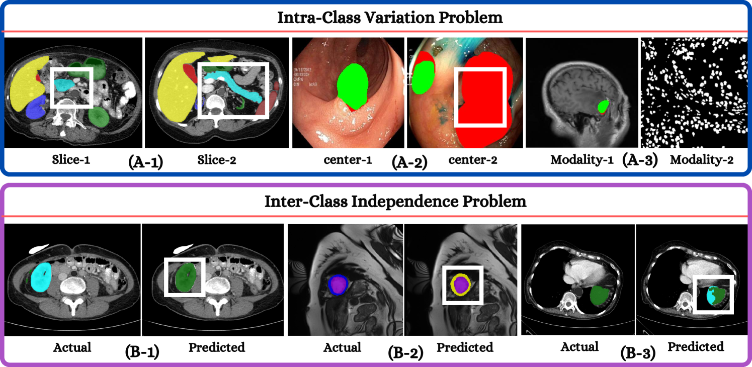
Deep learning has demonstrated remarkable achievements in medical image segmentation. However, prevailing deep learning models struggle with poor generalization due to (i) intra-class variations, where the same class appears differently in different samples, and (ii) inter-class independence, resulting in difficulties capturing intricate relationships between distinct objects, leading to higher false negative cases. This paper presents a novel approach that synergies spatial and spectral representations to enhance domain-generalized medical image segmentation. We introduce the innovative Spectral Correlation Coefficient objective to improve the model's capacity to capture middle-order features and contextual long-range dependencies. This objective complements traditional spatial objectives by incorporating valuable spectral information. Extensive experiments reveal that optimizing this objective with existing architectures like UNet and TransUNet significantly enhances generalization, interpretability, and noise robustness, producing more confident predictions. For instance, in cardiac segmentation, we observe a 0.81 pp and 1.63 pp (pp = percentage point) improvement in DSC over UNet and TransUNet, respectively. Our interpretability study demonstrates that, in most tasks, objectives optimized with UNet outperform even TransUNet by introducing global contextual information alongside local details. These findings underscore the versatility and effectiveness of our proposed method across diverse imaging modalities and medical domains.
Read more8/9/2024

0
Biomedical Image Segmentation: A Systematic Literature Review of Deep Learning Based Object Detection Methods
Fazli Wahid, Yingliang Ma, Dawar Khan, Muhammad Aamir, Syed U. K. Bukhari
Biomedical image segmentation plays a vital role in diagnosis of diseases across various organs. Deep learning-based object detection methods are commonly used for such segmentation. There exists an extensive research in this topic. However, there is no standard review on this topic. Existing surveys often lack a standardized approach or focus on broader segmentation techniques. In this paper, we conducted a systematic literature review (SLR), collected and analysed 148 articles that explore deep learning object detection methods for biomedical image segmentation. We critically analyzed these methods, identified the key challenges, and discussed the future directions. From the selected articles we extracted the results including the deep learning models, targeted imaging modalities, targeted diseases, and the metrics for the analysis of the methods. The results have been presented in tabular and/or charted forms. The results are presented in three major categories including two stage detection models, one stage detection models and point-based detection models. Each article is individually analyzed along with its pros and cons. Finally, we discuss open challenges, potential benefits, and future research directions. This SLR aims to provide the research community with a quick yet deeper understanding of these segmentation models, ultimately facilitating the development of more powerful solutions for biomedical image analysis.
Read more8/30/2024
🖼️
0
Comparative Analysis of Hyperspectral Image Reconstruction Using Deep Learning for Agricultural and Biological Applications
Md. Toukir Ahmed, Arthur Villordon, Mohammed Kamruzzaman
Hyperspectral imaging (HSI) has become a key technology for non-invasive quality evaluation in various fields, offering detailed insights through spatial and spectral data. Despite its efficacy, the complexity and high cost of HSI systems have hindered their widespread adoption. This study addressed these challenges by exploring deep learning-based hyperspectral image reconstruction from RGB (Red, Green, Blue) images, particularly for agricultural products. Specifically, different hyperspectral reconstruction algorithms, such as Hyperspectral Convolutional Neural Network - Dense (HSCNN-D), High-Resolution Network (HRNET), and Multi-Scale Transformer Plus Plus (MST++), were compared to assess the dry matter content of sweet potatoes. Among the tested reconstruction methods, HRNET demonstrated superior performance, achieving the lowest mean relative absolute error (MRAE) of 0.07, root mean square error (RMSE) of 0.03, and the highest peak signal-to-noise ratio (PSNR) of 32.28 decibels (dB). Some key features were selected using the genetic algorithm (GA), and their importance was interpreted using explainable artificial intelligence (XAI). Partial least squares regression (PLSR) models were developed using the RGB, reconstructed, and ground truth (GT) data. The visual and spectra quality of these reconstructed methods was compared with GT data, and predicted maps were generated. The results revealed the prospect of deep learning-based hyperspectral image reconstruction as a cost-effective and efficient quality assessment tool for agricultural and biological applications.
Read more6/4/2024