Harmonized Spatial and Spectral Learning for Robust and Generalized Medical Image Segmentation

0
Sign in to get full access
Overview
- This paper presents a new method for robust and generalized medical image segmentation.
- The proposed approach combines spatial and spectral learning to improve performance compared to previous techniques.
- Key innovations include a harmonized learning framework and leveraging both spatial and spectral information for segmentation.
Plain English Explanation
Medical image segmentation is the process of dividing an image into meaningful regions, such as organs or tumors. This is an important task for disease diagnosis and treatment planning. However, existing segmentation methods can struggle with complex medical images, especially when applied to new datasets or modalities.
The researchers in this paper developed a new approach called Harmonized Spatial and Spectral Learning to address these challenges. The core idea is to combine information about the spatial structure of the image (the shapes and locations of objects) with information about the spectral or color properties of the image (the distribution of pixel values).
By learning from both the spatial and spectral characteristics of the image, the model can build a more comprehensive understanding of the anatomy and better distinguish different tissues or structures. This harmonized learning framework allows the model to be more robust to variations in the input data and generalize better to new medical imaging scenarios.
The authors demonstrate the effectiveness of their approach through experiments on several medical image segmentation benchmarks. Their method outperforms previous state-of-the-art techniques, showing the value of fusing spatial and spectral information for this task.
Technical Explanation
The key innovation in this paper is the Harmonized Spatial and Spectral Learning (HSSL) framework for medical image segmentation. The authors hypothesize that incorporating both spatial and spectral information can lead to more robust and generalizable segmentation models.
The HSSL architecture consists of two parallel branches - one focusing on spatial learning and the other on spectral learning. The spatial branch uses standard convolutional layers to capture the spatial structure of the image, while the spectral branch leverages spectral-spatial features to encode the color/intensity distributions.
The outputs of these two branches are then harmonized through a series of fusion modules that aggregate the spatial and spectral features. This allows the model to learn a joint representation that encodes both the shape and appearance characteristics of the medical structures.
The authors evaluate their HSSL approach on several public medical image segmentation datasets, including Brain Tumor Segmentation and Cardiac Segmentation. The results show that HSSL outperforms previous state-of-the-art methods, achieving higher segmentation accuracy and better generalization to new data.
Critical Analysis
One of the key strengths of this research is the careful design of the HSSL architecture to leverage both spatial and spectral information for segmentation. The authors provide a thorough analysis of how this harmonized learning approach improves upon previous techniques that only consider one type of information.
However, the paper does not extensively discuss the computational complexity or inference speed of the HSSL model. As medical image analysis often requires real-time processing, the efficiency of the segmentation algorithm is an important practical consideration that could be explored further.
Additionally, the experiments are limited to a few commonly used medical image datasets. While the results are promising, it would be valuable to see how HSSL performs on a broader range of medical imaging modalities and anatomical structures, including more challenging cases that may arise in clinical practice.
Overall, this work demonstrates the benefits of incorporating multi-modal information for robust and generalizable medical image segmentation. The harmonized spatial-spectral learning approach proposed in this paper is a valuable contribution to the field and warrants further exploration and validation.
Conclusion
The "Harmonized Spatial and Spectral Learning for Robust and Generalized Medical Image Segmentation" paper presents a novel deep learning framework that fuses spatial and spectral information to achieve state-of-the-art performance on medical image segmentation tasks. By combining these complementary data sources, the proposed HSSL model is able to better capture the complex characteristics of anatomical structures, leading to improved segmentation accuracy and generalization.
This research highlights the importance of developing multi-modal learning techniques for medical image analysis, which could have significant implications for computer-aided diagnosis, surgical planning, and other clinical applications. While further validation is needed, the HSSL approach represents an important step forward in creating more robust and reliable medical image segmentation solutions.
This summary was produced with help from an AI and may contain inaccuracies - check out the links to read the original source documents!
Related Papers
0
Harmonized Spatial and Spectral Learning for Robust and Generalized Medical Image Segmentation
Vandan Gorade, Sparsh Mittal, Debesh Jha, Rekha Singhal, Ulas Bagci
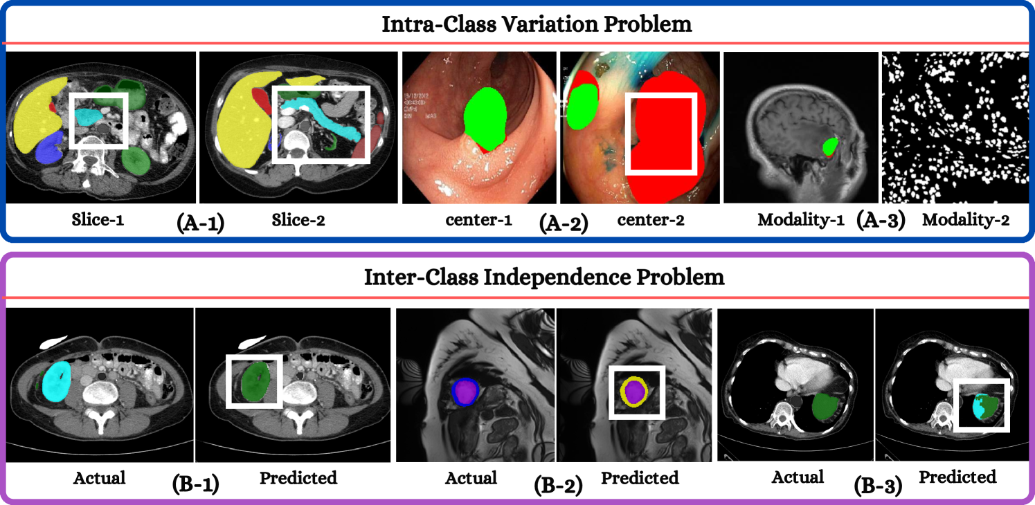
Deep learning has demonstrated remarkable achievements in medical image segmentation. However, prevailing deep learning models struggle with poor generalization due to (i) intra-class variations, where the same class appears differently in different samples, and (ii) inter-class independence, resulting in difficulties capturing intricate relationships between distinct objects, leading to higher false negative cases. This paper presents a novel approach that synergies spatial and spectral representations to enhance domain-generalized medical image segmentation. We introduce the innovative Spectral Correlation Coefficient objective to improve the model's capacity to capture middle-order features and contextual long-range dependencies. This objective complements traditional spatial objectives by incorporating valuable spectral information. Extensive experiments reveal that optimizing this objective with existing architectures like UNet and TransUNet significantly enhances generalization, interpretability, and noise robustness, producing more confident predictions. For instance, in cardiac segmentation, we observe a 0.81 pp and 1.63 pp (pp = percentage point) improvement in DSC over UNet and TransUNet, respectively. Our interpretability study demonstrates that, in most tasks, objectives optimized with UNet outperform even TransUNet by introducing global contextual information alongside local details. These findings underscore the versatility and effectiveness of our proposed method across diverse imaging modalities and medical domains.
Read more8/9/2024
✨
0
Advancements in Feature Extraction Recognition of Medical Imaging Systems Through Deep Learning Technique
Qishi Zhan, Dan Sun, Erdi Gao, Yuhan Ma, Yaxin Liang, Haowei Yang
This study introduces a novel unsupervised medical image feature extraction method that employs spatial stratification techniques. An objective function based on weight is proposed to achieve the purpose of fast image recognition. The algorithm divides the pixels of the image into multiple subdomains and uses a quadtree to access the image. A technique for threshold optimization utilizing a simplex algorithm is presented. Aiming at the nonlinear characteristics of hyperspectral images, a generalized discriminant analysis algorithm based on kernel function is proposed. In this project, a hyperspectral remote sensing image is taken as the object, and we investigate its mathematical modeling, solution methods, and feature extraction techniques. It is found that different types of objects are independent of each other and compact in image processing. Compared with the traditional linear discrimination method, the result of image segmentation is better. This method can not only overcome the disadvantage of the traditional method which is easy to be affected by light, but also extract the features of the object quickly and accurately. It has important reference significance for clinical diagnosis.
Read more6/28/2024

0
Spatial-Frequency Dual Progressive Attention Network For Medical Image Segmentation
Zhenhuan Zhou, Along He, Yanlin Wu, Rui Yao, Xueshuo Xie, Tao Li
In medical images, various types of lesions often manifest significant differences in their shape and texture. Accurate medical image segmentation demands deep learning models with robust capabilities in multi-scale and boundary feature learning. However, previous networks still have limitations in addressing the above issues. Firstly, previous networks simultaneously fuse multi-level features or employ deep supervision to enhance multi-scale learning. However, this may lead to feature redundancy and excessive computational overhead, which is not conducive to network training and clinical deployment. Secondly, the majority of medical image segmentation networks exclusively learn features in the spatial domain, disregarding the abundant global information in the frequency domain. This results in a bias towards low-frequency components, neglecting crucial high-frequency information. To address these problems, we introduce SF-UNet, a spatial-frequency dual-domain attention network. It comprises two main components: the Multi-scale Progressive Channel Attention (MPCA) block, which progressively extract multi-scale features across adjacent encoder layers, and the lightweight Frequency-Spatial Attention (FSA) block, with only 0.05M parameters, enabling concurrent learning of texture and boundary features from both spatial and frequency domains. We validate the effectiveness of the proposed SF-UNet on three public datasets. Experimental results show that compared to previous state-of-the-art (SOTA) medical image segmentation networks, SF-UNet achieves the best performance, and achieves up to 9.4% and 10.78% improvement in DSC and IOU. Codes will be released at https://github.com/nkicsl/SF-UNet.
Read more8/20/2024

0
New!Spectral U-Net: Enhancing Medical Image Segmentation via Spectral Decomposition
Yaopeng Peng, Milan Sonka, Danny Z. Chen
This paper introduces Spectral U-Net, a novel deep learning network based on spectral decomposition, by exploiting Dual Tree Complex Wavelet Transform (DTCWT) for down-sampling and inverse Dual Tree Complex Wavelet Transform (iDTCWT) for up-sampling. We devise the corresponding Wave-Block and iWave-Block, integrated into the U-Net architecture, aiming at mitigating information loss during down-sampling and enhancing detail reconstruction during up-sampling. In the encoder, we first decompose the feature map into high and low-frequency components using DTCWT, enabling down-sampling while mitigating information loss. In the decoder, we utilize iDTCWT to reconstruct higher-resolution feature maps from down-sampled features. Evaluations on the Retina Fluid, Brain Tumor, and Liver Tumor segmentation datasets with the nnU-Net framework demonstrate the superiority of the proposed Spectral U-Net.
Read more9/17/2024