AI-based Automatic Segmentation of Prostate on Multi-modality Images: A Review

0
🤷
Sign in to get full access
Overview
- Prostate cancer is a major health threat that requires early detection to reduce mortality.
- Computer-aided diagnosis (CAD) systems using multi-modal medical imaging (CT, MRI, ultrasound, etc.) can help with prostate cancer detection.
- However, accurately segmenting the prostate region in these images is challenging due to image imperfections and the complex prostate tissue structure.
- The rise of precision medicine and increased clinical capacity have spurred the need for data-driven tasks in medical imaging, including image segmentation.
- This paper proposes a new classification method that differentiates supervision types during the training phase for prostate segmentation.
- It also surveys AI-based automatic prostate segmentation methods, examines their advantages and limitations, and introduces variants of evaluation metrics.
- Finally, it discusses future research directions and development trends for high-precision prostate cancer detection and treatment.
Plain English Explanation
Prostate cancer is a serious health problem that can be deadly if not caught early. One way to improve early detection is to use computer-aided diagnosis (CAD) systems that analyze different types of medical scans, like CT, MRI, and ultrasound, of the prostate.
However, accurately identifying the prostate region in these scans is challenging. The images can have imperfections, and the prostate itself has a complex internal structure. As medicine becomes more personalized and hospitals have more data, there is a growing need for using artificial intelligence (AI) and data analysis to help with tasks like prostate segmentation (separating the prostate from the surrounding tissues).
This paper proposes a new way of training AI models to segment the prostate more accurately. It also reviews other AI-based prostate segmentation methods, looking at their strengths and weaknesses, and suggests new ways to evaluate how well these methods work.
Finally, the paper discusses future research directions, including using these advanced techniques to detect and treat prostate cancer with higher precision.
Technical Explanation
This paper introduces a new classification method that differentiates supervision types, either in number or kind, during the training phase for prostate segmentation. The authors conducted a survey on existing AI-based automatic prostate segmentation methods, examining the advantages and limitations of each approach.
The paper also introduces variants of evaluation metrics for verifying and assessing the performance of prostate segmentation methods. This includes measures beyond just overlap with the ground truth, such as considering the location and context of the segmented prostate.
Through their literature review, the authors identify several key challenges in prostate segmentation, including dealing with image imperfections and the complex prostate anatomy. They suggest that future research should focus on developing highly precise techniques for prostate cancer detection and treatment, leveraging the advancements in personalized medicine and increased clinical data availability.
Critical Analysis
The paper provides a comprehensive survey of AI-based prostate segmentation methods, highlighting both the progress made in this area and the persistent challenges. The authors' proposal of a new classification method that differentiates supervision types during training is an interesting approach, though its effectiveness would need to be empirically evaluated and compared to existing techniques.
One potential limitation of the study is the reliance on a literature review, which may not capture the full breadth of recent advancements in this rapidly evolving field. Additionally, the paper does not delve deeply into the specific architectures, training procedures, or detailed performance comparisons of the reviewed methods, which would be valuable for researchers and practitioners seeking to build upon this work.
The authors do acknowledge the need for further research to address the challenges of prostate segmentation, such as dealing with image imperfections and the complex prostate anatomy. Exploring domain transfer techniques and incorporating more advanced multi-modal fusion approaches may be promising avenues for future work in this area.
Overall, the paper provides a useful survey of the current state of AI-based prostate segmentation and highlights important directions for further research and development in this critical field of medical imaging.
Conclusion
This paper presents a comprehensive review of AI-based techniques for automatic prostate segmentation in medical imaging. The authors propose a new classification method that differentiates supervision types during the training phase, aiming to improve the accuracy of prostate segmentation.
The survey of existing AI-based prostate segmentation methods reveals both the progress made in this field and the ongoing challenges, such as dealing with image imperfections and the complex prostate anatomy. The authors also introduce variants of evaluation metrics to assess the performance of these segmentation algorithms more comprehensively.
Looking ahead, the paper suggests that future research should focus on developing highly precise techniques for prostate cancer detection and treatment, leveraging the advancements in personalized medicine and the increasing availability of clinical data. Exploring domain transfer and advanced multi-modal fusion approaches may be promising avenues to address the current limitations in this critical area of medical imaging.
This summary was produced with help from an AI and may contain inaccuracies - check out the links to read the original source documents!
Related Papers
🤷
0
AI-based Automatic Segmentation of Prostate on Multi-modality Images: A Review
Rui Jin, Derun Li, Dehui Xiang, Lei Zhang, Hailing Zhou, Fei Shi, Weifang Zhu, Jing Cai, Tao Peng, Xinjian Chen
Prostate cancer represents a major threat to health. Early detection is vital in reducing the mortality rate among prostate cancer patients. One approach involves using multi-modality (CT, MRI, US, etc.) computer-aided diagnosis (CAD) systems for the prostate region. However, prostate segmentation is challenging due to imperfections in the images and the prostate's complex tissue structure. The advent of precision medicine and a significant increase in clinical capacity have spurred the need for various data-driven tasks in the field of medical imaging. Recently, numerous machine learning and data mining tools have been integrated into various medical areas, including image segmentation. This article proposes a new classification method that differentiates supervision types, either in number or kind, during the training phase. Subsequently, we conducted a survey on artificial intelligence (AI)-based automatic prostate segmentation methods, examining the advantages and limitations of each. Additionally, we introduce variants of evaluation metrics for the verification and performance assessment of the segmentation method and summarize the current challenges. Finally, future research directions and development trends are discussed, reflecting the outcomes of our literature survey, suggesting high-precision detection and treatment of prostate cancer as a promising avenue.
Read more7/10/2024
0
How To Segment in 3D Using 2D Models: Automated 3D Segmentation of Prostate Cancer Metastatic Lesions on PET Volumes Using Multi-Angle Maximum Intensity Projections and Diffusion Models
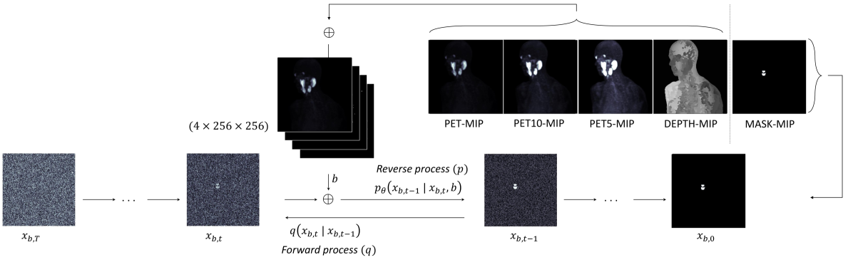
Amirhosein Toosi, Sara Harsini, Franc{c}ois B'enard, Carlos Uribe, Arman Rahmim
Prostate specific membrane antigen (PSMA) positron emission tomography/computed tomography (PET/CT) imaging provides a tremendously exciting frontier in visualization of prostate cancer (PCa) metastatic lesions. However, accurate segmentation of metastatic lesions is challenging due to low signal-to-noise ratios and variable sizes, shapes, and locations of the lesions. This study proposes a novel approach for automated segmentation of metastatic lesions in PSMA PET/CT 3D volumetric images using 2D denoising diffusion probabilistic models (DDPMs). Instead of 2D trans-axial slices or 3D volumes, the proposed approach segments the lesions on generated multi-angle maximum intensity projections (MA-MIPs) of the PSMA PET images, then obtains the final 3D segmentation masks from 3D ordered subset expectation maximization (OSEM) reconstruction of 2D MA-MIPs segmentations. Our proposed method achieved superior performance compared to state-of-the-art 3D segmentation approaches in terms of accuracy and robustness in detecting and segmenting small metastatic PCa lesions. The proposed method has significant potential as a tool for quantitative analysis of metastatic burden in PCa patients.
Read more7/29/2024

0
Automatic classification of prostate MR series type using image content and metadata
Deepa Krishnaswamy, B'alint Kov'acs, Stefan Denner, Steve Pieper, David Clunie, Christopher P. Bridge, Tina Kapur, Klaus H. Maier-Hein, Andrey Fedorov
With the wealth of medical image data, efficient curation is essential. Assigning the sequence type to magnetic resonance images is necessary for scientific studies and artificial intelligence-based analysis. However, incomplete or missing metadata prevents effective automation. We therefore propose a deep-learning method for classification of prostate cancer scanning sequences based on a combination of image data and DICOM metadata. We demonstrate superior results compared to metadata or image data alone, and make our code publicly available at https://github.com/deepakri201/DICOMScanClassification.
Read more8/1/2024

0
A Review of Image Processing Methods in Prostate Ultrasound
Haiqiao Wang, Hong Wu, Zhuoyuan Wang, Peiyan Yue, Dong Ni, Pheng-Ann Heng, Yi Wang
Prostate cancer (PCa) poses a significant threat to men's health, with early diagnosis being crucial for improving prognosis and reducing mortality rates. Transrectal ultrasound (TRUS) plays a vital role in the diagnosis and image-guided intervention of PCa.To facilitate physicians with more accurate and efficient computer-assisted diagnosis and interventions, many image processing algorithms in TRUS have been proposed and achieved state-of-the-art performance in several tasks, including prostate gland segmentation, prostate image registration, PCa classification and detection, and interventional needle detection.The rapid development of these algorithms over the past two decades necessitates a comprehensive summary. In consequence, this survey provides a systematic analysis of this field, outlining the evolution of image processing methods in the context of TRUS image analysis and meanwhile highlighting their relevant contributions. Furthermore, this survey discusses current challenges and suggests future research directions to possibly advance this field further.
Read more7/2/2024