How To Segment in 3D Using 2D Models: Automated 3D Segmentation of Prostate Cancer Metastatic Lesions on PET Volumes Using Multi-Angle Maximum Intensity Projections and Diffusion Models

0
Sign in to get full access
Overview
- This paper presents an automated method for 3D segmentation of prostate cancer metastatic lesions on PET volumes using multi-angle maximum intensity projections (MIPs) and diffusion models.
- The approach leverages 2D models to perform 3D segmentation, addressing the challenge of directly segmenting 3D PET volumes.
- Experiments on a dataset of prostate cancer patients demonstrate the effectiveness of the proposed technique.
Plain English Explanation
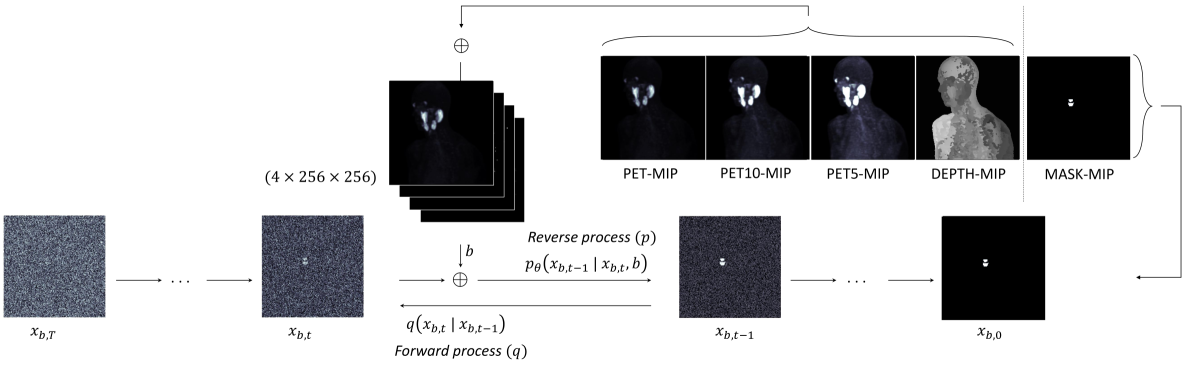
The paper describes a way to automatically identify and outline 3D regions of prostate cancer that have spread to other parts of the body, using PET scans. [PET scans measure radioactive tracer uptake in the body to detect cancer.]
The key idea is to use 2D models to do this 3D segmentation task, rather than trying to directly segment the 3D PET volumes. Specifically, the method generates 2D "maximum intensity projection" (MIP) images from different angles of the 3D PET data. These 2D MIP images are then processed by a segmentation model to identify the cancer regions. Finally, the 2D segmentations are combined to reconstruct the 3D shapes of the metastatic lesions.
This approach is advantageous because 2D segmentation models are generally easier to develop and more accurate than directly tackling the full 3D problem. The researchers show that their method performs well on a dataset of prostate cancer patients, providing an automated way to analyze PET scans and identify the locations of metastatic disease.
Technical Explanation
The paper presents an automated 3D segmentation approach for identifying prostate cancer metastatic lesions on PET volumes. The key innovation is the use of 2D models to perform this inherently 3D task.
Specifically, the method generates multi-angle maximum intensity projection (MIP) images from the 3D PET data. MIPs represent the maximum intensity values along the projection axis, effectively collapsing the 3D volume into a 2D representation. These 2D MIP images are then processed by a segmentation model to identify the cancer regions.
The 2D segmentations are subsequently combined to reconstruct the 3D shapes of the metastatic lesions. This approach leverages the advantages of 2D models, which are generally more accurate and easier to train than direct 3D segmentation methods.
The proposed technique was evaluated on a dataset of prostate cancer patients with PET/CT imaging. The experiments demonstrate the effectiveness of the multi-angle MIP and diffusion modeling approach, achieving promising results for automated 3D segmentation of prostate cancer metastases.
Critical Analysis
The paper presents a novel and promising approach for the challenging problem of 3D segmentation of prostate cancer lesions from PET data. The use of 2D models to tackle this inherently 3D task is a clever strategy that capitalizes on the strengths of 2D segmentation methods.
However, the authors acknowledge several limitations and areas for further research. For example, the dataset used in the experiments is relatively small, and the researchers note the need for larger-scale validation of the method. Additionally, the performance of the 2D segmentation models may be sensitive to factors like image resolution and noise levels in the PET data.
Furthermore, the paper does not provide a detailed analysis of the failure cases or potential biases in the segmentation results. Understanding the types of errors or systematic issues with the approach could help guide future improvements.
Overall, the proposed technique represents an interesting and potentially valuable contribution to the field of medical image analysis. However, further research is needed to fully assess the robustness and generalizability of the method, as well as explore ways to integrate the 2D and 3D components more seamlessly.
Conclusion
This paper introduces an automated 3D segmentation approach for identifying prostate cancer metastatic lesions on PET volumes. By leveraging 2D models to perform the inherently 3D task, the method addresses the challenges of directly segmenting 3D PET data.
The experiments on a dataset of prostate cancer patients demonstrate the effectiveness of the multi-angle MIP and diffusion modeling strategy, providing a promising avenue for automated analysis of PET scans and detection of metastatic disease. While the approach has some limitations, the paper presents an innovative solution to a clinically relevant problem in medical imaging.
This summary was produced with help from an AI and may contain inaccuracies - check out the links to read the original source documents!
Related Papers
0
How To Segment in 3D Using 2D Models: Automated 3D Segmentation of Prostate Cancer Metastatic Lesions on PET Volumes Using Multi-Angle Maximum Intensity Projections and Diffusion Models
Amirhosein Toosi, Sara Harsini, Franc{c}ois B'enard, Carlos Uribe, Arman Rahmim
Prostate specific membrane antigen (PSMA) positron emission tomography/computed tomography (PET/CT) imaging provides a tremendously exciting frontier in visualization of prostate cancer (PCa) metastatic lesions. However, accurate segmentation of metastatic lesions is challenging due to low signal-to-noise ratios and variable sizes, shapes, and locations of the lesions. This study proposes a novel approach for automated segmentation of metastatic lesions in PSMA PET/CT 3D volumetric images using 2D denoising diffusion probabilistic models (DDPMs). Instead of 2D trans-axial slices or 3D volumes, the proposed approach segments the lesions on generated multi-angle maximum intensity projections (MA-MIPs) of the PSMA PET images, then obtains the final 3D segmentation masks from 3D ordered subset expectation maximization (OSEM) reconstruction of 2D MA-MIPs segmentations. Our proposed method achieved superior performance compared to state-of-the-art 3D segmentation approaches in terms of accuracy and robustness in detecting and segmenting small metastatic PCa lesions. The proposed method has significant potential as a tool for quantitative analysis of metastatic burden in PCa patients.
Read more7/29/2024
✨
0
Segmentation of Prostate Tumour Volumes from PET Images is a Different Ball Game
Shrajan Bhandary, Dejan Kuhn, Zahra Babaiee, Tobias Fechter, Simon K. B. Spohn, Constantinos Zamboglou, Anca-Ligia Grosu, Radu Grosu
Accurate segmentation of prostate tumours from PET images presents a formidable challenge in medical image analysis. Despite considerable work and improvement in delineating organs from CT and MR modalities, the existing standards do not transfer well and produce quality results in PET related tasks. Particularly, contemporary methods fail to accurately consider the intensity-based scaling applied by the physicians during manual annotation of tumour contours. In this paper, we observe that the prostate-localised uptake threshold ranges are beneficial for suppressing outliers. Therefore, we utilize the intensity threshold values, to implement a new custom-feature-clipping normalisation technique. We evaluate multiple, established U-Net variants under different normalisation schemes, using the nnU-Net framework. All models were trained and tested on multiple datasets, obtained with two radioactive tracers: [68-Ga]Ga-PSMA-11 and [18-F]PSMA-1007. Our results show that the U-Net models achieve much better performance when the PET scans are preprocessed with our novel clipping technique.
Read more7/16/2024
🤷
0
AI-based Automatic Segmentation of Prostate on Multi-modality Images: A Review
Rui Jin, Derun Li, Dehui Xiang, Lei Zhang, Hailing Zhou, Fei Shi, Weifang Zhu, Jing Cai, Tao Peng, Xinjian Chen
Prostate cancer represents a major threat to health. Early detection is vital in reducing the mortality rate among prostate cancer patients. One approach involves using multi-modality (CT, MRI, US, etc.) computer-aided diagnosis (CAD) systems for the prostate region. However, prostate segmentation is challenging due to imperfections in the images and the prostate's complex tissue structure. The advent of precision medicine and a significant increase in clinical capacity have spurred the need for various data-driven tasks in the field of medical imaging. Recently, numerous machine learning and data mining tools have been integrated into various medical areas, including image segmentation. This article proposes a new classification method that differentiates supervision types, either in number or kind, during the training phase. Subsequently, we conducted a survey on artificial intelligence (AI)-based automatic prostate segmentation methods, examining the advantages and limitations of each. Additionally, we introduce variants of evaluation metrics for the verification and performance assessment of the segmentation method and summarize the current challenges. Finally, future research directions and development trends are discussed, reflecting the outcomes of our literature survey, suggesting high-precision detection and treatment of prostate cancer as a promising avenue.
Read more7/10/2024

0
Towards AI Lesion Tracking in PET/CT Imaging: A Siamese-based CNN Pipeline applied on PSMA PET/CT Scans
Stefan P. Hein, Manuel Schultheiss, Andrei Gafita, Raphael Zaum, Farid Yagubbayli, Robert Tauber, Isabel Rauscher, Matthias Eiber, Franz Pfeiffer, Wolfgang A. Weber
Assessing tumor response to systemic therapies is one of the main applications of PET/CT. Routinely, only a small subset of index lesions out of multiple lesions is analyzed. However, this operator dependent selection may bias the results due to possible significant inter-metastatic heterogeneity of response to therapy. Automated, AI based approaches for lesion tracking hold promise in enabling the analysis of many more lesions and thus providing a better assessment of tumor response. This work introduces a Siamese CNN approach for lesion tracking between PET/CT scans. Our approach is applied on the laborious task of tracking a high number of bone lesions in full-body baseline and follow-up [68Ga]Ga- or [18F]F-PSMA PET/CT scans after two cycles of [177Lu]Lu-PSMA therapy of metastatic castration resistant prostate cancer patients. Data preparation includes lesion segmentation and affine registration. Our algorithm extracts suitable lesion patches and forwards them into a Siamese CNN trained to classify the lesion patch pairs as corresponding or non-corresponding lesions. Experiments have been performed with different input patch types and a Siamese network in 2D and 3D. The CNN model successfully learned to classify lesion assignments, reaching a lesion tracking accuracy of 83 % in its best configuration with an AUC = 0.91. For remaining lesions the pipeline accomplished a re-identification rate of 89 %. We proved that a CNN may facilitate the tracking of multiple lesions in PSMA PET/CT scans. Future clinical studies are necessary if this improves the prediction of the outcome of therapies.
Read more7/9/2024