AResNet-ViT: A Hybrid CNN-Transformer Network for Benign and Malignant Breast Nodule Classification in Ultrasound Images

0
🌐
Sign in to get full access
Overview
- Researchers propose a deep learning network that integrates Convolutional Neural Networks (CNNs) and Vision Transformers (ViTs) to classify benign and malignant breast lesions in ultrasound images.
- The network uses a dual-branch architecture to extract local and global features, leveraging the strengths of CNNs and ViTs.
- The goal is to address challenges in accurately classifying breast lesions, such as similarity between lesions and surrounding tissues, overlapping appearances of partially benign and malignant nodules, and difficulty in classification.
Plain English Explanation
The researchers have developed a new deep learning system to help doctors better identify whether breast lumps or "nodules" are benign (non-cancerous) or malignant (cancerous). This is an important problem because sometimes it can be hard to tell the difference just by looking at the nodule and the surrounding tissue in medical scans like ultrasound images.
The new system uses a combination of two powerful AI techniques - Convolutional Neural Networks (CNNs) and Vision Transformers (ViTs). CNNs are good at capturing the local details and textures of the nodule itself, while ViTs can understand the overall shape, edges, and relationship of the nodule to the surrounding tissue.
By using both of these approaches together in a dual-branch architecture, the researchers' system can get a more complete understanding of the nodule and make more accurate classifications of whether it is benign or malignant. This helps address the challenges of similarity between lesions, overlapping appearances, and difficulty in classification that doctors sometimes face.
Technical Explanation
The proposed deep learning network integrates CNNs and ViTs to perform the classification of benign and malignant breast lesions in ultrasound images. The network adopts a dual-branch architecture for local and global feature extraction.
The local feature extraction branch employs a residual network with multiple attention-guided modules, which effectively captures the local details and texture features of breast nodules. This helps the network be sensitive to subtle changes within the nodules, aiding accurate classification of their benign or malignant status.
The global feature extraction branch utilizes a multi-head self-attention ViT network, which can capture the overall shape, boundary, and relationship of the nodule with the surrounding tissues. This enhances the understanding and modeling of both nodule and global image features.
Experimental results on a public ultrasound breast nodule dataset show that the proposed method outperforms other comparison networks. This indicates that the fusion of CNN and Transformer networks can effectively improve the performance of the classification model, providing a powerful solution for the benign-malignant classification of breast ultrasound images.
Critical Analysis
The paper presents a novel and promising approach to address the challenge of accurately classifying breast lesions in ultrasound images. The integration of CNN and Transformer architectures is a sensible strategy, as it leverages the complementary strengths of these techniques to capture both local and global features.
One potential limitation is the reliance on a single public dataset for evaluation. While the results are encouraging, it would be valuable to validate the performance of the model on additional independent datasets to ensure its robustness and generalizability.
Additionally, the paper does not provide much insight into the computational complexity or inference time of the proposed network. This information would be helpful for assessing the practicality of deploying such a system in real-world clinical settings, where efficient processing of images is crucial.
Further research could also explore the interpretability of the model's decision-making process. Providing clinicians with explanations for the model's classifications could increase trust and facilitate the integration of such AI-powered tools into the medical decision-making workflow.
Conclusion
The researchers have developed a novel deep learning network that combines CNNs and Transformers to classify benign and malignant breast lesions in ultrasound images. By leveraging the complementary strengths of these architectures, the proposed system demonstrates improved performance compared to other models, addressing key challenges in this domain.
While further validation and optimization may be needed, this work represents a significant step forward in the development of AI-powered tools to assist medical professionals in the accurate diagnosis of breast lesions. If successfully deployed, such systems could potentially improve patient outcomes by facilitating earlier detection and more informed treatment decisions.
This summary was produced with help from an AI and may contain inaccuracies - check out the links to read the original source documents!
Related Papers
🌐
0
AResNet-ViT: A Hybrid CNN-Transformer Network for Benign and Malignant Breast Nodule Classification in Ultrasound Images
Xin Zhao, Qianqian Zhu, Jialing Wu
To address the challenges of similarity between lesions and surrounding tissues, overlapping appearances of partially benign and malignant nodules, and difficulty in classification, a deep learning network that integrates CNN and Transformer is proposed for the classification of benign and malignant breast lesions in ultrasound images. This network adopts a dual-branch architecture for local-global feature extraction, making full use of the advantages of CNN in extracting local features and the ability of ViT to extract global features to enhance the network's feature extraction capabilities for breast nodules. The local feature extraction branch employs a residual network with multiple attention-guided modules, which can effectively capture the local details and texture features of breast nodules, enhance sensitivity to subtle changes within the nodules, and thus can aid in accurate classification of their benign and malignancy. The global feature extraction branch utilizes the multi-head self-attention ViT network, which can capture the overall shape, boundary, and relationship with surrounding tissues, and thereby enhancing the understanding and modeling of both nodule and global image features. Experimental results on a public ultrasound breast nodule data set show that the proposed method is better than other comparison networks, This indicates that the fusion of CNN and Transformer networks can effectively improve the performance of the classification model and provide a powerful solution for the benign-malignant classification of ultrasound breast.
Read more7/30/2024

0
A Comparative Study of CNN, ResNet, and Vision Transformers for Multi-Classification of Chest Diseases
Ananya Jain, Aviral Bhardwaj, Kaushik Murali, Isha Surani
Large language models, notably utilizing Transformer architectures, have emerged as powerful tools due to their scalability and ability to process large amounts of data. Dosovitskiy et al. expanded this architecture to introduce Vision Transformers (ViT), extending its applicability to image processing tasks. Motivated by this advancement, we fine-tuned two variants of ViT models, one pre-trained on ImageNet and another trained from scratch, using the NIH Chest X-ray dataset containing over 100,000 frontal-view X-ray images. Our study evaluates the performance of these models in the multi-label classification of 14 distinct diseases, while using Convolutional Neural Networks (CNNs) and ResNet architectures as baseline models for comparison. Through rigorous assessment based on accuracy metrics, we identify that the pre-trained ViT model surpasses CNNs and ResNet in this multilabel classification task, highlighting its potential for accurate diagnosis of various lung conditions from chest X-ray images.
Read more6/4/2024

0
Prototype Learning Guided Hybrid Network for Breast Tumor Segmentation in DCE-MRI
Lei Zhou, Yuzhong Zhang, Jiadong Zhang, Xuejun Qian, Chen Gong, Kun Sun, Zhongxiang Ding, Xing Wang, Zhenhui Li, Zaiyi Liu, Dinggang Shen
Automated breast tumor segmentation on the basis of dynamic contrast-enhancement magnetic resonance imaging (DCE-MRI) has shown great promise in clinical practice, particularly for identifying the presence of breast disease. However, accurate segmentation of breast tumor is a challenging task, often necessitating the development of complex networks. To strike an optimal trade-off between computational costs and segmentation performance, we propose a hybrid network via the combination of convolution neural network (CNN) and transformer layers. Specifically, the hybrid network consists of a encoder-decoder architecture by stacking convolution and decovolution layers. Effective 3D transformer layers are then implemented after the encoder subnetworks, to capture global dependencies between the bottleneck features. To improve the efficiency of hybrid network, two parallel encoder subnetworks are designed for the decoder and the transformer layers, respectively. To further enhance the discriminative capability of hybrid network, a prototype learning guided prediction module is proposed, where the category-specified prototypical features are calculated through on-line clustering. All learned prototypical features are finally combined with the features from decoder for tumor mask prediction. The experimental results on private and public DCE-MRI datasets demonstrate that the proposed hybrid network achieves superior performance than the state-of-the-art (SOTA) methods, while maintaining balance between segmentation accuracy and computation cost. Moreover, we demonstrate that automatically generated tumor masks can be effectively applied to identify HER2-positive subtype from HER2-negative subtype with the similar accuracy to the analysis based on manual tumor segmentation. The source code is available at https://github.com/ZhouL-lab/PLHN.
Read more8/13/2024
0
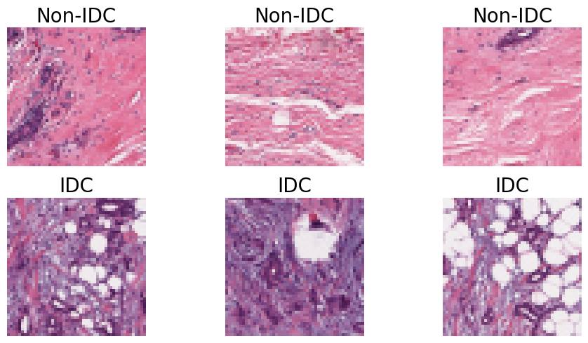
Comparative Analysis of Transfer Learning Models for Breast Cancer Classification
Sania Eskandari, Ali Eslamian, Qiang Cheng
The classification of histopathological images is crucial for the early and precise detection of breast cancer. This study investigates the efficiency of deep learning models in distinguishing between Invasive Ductal Carcinoma (IDC) and non-IDC in histopathology slides. We conducted a thorough comparison examination of eight sophisticated models: ResNet-50, DenseNet-121, ResNeXt-50, Vision Transformer (ViT), GoogLeNet (Inception v3), EfficientNet, MobileNet, and SqueezeNet. This analysis was carried out using a large dataset of 277,524 image patches. Our research makes a substantial contribution to the field by offering a comprehensive assessment of the performance of each model. We particularly highlight the exceptional efficacy of attention-based mechanisms in the ViT model, which achieved a remarkable validation accuracy of 93%, surpassing conventional convolutional networks. This study highlights the promise of advanced machine learning approaches in clinical settings, offering improved precision as well as efficiency in breast cancer diagnosis.
Read more9/2/2024