Prototype Learning Guided Hybrid Network for Breast Tumor Segmentation in DCE-MRI

0

Sign in to get full access
Overview
- The paper discusses a novel deep learning model called the Prototype Learning Guided Hybrid Network (PLG-HybridNet) for segmenting breast tumors in Dynamic Contrast-Enhanced Magnetic Resonance Imaging (DCE-MRI) scans.
- The proposed model combines the strengths of Convolutional Neural Networks (CNNs) and Transformer architectures to improve the accuracy and robustness of breast tumor segmentation.
- The key innovations include a prototype learning module that captures the distinctive features of different tumor types and a hybrid network architecture that integrates CNN and Transformer components.
Plain English Explanation
The paper presents a new deep learning system for analyzing medical images of the breast to identify and outline tumors. This is an important task, as accurately detecting and delineating breast tumors in MRI scans can help doctors better understand the size, shape, and location of any abnormal growths, which is crucial for diagnosis and treatment planning.
The researchers developed a [object Object] that combines two powerful machine learning techniques: Convolutional Neural Networks (CNNs) and Transformers. CNNs are well-suited for processing visual data like medical images, as they can effectively capture local spatial features. Transformers, on the other hand, are adept at modeling long-range dependencies and global context, which can be valuable for segmentation tasks.
The key innovation in this work is the [object Object], which learns to identify the distinctive features of different tumor types. This helps the model better recognize and segment various kinds of breast tumors, even ones it may not have seen during training. The researchers also designed a [object Object] that integrates the strengths of CNNs and Transformers to capture both local and global information for improved segmentation accuracy.
Overall, this work demonstrates how combining advanced deep learning techniques can lead to more robust and effective medical image analysis tools, which could ultimately help clinicians provide better care for patients with breast cancer.
Technical Explanation
The [object Object] proposed in this paper consists of three key components:
-
Backbone CNN: This serves as the base feature extractor, using a standard CNN architecture like [object Object] to capture local spatial features from the input MRI images.
-
Prototype Learning Module: This module learns a set of prototypes that represent the distinctive features of different tumor types. By comparing the input features to these prototypes, the model can better identify and segment various kinds of breast tumors.
-
Transformer Encoder: This component processes the features from the backbone CNN and the prototype learning module to capture long-range dependencies and global context, which can further improve segmentation performance.
The researchers trained and evaluated their PLG-HybridNet model on a dataset of breast DCE-MRI scans, and compared its performance to several state-of-the-art segmentation approaches. The results showed that the proposed [object Object] outperformed the baselines, demonstrating the value of integrating prototype learning and transformer-based reasoning for this task.
Critical Analysis
The paper presents a well-designed and thoroughly evaluated deep learning model for breast tumor segmentation in DCE-MRI scans. The key strengths of the work include:
- Robust Tumor Representation: The prototype learning module allows the model to capture the distinctive features of different tumor types, improving its ability to segment a wide variety of abnormal growths.
- Effective Hybrid Architecture: The integration of CNN and Transformer components helps the model leverage both local and global information for accurate segmentation.
- Comprehensive Evaluation: The researchers conducted experiments on a real-world dataset and compared their approach to multiple state-of-the-art baselines, demonstrating the benefits of their proposed method.
However, the paper also acknowledges some limitations and areas for future work:
- Dataset Size: The evaluation was conducted on a relatively small dataset, so further testing on larger, more diverse medical imaging repositories would be valuable to assess the model's generalization capabilities.
- Interpretability: The inner workings of the prototype learning module and hybrid network are complex, so additional analysis to improve the interpretability of the model's decisions could enhance trust and adoption in clinical settings.
- Real-Time Performance: The computational requirements of the proposed approach were not thoroughly explored, and its suitability for real-time applications, such as intraoperative tumor visualization, remains to be investigated.
Overall, this paper presents a promising deep learning solution for breast tumor segmentation that merits further research and development to address these remaining challenges.
Conclusion
The [object Object] introduced in this paper demonstrates the potential of combining advanced deep learning techniques, such as prototype learning and hybrid CNN-Transformer architectures, to tackle the challenging task of breast tumor segmentation in DCE-MRI scans. By capturing the distinctive features of different tumor types and integrating local and global information, the proposed model achieved state-of-the-art performance, showcasing the value of this approach for improving medical image analysis and, ultimately, supporting more accurate diagnosis and treatment planning for breast cancer patients.
This summary was produced with help from an AI and may contain inaccuracies - check out the links to read the original source documents!
Related Papers
0
Prototype Learning Guided Hybrid Network for Breast Tumor Segmentation in DCE-MRI
Lei Zhou, Yuzhong Zhang, Jiadong Zhang, Xuejun Qian, Chen Gong, Kun Sun, Zhongxiang Ding, Xing Wang, Zhenhui Li, Zaiyi Liu, Dinggang Shen
Automated breast tumor segmentation on the basis of dynamic contrast-enhancement magnetic resonance imaging (DCE-MRI) has shown great promise in clinical practice, particularly for identifying the presence of breast disease. However, accurate segmentation of breast tumor is a challenging task, often necessitating the development of complex networks. To strike an optimal trade-off between computational costs and segmentation performance, we propose a hybrid network via the combination of convolution neural network (CNN) and transformer layers. Specifically, the hybrid network consists of a encoder-decoder architecture by stacking convolution and decovolution layers. Effective 3D transformer layers are then implemented after the encoder subnetworks, to capture global dependencies between the bottleneck features. To improve the efficiency of hybrid network, two parallel encoder subnetworks are designed for the decoder and the transformer layers, respectively. To further enhance the discriminative capability of hybrid network, a prototype learning guided prediction module is proposed, where the category-specified prototypical features are calculated through on-line clustering. All learned prototypical features are finally combined with the features from decoder for tumor mask prediction. The experimental results on private and public DCE-MRI datasets demonstrate that the proposed hybrid network achieves superior performance than the state-of-the-art (SOTA) methods, while maintaining balance between segmentation accuracy and computation cost. Moreover, we demonstrate that automatically generated tumor masks can be effectively applied to identify HER2-positive subtype from HER2-negative subtype with the similar accuracy to the analysis based on manual tumor segmentation. The source code is available at https://github.com/ZhouL-lab/PLHN.
Read more8/13/2024
0
D-TrAttUnet: Toward Hybrid CNN-Transformer Architecture for Generic and Subtle Segmentation in Medical Images
Fares Bougourzi, Fadi Dornaika, Cosimo Distante, Abdelmalik Taleb-Ahmed
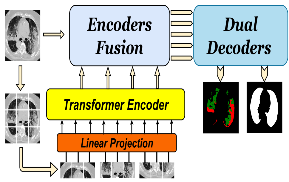
Over the past two decades, machine analysis of medical imaging has advanced rapidly, opening up significant potential for several important medical applications. As complicated diseases increase and the number of cases rises, the role of machine-based imaging analysis has become indispensable. It serves as both a tool and an assistant to medical experts, providing valuable insights and guidance. A particularly challenging task in this area is lesion segmentation, a task that is challenging even for experienced radiologists. The complexity of this task highlights the urgent need for robust machine learning approaches to support medical staff. In response, we present our novel solution: the D-TrAttUnet architecture. This framework is based on the observation that different diseases often target specific organs. Our architecture includes an encoder-decoder structure with a composite Transformer-CNN encoder and dual decoders. The encoder includes two paths: the Transformer path and the Encoders Fusion Module path. The Dual-Decoder configuration uses two identical decoders, each with attention gates. This allows the model to simultaneously segment lesions and organs and integrate their segmentation losses. To validate our approach, we performed evaluations on the Covid-19 and Bone Metastasis segmentation tasks. We also investigated the adaptability of the model by testing it without the second decoder in the segmentation of glands and nuclei. The results confirmed the superiority of our approach, especially in Covid-19 infections and the segmentation of bone metastases. In addition, the hybrid encoder showed exceptional performance in the segmentation of glands and nuclei, solidifying its role in modern medical image analysis.
Read more5/8/2024

0
Progressive Dual Priori Network for Generalized Breast Tumor Segmentation
Li Wang, Lihui Wang, Zixiang Kuai, Lei Tang, Yingfeng Ou, Chen Ye, Yuemin Zhu
To promote the generalization ability of breast tumor segmentation models, as well as to improve the segmentation performance for breast tumors with smaller size, low-contrast and irregular shape, we propose a progressive dual priori network (PDPNet) to segment breast tumors from dynamic enhanced magnetic resonance images (DCE-MRI) acquired at different centers. The PDPNet first cropped tumor regions with a coarse-segmentation based localization module, then the breast tumor mask was progressively refined by using the weak semantic priori and cross-scale correlation prior knowledge. To validate the effectiveness of PDPNet, we compared it with several state-of-the-art methods on multi-center datasets. The results showed that, comparing against the suboptimal method, the DSC and HD95 of PDPNet were improved at least by 5.13% and 7.58% respectively on multi-center test sets. In addition, through ablations, we demonstrated that the proposed localization module can decrease the influence of normal tissues and therefore improve the generalization ability of the model. The weak semantic priors allow focusing on tumor regions to avoid missing small tumors and low-contrast tumors. The cross-scale correlation priors are beneficial for promoting the shape-aware ability for irregular tumors. Thus integrating them in a unified framework improved the multi-center breast tumor segmentation performance. The source code and open data can be accessed at https://github.com/wangli100209/PDPNet.
Read more6/18/2024

0
TBConvL-Net: A Hybrid Deep Learning Architecture for Robust Medical Image Segmentation
Shahzaib Iqbal, Tariq M. Khan, Syed S. Naqvi, Asim Naveed, Erik Meijering
Deep learning has shown great potential for automated medical image segmentation to improve the precision and speed of disease diagnostics. However, the task presents significant difficulties due to variations in the scale, shape, texture, and contrast of the pathologies. Traditional convolutional neural network (CNN) models have certain limitations when it comes to effectively modelling multiscale context information and facilitating information interaction between skip connections across levels. To overcome these limitations, a novel deep learning architecture is introduced for medical image segmentation, taking advantage of CNNs and vision transformers. Our proposed model, named TBConvL-Net, involves a hybrid network that combines the local features of a CNN encoder-decoder architecture with long-range and temporal dependencies using biconvolutional long-short-term memory (LSTM) networks and vision transformers (ViT). This enables the model to capture contextual channel relationships in the data and account for the uncertainty of segmentation over time. Additionally, we introduce a novel composite loss function that considers both the segmentation robustness and the boundary agreement of the predicted output with the gold standard. Our proposed model shows consistent improvement over the state of the art on ten publicly available datasets of seven different medical imaging modalities.
Read more9/6/2024