Automatic Organ and Pan-cancer Segmentation in Abdomen CT: the FLARE 2023 Challenge

0

Sign in to get full access
Overview
- This paper presents the results of the "Automatic Organ and Pan-cancer Segmentation in Abdomen CT: the FLARE 2023 Challenge".
- The challenge aimed to develop automated methods for segmenting organs and detecting cancerous lesions in abdominal CT scans.
- Multiple research teams participated in the challenge, and the paper summarizes the approaches and findings.
Plain English Explanation
The research described in this paper focused on developing artificial intelligence (AI) systems that can automatically analyze abdominal CT scans. The goal was to create algorithms that could accurately identify and outline the different organs, as well as detect any cancerous growths, in these medical images.
This is an important task because manually reviewing all the details in CT scans can be time-consuming for radiologists. Automated systems could help streamline the process and potentially improve patient care. The researchers organized a challenge, inviting teams from around the world to submit their best machine learning algorithms for this task.
The teams used a variety of deep learning techniques, including convolutional neural networks and other advanced computer vision models, to analyze the CT scans. The performance of these algorithms was then evaluated, and the results are summarized in this paper.
Technical Explanation
The researchers organized the "Automatic Organ and Pan-cancer Segmentation in Abdomen CT: the FLARE 2023 Challenge" to advance the state-of-the-art in automated analysis of abdominal CT scans. Participants were tasked with developing algorithms that could accurately segment organs and detect cancerous lesions in these medical images.
The challenge dataset consisted of abdominal CT scans from multiple institutions, with detailed annotations provided for the organs and any cancerous growths. Participants used a variety of deep learning techniques, such as convolutional neural networks and other computer vision models, to analyze the images and produce their segmentation and detection results.
The algorithms were evaluated based on their ability to accurately identify the organs and lesions, as measured by metrics like Dice similarity coefficient and precision-recall curves. The top-performing teams demonstrated impressive results, showcasing the potential of these machine learning techniques for assisting radiologists in their clinical workflow.
Critical Analysis
The paper provides a comprehensive overview of the FLARE 2023 Challenge and the various approaches taken by the participating teams. The use of a standardized dataset and evaluation metrics helps to ensure the comparability of the results.
However, the paper does not delve into the specific architectural details or hyperparameter choices of the individual models, which could limit the ability to replicate or build upon the findings. Additionally, the paper does not discuss any potential biases or limitations in the dataset, which could affect the generalizability of the models.
Further research is needed to explore the clinical implementation and real-world performance of these automated organ segmentation and cancer detection algorithms. Factors such as integration with existing healthcare workflows, interpretability of the models, and long-term patient outcomes should be considered.
Conclusion
The FLARE 2023 Challenge has demonstrated the impressive capabilities of deep learning and computer vision techniques for automating the analysis of abdominal CT scans. The top-performing algorithms were able to accurately segment organs and detect cancerous lesions, suggesting that these technologies could potentially assist radiologists in their clinical practice.
However, further research is needed to address the limitations and ensure the safe and effective deployment of these systems in healthcare settings. Ongoing collaboration between the AI research community and medical professionals will be crucial for advancing this field and harnessing the full potential of these artificial intelligence tools to improve patient outcomes.
This summary was produced with help from an AI and may contain inaccuracies - check out the links to read the original source documents!
Related Papers
0
Automatic Organ and Pan-cancer Segmentation in Abdomen CT: the FLARE 2023 Challenge
Jun Ma, Yao Zhang, Song Gu, Cheng Ge, Ershuai Wang, Qin Zhou, Ziyan Huang, Pengju Lyu, Jian He, Bo Wang
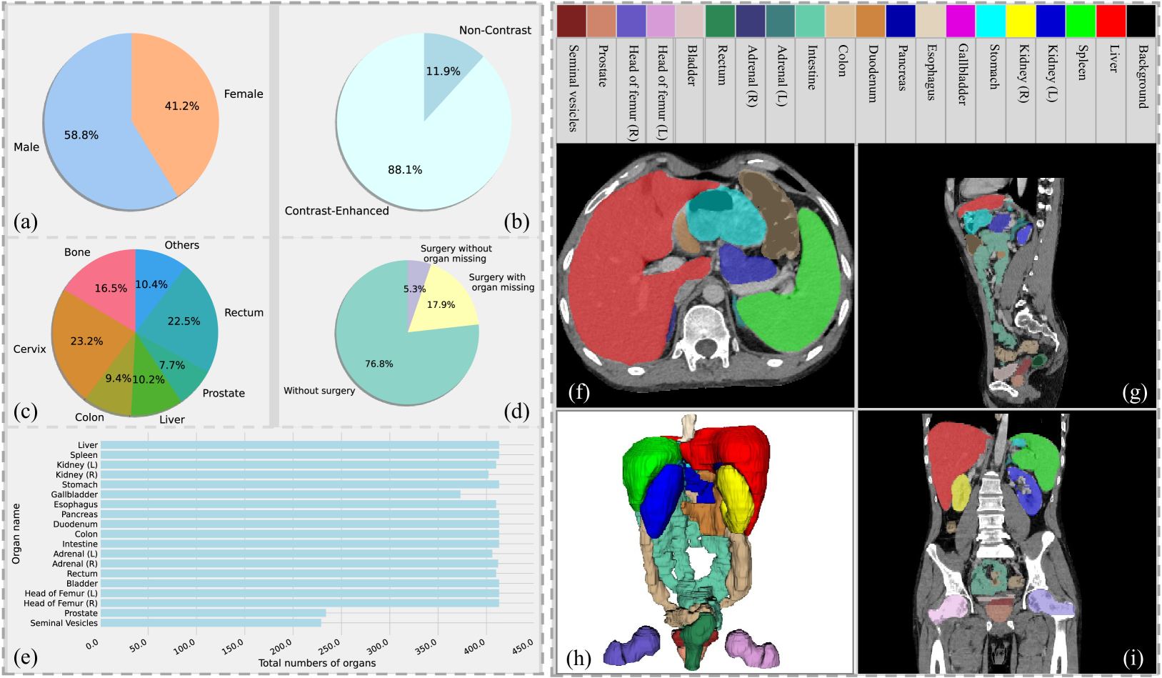
Organ and cancer segmentation in abdomen Computed Tomography (CT) scans is the prerequisite for precise cancer diagnosis and treatment. Most existing benchmarks and algorithms are tailored to specific cancer types, limiting their ability to provide comprehensive cancer analysis. This work presents the first international competition on abdominal organ and pan-cancer segmentation by providing a large-scale and diverse dataset, including 4650 CT scans with various cancer types from over 40 medical centers. The winning team established a new state-of-the-art with a deep learning-based cascaded framework, achieving average Dice Similarity Coefficient scores of 92.3% for organs and 64.9% for lesions on the hidden multi-national testing set. The dataset and code of top teams are publicly available, offering a benchmark platform to drive further innovations https://codalab.lisn.upsaclay.fr/competitions/12239.
Read more8/23/2024
0
Rethinking Abdominal Organ Segmentation (RAOS) in the clinical scenario: A robustness evaluation benchmark with challenging cases
Xiangde Luo, Zihan Li, Shaoting Zhang, Wenjun Liao, Guotai Wang
Deep learning has enabled great strides in abdominal multi-organ segmentation, even surpassing junior oncologists on common cases or organs. However, robustness on corner cases and complex organs remains a challenging open problem for clinical adoption. To investigate model robustness, we collected and annotated the RAOS dataset comprising 413 CT scans ($sim$80k 2D images, $sim$8k 3D organ annotations) from 413 patients each with 17 (female) or 19 (male) labelled organs, manually delineated by oncologists. We grouped scans based on clinical information into 1) diagnosis/radiotherapy (317 volumes), 2) partial excision without the whole organ missing (22 volumes), and 3) excision with the whole organ missing (74 volumes). RAOS provides a potential benchmark for evaluating model robustness including organ hallucination. It also includes some organs that can be very hard to access on public datasets like the rectum, colon, intestine, prostate and seminal vesicles. We benchmarked several state-of-the-art methods in these three clinical groups to evaluate performance and robustness. We also assessed cross-generalization between RAOS and three public datasets. This dataset and comprehensive analysis establish a potential baseline for future robustness research: url{https://github.com/Luoxd1996/RAOS}.
Read more6/21/2024

0
Universal and Extensible Language-Vision Models for Organ Segmentation and Tumor Detection from Abdominal Computed Tomography
Jie Liu, Yixiao Zhang, Kang Wang, Mehmet Can Yavuz, Xiaoxi Chen, Yixuan Yuan, Haoliang Li, Yang Yang, Alan Yuille, Yucheng Tang, Zongwei Zhou
The advancement of artificial intelligence (AI) for organ segmentation and tumor detection is propelled by the growing availability of computed tomography (CT) datasets with detailed, per-voxel annotations. However, these AI models often struggle with flexibility for partially annotated datasets and extensibility for new classes due to limitations in the one-hot encoding, architectural design, and learning scheme. To overcome these limitations, we propose a universal, extensible framework enabling a single model, termed Universal Model, to deal with multiple public datasets and adapt to new classes (e.g., organs/tumors). Firstly, we introduce a novel language-driven parameter generator that leverages language embeddings from large language models, enriching semantic encoding compared with one-hot encoding. Secondly, the conventional output layers are replaced with lightweight, class-specific heads, allowing Universal Model to simultaneously segment 25 organs and six types of tumors and ease the addition of new classes. We train our Universal Model on 3,410 CT volumes assembled from 14 publicly available datasets and then test it on 6,173 CT volumes from four external datasets. Universal Model achieves first place on six CT tasks in the Medical Segmentation Decathlon (MSD) public leaderboard and leading performance on the Beyond The Cranial Vault (BTCV) dataset. In summary, Universal Model exhibits remarkable computational efficiency (6x faster than other dataset-specific models), demonstrates strong generalization across different hospitals, transfers well to numerous downstream tasks, and more importantly, facilitates the extensibility to new classes while alleviating the catastrophic forgetting of previously learned classes. Codes, models, and datasets are available at https://github.com/ljwztc/CLIP-Driven-Universal-Model
Read more5/29/2024

0
AbdomenAtlas: A Large-Scale, Detailed-Annotated, & Multi-Center Dataset for Efficient Transfer Learning and Open Algorithmic Benchmarking
Wenxuan Li, Chongyu Qu, Xiaoxi Chen, Pedro R. A. S. Bassi, Yijia Shi, Yuxiang Lai, Qian Yu, Huimin Xue, Yixiong Chen, Xiaorui Lin, Yutong Tang, Yining Cao, Haoqi Han, Zheyuan Zhang, Jiawei Liu, Tiezheng Zhang, Yujiu Ma, Jincheng Wang, Guang Zhang, Alan Yuille, Zongwei Zhou
We introduce the largest abdominal CT dataset (termed AbdomenAtlas) of 20,460 three-dimensional CT volumes sourced from 112 hospitals across diverse populations, geographies, and facilities. AbdomenAtlas provides 673K high-quality masks of anatomical structures in the abdominal region annotated by a team of 10 radiologists with the help of AI algorithms. We start by having expert radiologists manually annotate 22 anatomical structures in 5,246 CT volumes. Following this, a semi-automatic annotation procedure is performed for the remaining CT volumes, where radiologists revise the annotations predicted by AI, and in turn, AI improves its predictions by learning from revised annotations. Such a large-scale, detailed-annotated, and multi-center dataset is needed for two reasons. Firstly, AbdomenAtlas provides important resources for AI development at scale, branded as large pre-trained models, which can alleviate the annotation workload of expert radiologists to transfer to broader clinical applications. Secondly, AbdomenAtlas establishes a large-scale benchmark for evaluating AI algorithms -- the more data we use to test the algorithms, the better we can guarantee reliable performance in complex clinical scenarios. An ISBI & MICCAI challenge named BodyMaps: Towards 3D Atlas of Human Body was launched using a subset of our AbdomenAtlas, aiming to stimulate AI innovation and to benchmark segmentation accuracy, inference efficiency, and domain generalizability. We hope our AbdomenAtlas can set the stage for larger-scale clinical trials and offer exceptional opportunities to practitioners in the medical imaging community. Codes, models, and datasets are available at https://www.zongweiz.com/dataset
Read more7/24/2024